Chapter 51
Surgical Anatomy of Vitreous and the Vitreoretinal Interface
J. SEBAG
Main Menu   Table Of Contents

Search

HISTORICAL PERSPECTIVE
VITREOUS EXAMINATION TECHNIQUES
MOLECULAR STRUCTURE
CORPUS VITREOUS
VITREOUS BASE
VITREOUS CORTEX
ZONULES
SURGICAL ANATOMY OF ANOMALOUS POSTERIOR VITREOUS DETACHMENT
REFERENCES

HISTORICAL PERSPECTIVE
Duke-Elder1 claimed that the first descriptions of vitreous structure proposed that vitreous was composed of “loose and delicate filaments surrounded by fluid.” During the 18th and 19th centuries there prevailed no fewer than four major theories of vitreous structure: the “alveolar theory,”2 the “lamellar theory,”3 the “radial sector theory,”4 and the “fibrillar theory.”5,6 The elegant studies of Szent-Gyorgi7 supported the “fibrillar” descriptions of Retzius and introduced the concept that vitreous structure changes with age. The work of Baurmann,8 Stroemberg,9 and Redslob10 showed that many of the early studies were flawed by artifacts resulting from the use of tissue fixatives. Thus, it was anticipated that the use of slit lamp biomicroscopy to study vitreous structure would eliminate this problem since investigations could be performed without introducing artifacts. Yet, the use of in vivo slit lamp biomicroscopy spawned an equally varied set of descriptions: Gullstrand11 saw membranes, Koeppe12 described vertical and horizontal fibers, and Baurmann13 saw a grill-like pattern of darker and lighter bands resembling several layers of chain-linked fences. Even the postmortem use of darkfield microscopy resulted in various interpretations ranging from Goedbloed's14 description of fibrillar structures to Friedenwald and Stiehler's15 description of concentric sheets and Eisner's16 observation of “membranelles.” In Eisner's meticulous studies, dissections of human vitreous were observed to contain membranous structures that coursed from the region about the lens in a circumferential pattern, parallel with the vitreous cortex, to insert at the posterior pole. Eisner has described these “membranelles” as funnels that are packed into one another and diverge outward and anteriorly from the prepapillary vitreous. He named these membranelles “tractae” according to their location. Worst17 has also studied preparations of dissected human vitreous and has described that the “tracts” of Eisner constitute the walls of “cisterns” within the vitreous. In Worst's studies these cisterns are visualized by filling with white India ink. He has studied the premacular vitreous in great detail and has proposed the existence of a “bursa premacularis,” which he described as a pear-shaped space that is connected to the cisternal system in front of the ciliary body. In a more recent study a different aqueous dye (fluorescein) was used to demonstrate this same premacular region of liquefied vitreous.18
Back to Top
VITREOUS EXAMINATION TECHNIQUES
Beginning with the invention of the ophthalmoscope by Helmholz, examiners of the eye have long benefited from advancing technology and the development of ever more powerful examination tools. Indeed, today one can evaluate not only morphologic but also functional and physiologic aspects of the eye with the aid of lasers.20 However, of all the different parts of the eye, evaluation of vitreous has advanced least in terms of the development of accurate, reproducible clinical examination techniques. This is evident in the multitude of descriptions of vitreous structure that resulted after the introduction of slit lamp biomicroscopy and the various different interpretations that exist to this day.

This controversy stems from the fact that vitreous examination essentially consists of visualizing a structure intended to be virtually invisible. The Tyndall phenomenon forms the basis of vitreous examination. This can be defined as the scattering of incident light by opaque structures or particles in an otherwise transparent medium. Essential to the success of achieving an adequate Tyndall effect clinically is maximizing pupil dilation in the patient and dark adaptation in the examiner.21 Since the Tyndall effect increases with an increasingly large subtended angle between the axis of illumination and the line of observation (up to a maximum of 90°), a widely dilated pupil enhances this effect. Some observers22 propose that green light further enhances the Tyndall effect. This concept has been incorporated in a biomicroscope that utilizes a green laser to enhance visualization of the vitreoretinal interface.23

Before examining the individual parts of vitreous, it is useful to consider the overall shape of the entire vitreous as a clue to characterizing vitreoretinal pathology. Charles24 has described a “problem-oriented” approach to preoperative vitreous examination in which an appreciation of the configuration and mobility of the entire vitreous can guide the operative approach. For example, the identification of a “cone” in a partial posterior vitreous detachment (PVD) and the characterization of the locations and number of apices in this cone can help in the accurate diagnosis of vitreoretinal pathology and determine the correct surgical approach.

ANTERIOR VITREOUS

It is probably easiest to examine the anterior vitreous first, since this can be done at the slit lamp immediately after examination of the anterior segment and does not require gel solutions or a contact or preset Hruby type lens, which are used in examination of the posterior vitreous. In the absence of a crystalline or implanted intraocular lens, vitreous prolapse into the anterior chamber could be an important finding in terms of vitreocorneal touch and the risks of corneal endothelial cell dysfunction.25 Vitreous adhesions to a cataract wound or to the iris may be important in the pathogenesis of postoperative cystoid macular edema. Particulate opacities in the anterior vitreous can be seen at the slit lamp and can give important clues as to the possible presence of posterior pathology. For example, particulate opacities in vitreous can be the earliest pathologic change in retinitis pigmentosa, and as such would provide an important aid to diagnosis.21 Nonpigmented cells can be indicative of inflammation at the vitreous base or infections in the retina. Pigmented cells should raise the examiner's index of suspicion for peripheral retinal tears and/or detachment. Lacqua and Machemer,26 as well as Scott,27 have described that an increase in the number and size of pigmented cells in vitreous of patients with retinal detachment (preoperatively or postoperatively) heralds the development of proliferative vitreoretinopathy (PVR). Bleeding can be associated with red blood cells in the anterior vitreous. Various neoplastic diseases, such as endophytic retinoblastoma, choroidal melanoma, and reticulum cell sarcoma, can result in anterior vitreous cells.

Anterior vitreous structures such as Mittendorf's dot, a remnant of embryonic hyaloid vessel regression, can be seen at the slit lamp and should alert the examiner to the possibility of other developmental disorders, such as persistent hyperplastic primary vitreous in the fellow eye.28

CENTRAL AND POSTERIOR VITREOUS

Examination of the central and posterior vitreous can rarely be achieved without the use of either a preset or a contact lens. Preset lens biomicroscopy can be performed either with a plano concave lens (e.g., -55- to -58.6-D lenses of Hruby) or various convex lenses (e.g., +32 D, +58.6 D, or +60 D). The plano concave lenses produce a highly magnified, narrow-field, erect image with visualization of the posterior pole, although it is difficult to achieve an adequate illumination-observation angle. Peripheral examination can only be performed by varying the position of the fixation point of the eye, and the quality of the image is reduced by optical distortions.

El-Bayadi29 first proposed the use of a +55-D preset lens and advised maintaining at least a 10° illumination-observation angle. The resultant image is inverted and can be photographed.30 The advantage offered by this form of posterior vitreous biomicroscopy is that the avoidance of a contact lens facilitates the “ascension/descension” gaze technique whereby eye movements are used to displace the corpus vitreous.31 This can be helpful in visualizing structures such as an operculum or the prepapillary ring in the posterior vitreous cortex that may have descended inferiorly in the presence of a PVD with vitreous syneresis (collapse). It is principally this feature that makes the preset approach superior to contact lens systems for examination of the posterior vitreous.32 Indeed it is now common practice to use a +60- or +90-D biconcave hand-held lens that at the slit lamp provides a stereoscopic, inverted image of high quality that also enables the performance of the ascension/descension examination technique.

PERIPHERAL VITREOUS

The major difficulty in examining the peripheral vitreous arises from a loss of stereopsis.31 This is because when examining the periphery the circular pupil becomes an elliptic aperture, making it difficult to obtain an adequate view with both of the observer's eyes. This is more of a problem in the horizontal than vertical meridians, since the examiner's two eyes are positioned horizontally. Schepens31 suggests reducing the illumination-observation angle, rotating the slit beam to the axis of the meridian being observed, positioning both arms of the biomicroscope opposite the meridian under observation, and reducing the interpupillary distance of the slit lamp eyepieces as ways of minimizing the loss of stereopsis.

A contact lens is often required for adequate visualization of the periphery. Traditionally, the Goldmann 3-mirror lens has been most popular as a tool for peripheral vitreous and retinal examination.33 Jaffe34 has described a plan for vitreous examination with a contact lens beginning with the central region, followed by establishing the presence or absence of a PVD and identifying the extent of detachment by following the posterior vitreous cortex peripherally. He recommends using a widened slit lamp beam and “rocking” the joystick to enhance visualization of the posterior cortex. Schepens31 described this maneuver as the “oscillation technique” in which the position of the slit lamp beam is moved side to side (for a vertical slit) or up and down (for a horizontal slit). The two positions of the beam are altered so as to alternate between direct and retro illumination of a structure or point of interest. Retro illumination is especially useful for identification of particulate or cellular opacities in the posterior vitreous. Schepens31 further describes the use of a tilted slit lamp column to enhance visualization of the peripheral vitreous. Eisner16 has devised a cone-shaped apparatus that fits onto a Goldmann 3-mirror lens and enables peripheral scleral indentation during slit lamp biomicroscopy with a contact lens. This can facilitate visualization of the peripheral retina, ora serrata, and posterior pars plana.

The recent development of inverted-image contact lenses with various-sized fields has greatly enhanced stereoscopic examination of the fundus and vitreous and are now routinely used for laser photocoagulation therapy of the fundus. The Mainster Retina Laser Lens is a +61-D convex aspheric contact lens that produces a real inverted image of about 45° degrees of the posterior fundus with excellent stereopsis.35 The “panfunduscopic” lens is a +85-D convex spheric lens that provides less magnification and image clarity but has a wider angle view and can be useful for peripheral examination to 60° or 70° with a 15° tilt.

OPACIFIED VITREOUS

When examination of vitreous is made difficult by opacification of the ocular media, there can nevertheless be worthwhile information garnered from careful study. As pointed out by Charles,24 much can be learned from studying the geometric configuration of a partly opaque or opacified vitreous. When opacification is advanced, however, real-time ultrasonography can be helpful in defining the consistency of the opacification, the three-dimensional configuration of the opaque vitreous, mobility, and the presence or absence of structural pathology behind the corpus vitreous. Fischer and colleagues36 demonstrated that there was excellent agreement between kinetic ultrasonography and conventional clinical examination techniques in subjects with clear ocular media. Arzabe and associates37 had similar findings in patients with proliferative diabetic retinopathy.

Back to Top
MOLECULAR STRUCTURE
Although vitreous is 98% water, this tissue is gel-like and has viscoelasticity. These properties result from collagen and hyaluronan, which are the major structural components of vitreous.

COLLAGEN

Before the turn of the 20th century it was already suspected that vitreous is, to some extent, collagenous. Morner38 found a protein residue that was retained by a filter after passage of fresh vitreous and termed it “residual protein.” Both he and Young39 considered the residue, also named “vitrosin,” to be collagenous since it dissolved when boiled and gelled on cooling. Much later, Pirie and co-workers40 documented the presence of collagen by chemical analysis. Using phase-contrast microscopy, Bembridge and colleagues41 demonstrated the existence of fibrils in vitreous of several species including humans. Matoltsky and associates42 and later Schwarz43 described the ultrastructure of vitreous as a random network of thin, uniform filaments and confirmed the collagenous nature of these filaments using electron microscopy and x-ray diffraction studies. Young and Williams44 determined that “vitrosin” contained 18% glycine, 8.4% proline, 15.4% hydroxyproline, and small amounts of cysteine. Swann,45 among others, later ascertained the more specific chemical characteristics of vitreous collagen, work that must be considered in progress, as the various subtypes of collagen and associated extracellular matrix components are still being defined.46

In humans, vitreous collagen is organized as thin fibrils 10 to 25 nm in diameter with cross-striations. Snowden and Swann47 identified a major period in the cross-striations of 62 nm (unfixed, dried bovine vitreous), while others48–50 describe a banding pattern with a periodicity of 12 to 25 nm. Vitreous collagen fibrils are unbranched51,52 and in the normal state are not cross-linked.53 This is supported by studies of the mechanical properties of vitreous54 demonstrating a softening of the spring constant with increased elongation, suggesting that vitreous collagen is organized in a network where fibrils can slip alongside each other. This would, however, require some breakdown of the proteoglycans in the interfibrillar space.

There is heterogeneity in the arrangement of collagen distribution throughout the corpus vitreous. Chemical51,55 and light-scattering studies56 have shown that the highest density of collagen fibrils is present in the vitreous base, followed by the posterior vitreous cortex anterior to the retina and then the anterior vitreous cortex behind the posterior chamber and lens. The lowest density is found in the central vitreous and adjacent to the anterior vitreous cortex.

Vitreous collagen fibrils appear to be continuous and unbroken from the anterior peripheral vitreous to the posterior vitreous. If retinal Müller cells are primarily responsible for vitreous collagen synthesis, assembly of procollagen molecules into newly synthesized collagen can be achieved by adding to existing fibrils at their most posterior end, nearest the Müller cell. Alternatively, if fibroblasts are responsible for vitreous collagen synthesis and assembly into fibrils, the orientation of vitreous collagen fibrils could be the consequence of relatively large numbers of fibroblasts in the primary vitreous that migrate during secondary vitreous formation to the vitreous base and posterior vitreous. While migrating to their final destinations in the anterior and posterior vitreous, fibroblasts assemble collagen fibrils in their wake.57 Thus, the continuity of these fibrils and their course through the corpus vitreous could result from the path of fibroblast migration during embryogenesis.

HYALURONAN

Although hyaluronan (HA) is found throughout the body, it was from the bovine vitreous that Meyer and Palmer58 first isolated this macromolecule. Consequently, HA's more common name, “hyaluronic acid,” is derived from the fact that it was first discovered in the clear, colorless vitreous (hyalos, meaning “glass”) and that it contains uronic acid. Subsequent studies by Meyer,59,60 Comper and Laurent,61 and others characterized this macromolecule and its configuration within vitreous.

The volume of the unhydrated HA molecule is about 0.66 cc/g, whereas the hydrated specific volume is 2 to 3000 cc/g.62 Thus, the degree of hydration (and any pathologic condition that alters hydration) can have a significant influence on the size and configuration of the HA molecular network in vitreous. Because the solution domains are so large, the long unbranched HA chains form widely open coils that at concentrations greater than 1 mg/cc become highly entangled.61 The large domain of HA spreads the anionic charge of the molecule over a wide space. Because of its entanglement and relative immobilization in vitreous, HA acts much like an ion-exchange resin in that an electrostatic interaction occurs between the small charges of mobile ions in vitreous and the electrostatic envelope of this stationary polyelectrolyte. This electrostatic interaction forms the basis for various properties of HA, including its influence on osmotic pressure, ion transport and distribution, and electric potentials within vitreous.61

Based on x-ray diffractograms, Atkins and Sheehan63 described HA as a linear helix. Further studies64 demonstrated a left-handed, threefold helix with a rise per disaccharide on the helix axis of 0.98 nm. As described in a detailed review by Chakrabarti and Park,65 this periodicity can vary depending on whether the helix is in a “compressed” or “extended” configuration. The existence of the molecule in either of these two states can greatly influence an HA molecule's interactions with neighboring molecules. A compressed HA chain has extensive “interdigitations” since it interacts with nearest antiparallel as well as parallel neighbors (totaling eight molecules), whereas extended forms only interact with three antiparallel neighbors. It is known that changes in the microenvironment and in the types of surrounding counter-ions can cause changes in the conformation of the HA polyanion.61 For example, a decrease in ionic strength can cause the anionic charges on the polysaccharide backbone to repel one another and result in an extended configuration of the macromolecule. Thus, changes in the ionic milieu surrounding vitreous HA may be converted to mechanical energy on extension or contraction of the HA macromolecule and, in turn, swelling or shrinkage of vitreous. This can be important in certain pathologic conditions, such as diabetes. Early studies by Christiansson66 showed that alloxan-induced experimental diabetes in rabbits resulted in increased glucosamine content and viscosity of vitreous and a decrease in vitreous volume. In another study67 a slight increase was shown in the tonicity of diabetic human vitreous (324 ± 23 mOsm vs 316 ± 21 mOsm in controls). In diabetes there can be significant fluctuations in the systemic concentrations of a variety of molecules that can alter the ionic milieu of vitreous. Such shifts could induce structural and volumetric changes on a molecular level. Furthermore, shifts in systemic metabolism and in turn osmolarity and hydration of vitreous could result in periodic swelling and contraction of the entire vitreous with resultant traction on structures attached to the vitreous cortex such as new blood vessels. These events could influence the course of diabetic retinopathy by contributing to the proliferation of neovascular fronds68 and perhaps even by inducing rupture of the new vessels, causing vitreous hemorrhage. Indeed, Tasman69 has found that in 53 cases of vitreous hemorrhage due to proliferative diabetic retinopathy, 62.3% of bleeding episodes occurred between midnight and 6 [a 12]am[rt] while the remaining parts of the day had only an 11% to 13% incidence. Although Tasman speculated that this could be due to nocturnal hypoglycemia, other metabolic or hormonal fluctuations could influence vitreous in the ways described previously and lead to vitreous hemorrhage.

HA molecules have a different concentration gradient in the corpus vitreous than collagen fibrils. HA is most abundant in the posterior cortical gel with a gradient of decreasing concentration more centrally and anteriorly.51, 70–73 Balazs74 has hypothesized that this is due to the inability of HA synthesized by hyalocytes in the posterior vitreous cortex to traverse the internal limiting lamina of the retina. HA leaves vitreous to enter the posterior chamber by way of the annulus of anterior vitreous cortex about the lens that is not adjacent to a basal lamina. Bound water (which is nonfreezable) has a similar distribution within vitreous to that of HA,75 presumably due to binding by HA.

COLLAGEN-HYALURONAN INTERACTION

Schwarz76 observed that after staining with ruthenium red, aggregates of precipitated HA molecules can be seen on electron microscopy as 8-nm granules situated in the interfibrillar space. Streeten77 has claimed that large polymeric aggregates of HA “coat” the collagen fibrils supporting this postulate. Indeed, using ruthenium red, Asakura78 found HA aggregates on collagen fibrils. These appearances, however, could be the result of artifact related to ruthenium red staining.

Balazs74 described that HA molecules fill the spaces between the collagen fibrils and provide a “stabilizing effect” on the collagen network. In this regard there are two important functions provided by this molecular arrangement. First, the large domains of the HA molecules spread apart the collagen fibrils and minimize light-scattering by these structures, thereby contributing to the transparency of vitreous. Second, the viscoelastic properties and mechanical functions79 of vitreous are the result of the presence of both HA and collagen and are very likely related to their association on a molecular level. Adjacent collagen fibrils would tend to cross link and alter these properties. Consequently, the presence of HA molecules to spread apart the collagen fibrils not only achieves transparency but also “stabilizes” the viscoelasticity of vitreous.

It is likely that other proteoglycans also interact with vitreous collagen. Either chondroitin sulfate, as suggested by Asakura,78 or keratan sulfate, or both, as has been described in the cornea by Scott,80 bind to collagen fibrils by means of the protein cores of these proteoglycans. Organized in a manner that keeps vitreous collagen fibrils at a critical distance apart,81 this “network” of different extracellular matrix molecules allows light to pass with little or no scattering, yet has a gel-like structure and viscoelasticity. Alterations in the interaction between these structural components with, for example, aging82 or diabetes,83 could influence these important physical properties of vitreous. Indeed, a breakdown in the association of these macromolecules is very likely at the root of the phenomenon of vitreous liquefaction in both aging and disease. An alternative hypothesis84 states that vitreous liquefaction and syneresis are related only to decreased HA concentrations.

The topographic heterogeneity in the distribution and density of both collagen and proteoglycans (especially HA) molecules creates a heterogeneous molecular network. This inhomogeneity renders an optical anisotropy to vitreous that has been measured using laser light-scattering techniques.56 The phenomenon of quasi-elastic light-scattering in human vitreous has also been measured in vitro (Sebag and Libondi, unpublished data) and may provide a noninvasive means to measure the viscoelastic properties of vitreous.

Back to Top
CORPUS VITREOUS
The corpus vitreous (vitreous body) is the fully developed form of the “secondary vitreous.” In an emmetropic adult human eye it is approximately 16.5 mm in axial length with a depression anteriorly just behind the lens (patellar fossa). Various structures and regions within the corpus vitreous are named after the anatomists and histologists who first described them (Fig. 1). The hyaloideocapsular ligament (of Weiger) is the annular region 1 to 2 mm in width and 8 to 9 mm in diameter where the corpus vitreous is attached to the posterior aspect of the lens. “Erggelet's” or “Berger's” space is at the center of the hyaloideocapsular ligament. Arising from this space and coursing posteriorly through the central vitreous is the canal of Cloquet (see Figs. 1, 4H, AND 8B), which is the former site of the hyaloid artery in the primary vitreous. The former lumen of the artery is an area devoid of vitreous collagen fibrils, surrounded by multifenestrated sheaths that were previously the basal laminae of the hyaloid artery wall.85,86 Posteriorly, Cloquet's canal opens into a funnel-shaped region anterior to the optic disc known as the area Martegiani.

Fig. 1. Schematic diagram of corpus vitreous. (Schepens CL, Neetens AE [eds]: The Vitreous and Vitreoretinal Interface, p 20. New York, Springer-Verlag, 1987)

Fig. 4. Human vitreous structure is visualized by darkfield slit microscopy. All photographs are oriented with the anterior segment below and the posterior pole above. Photographs are sequential, beginning in the upper left hand corner and moving left to right. A. Posterior vitreous in the left eye of a 52-year-old man. The corpus vitreous is enclosed by the vitreous cortex. There is a hole in the prepapillary (small, to the left) vitreous cortex. B. Posterior vitreous in a 57-year-old man. A large bundle of prominent fibers is seen coursing anteroposteriorly and entering the retrohyaloid space through the premacular vitreous cortex. C. Same view as B at higher magnification. D. Posterior vitreous in the right eye of a 53-year-old woman. There is posterior extrusion of vitreous out the prepapillary hole (to the right) and premacular (large extrusion to the left) vitreous cortex. Fibers course anteroposteriorly in the central vitreous and out into the retrocortical (formerly preretinal, before dissection) space. E. Horizontal optical section of the same specimen as D at a different level. A large fiber courses posteriorly from the central vitreous and inserts into the premacular vitreous cortex. F. Same view as E at higher magnification. The large fiber has a curvilinear appearance because of traction by vitreous extruding into the retrocortical space. However, because of its attachment to the posterior vitreous cortex the fiber arcs back to its point of insertion. G. Anterior and central vitreous in a 33-year-old woman. Cloquet's canal is seen forming the retrolental space of Berger. H. Anterior and peripheral vitreous in a 57-year-old man. The specimen is tilted forward to enable visualization of the posterior aspect of the lens and the peripheral anterior vitreous. Behind and to the right of the lens there are fibers coursing anteroposteriorly that insert into the vitreous base. These fibers “splay out” to insert anterior and posterior to the ora serrata. (A, E, and F: Sebag J, Balazs EA: Pathogenesis of cystoid macular edema: An anatomic consideration of vitreoretinal adhesions. Surv Ophthalmol 28[suppl]:493, 1984; B and C: Sebag J, Balazs EA: Morphology and ultrastructure of human vitreous fibers. Invest Ophthalmol Vis Sci 30:187, 1989)

Investigations of vitreous structure have long been hampered by transparency within the corpus vitreous and the absence of easily recognized landmarks. Without landmarks, removal of the corpus vitreous from the eye results in a loss of orientation. The transparency of vitreous renders observation in conventional diffuse light unrewarding. Attempts to study vitreous structure with opaque dyes do visualize the areas filled by dye but obscure the appearance of adjacent structures. The use of the contrast-enhancing techniques of histology usually involves tissue fixation, which includes dehydration. Since vitreous is 98% water, dehydration induces profound alteration of internal morphology. Consequently, any investigation of vitreous structure must overcome these difficulties.

Dissection of the outer layers of the eye (sclera, choroid, and retina) can be performed and the “naked” vitreous body can be maintained intact and attached to the anterior segment of the eye (Fig. 2). This enables study of internal vitreous morphology without a loss of intraocular orientation. However, depending on the age of the subject and consequently the degree of vitreous liquefaction,87 the dissected vitreous will remain solid and intact (young individuals, see Fig. 2) or will be flaccid and collapse. The latter is most often the case in specimens from older adults, and consequently vitreous turgescence must be maintained so as to avoid distortion of intravitreal morphology. Immersion of a dissected vitreous specimen that is still attached to the anterior segment into a physiologic solution maintains vitreous turgescence and avoids structural distortion.

Fig. 2. Human vitreous dissection. A. Vitreous obtained at autopsy from a 9-month-old child. The sclera, choroid, and retina were dissected off the vitreous, which remains attached to the anterior segment. A band of gray tissue can be seen posterior to the ora serrata. This is neural retina that was firmly adherent to the vitreous base and could not be dissected. Because of the young age of the donor, the corpus vitreous is almost entirely gel. Thus, it is solid and maintains its shape, although situated on a surgical towel exposed to room air. (Sebag J: The Vitreous--Structure, Function and Pathobiology. New York, Springer-Verlag, 1989) B. Human vitreous with the sclera, choroid, and retina dissected away; the corpus vitreous is still attached to the anterior segment. The specimen is mounted on a Lucite frame using sutures through the limbus and then immersed in a Lucite chamber containing an isotonic, physiologic solution. This maintains vitreous turgescence and avoids collapse and artifactual distortion of vitreous structure. (Sebag J, Balazs EA: Pathogenesis of cystoid macular edema: An anatomic consideration of vitreoretinal adhesions. Survey Ophthalmol 28[suppl]:493, 1984)

The limitations induced by vitreous transparency were first overcome by Goedbloed14 and then by Friedenwald and Stiehler15 and by Eisner,16 who employed darkfield slit illumination of the corpus vitreous to achieve visualization of intravitreal morphology. Illumination with a slit lamp beam directed into the corpus vitreous from the side and visualization of the illuminated portion from above produces an optical horizontal section (Fig. 3). The thickness of this cross section can be varied, as can the vertical position (level) of the cross section. This enables visualization of any portion of the corpus vitreous that is of interest. The illumination-observation angle of 90° that is achieved using this technique maximizes the Tyndall effect and thus overcomes the limitations induced by vitreous transparency. Furthermore, the avoidance of any tissue fixation, contrast solutions, or dyes eliminates the introduction of many of the artifacts that flawed earlier investigations.

Fig. 3. Schematic diagram of darkfield illumination system for the study of vitreous structure. The dissected specimen is mounted and immersed in a Lucite chamber containing a physiologic solution (see Fig. 2B). A slit lamp beam is shown from the side through the intact vitreous. The beam enters and exits laterally, avoiding any scattering of light by the structures of the anterior segment. The position of the beam can be raised or lowered, and the thickness of the illuminated portion can be regulated. Observation is performed from above, perpendicular to the plane of illumination. This achieves a 90° illumination/observation angle and thus maximizes the Tyndall effect. (Sebag J: The Vitreous--Structure, Function and Pathobiology. New York, Springer-Verlag, 1989)

Researchers88–91have used these techniques to study human vitreous structure. Within the adult human vitreous there are fine, parallel fibers coursing in an anteroposterior direction, as shown in Figs. 4B AND C, 5, and 6. The fibers attach into the vitreous base (see Figs. 4H and 5) where they splay out anterior and posterior to the ora serrata. As the peripheral fibers course posteriorly they are circumferential with the vitreous cortex, while central fibers “undulate” in a configuration parallel with Cloquet's canal.6 The fibers are continuous and do not branch. Posteriorly, these fibers attach into the vitreous cortex (see Fig. 4E and F), but not the internal limiting lamina of the retina.

Fig. 5. Posterior and central vitreous of a 59-year-old man. Fibers course anteroposteriorly in the center of the corpus vitreous and enter the retrocortical space through the premacular region of the vitreous cortex (to the top at the center). Within the cortex are many small “dots” that scatter light intensely (white arrows). The larger, irregular dots are debris. The smaller dots are hyalocytes. (Sebag J: The Vitreous--Structure, Function and Pathobiology. New York, Springer-Verlag, 1989)

Fig. 6. Vitreous structure in a 58-year-old woman. Fibers course anteroposteriorly in the central and peripheral vitreous. Posteriorly, fibers orient to the premacular region (top). Anteriorly, the fibers “splay out” to insert into the vitreous base (bottom right). (Sebag J: The Vitreous--Structure, Function and Pathobiology. New York, Springer-Verlag, 1989)

Ultrastructural studies90 have demonstrated that collagen organized in bundles of packed, parallel fibrils (Fig. 7) is the only microscopic structure that could correspond to these fibers. It has been hypothesized that the visible vitreous fibers form when HA molecules no longer separate the microscopic collagen fibrils, resulting in the aggregation of collagen fibrils into bundles from which HA molecules are excluded.82,92 Eventually the aggregates of collagen fibrils attain sufficiently large proportions that can be visualized in vitro (see Figs. 4 THROUGH 6) and clinically. The areas adjacent to these large fibers have a low density of collagen fibrils in association with HA molecules and therefore do not scatter light as intensely as the larger bundles of aggregated collagen fibrils. Furthermore, these adjacent areas offer relatively little resistance to bulk flow through vitreous, since they are largely occupied by hydrated HA.

Fig. 7. Although centrifuged to concentrate structural elements, this human vitreous specimen contains no membranes or membranous structures. Only collagen fibrils were detected. There were also bundles of parallel collagen fibrils such as the one shown here in cross section (arrow). (Sebag J: The Vitreous--Structure, Function and Pathobiology. New York, Springer-Verlag, 1989)

AGE-RELATED CHANGES

There are changes that occur in vitreous structure throughout life.82,92 Figure 8 demonstrates typical vitreous structure during late prenatal stages. Within the corpus vitreous there are no structures other than the remnants of the hyaloid artery oriented toward the prepapillary region. The corpus vitreous is relatively small and has an overall dense appearance with marked density at the outermost “shell” corresponding to the vitreous cortex. The generalized density of the corpus vitreous likely relates to the fact that at this stage of development, collagen and proteoglycan(s) other than HA are the principal structural components.62,79 HA synthesis begins after birth, increasing transparency by the aforementioned mechanisms.

Fig. 8. Vitreous morphology in human embryo. The posterior aspect of the lens is seen below. The corpus vitreous is enclosed by the dense, highly light-scattering vitreous cortex. Within the corpus vitreous, Cloquet's canal arcs from the prepapillary vitreous cortex toward the lens. Because its course undulates through the central vitreous, not all of Cloquet's canal can be visualized in a single horizontal section. (Sebag J: Age-related changes in human vitreous structure. Graefes Arch Clin Exp Ophthalmol 225:89, 1987)

During childhood only the vitreous cortex scatters incident light and thus appears dense (Fig. 9). There are no visible fibers within the corpus vitreous until middle age (see Figs. 4 THROUGH 6). During old age these fibers become thickened and tortuous, associated with many pockets of liquid vitreous and a collapsed (syneretic) appearance (Fig. 10). These changes are the result of age-related biochemical alterations in the composition and organization of the molecular components that simultaneously result in vitreous liquefaction and fiber formation. Pockets of liquid vitreous have classically been called “lacunae.” In addition to having a low density of collagen during youth, the central vitreous is the first region to undergo liquefaction during middle age.16 A report by Kishi and Shimizu18 described the presence of a “posterior vitreous pocket” that the authors interpreted to represent an anatomic entity. However, over 95% of the eyes examined in that study were from persons aged 65 years or older. Thus, these findings represent the result of age-related vitreous liquefaction in the central precortical posterior vitreous.93 Such changes could also explain the preferential pooling of aqueous dyes such as India ink17 placed onto the anterior vitreous and allowed to collect anterior to the macula in what appears as a “bursa.”17,94,95 The use of fluorescein by Kishi and Shimizu18 represents but another way of demonstrating the phenomenon of vitreous liquefaction in this region.

Fig. 9. This view of the posterior and central vitreous from a 4-year-old child demonstrates a dense vitreous cortex (arrows) with hyalocytes (small retractile “spots” in cortex) and no intravitreal fibrous structures. The lens (L) is seen below.

Fig. 10. Human vitreous morphology in old age. The central vitreous has thickened, tortuous fibers. The peripheral vitreous has pockets that do not scatter light. These areas contain liquid vitreous and correspond to lacunae as seen clinically on biomicroscopy. (Sebag J, Balazs EA: Human vitreous fibres and vitreoretinal disease. Trans Ophthalmol Soc UK 104:123, 1984)

In a large autopsy study of formalin-fixed human eyes, O'Malley96 provided quantitative confirmation of these observations. He found that more than half of the corpus vitreous was liquefied in 25% of persons aged 40 to 49 and that this increased to 62% of individuals aged 80 to 89. Oksala97 used ultrasonography in vivo to detect echoes from gel-liquid interfaces in 444 normal human eyes and observed echoes in 5% of young persons, in more than half of those aged 51 to 60, and in more than 80% of persons older than 60. Vitreous liquefaction actually begins much earlier than the ages at which clinical examination or ultrasonography detect changes. Balazs and Flood87 found evidence of liquid vitreous after the age of 4 and observed that by the time the human eye reaches its adult size (ages 14 to 18) approximately 20% of the total vitreous volume consists of liquid vitreous. In these postmortem studies of fresh, unfixed human eyes it was observed that after the age of 40 there is a steady increase in liquid vitreous that occurs concomitantly with a decrease in gel volume. By the ages of 80 to 90 years more than half the corpus vitreous is liquid. The finding that the central vitreous is where fibers are first observed is consistent with the observation that the central vitreous is the first area to undergo liquefaction, supporting the concept that dissolution of the HA-collagen complex results in the simultaneous formation of liquid vitreous and aggregation of collagen fibrils into bundles of parallel fibrils. A large pocket of liquid vitreous could be clinically mistaken as a PVD. At surgery one could mistake entry into a large pocket of liquid vitreous as entry into the retrocortical space created after PVD. If there is a split in the posterior vitreous cortex (“vitreoschisis”), entry into the schisis cavity could likewise be misinterpreted as entry behind the posterior vitreous cortex. When the entire posterior vitreous cortex detaches from the retina there is an overall reduction in the size of the corpus vitreous owing to the collapse (syneresis) of the corpus vitreous that occurs when liquid vitreous enters the space behind the posterior vitreous cortex, anterior to the retina. This displacement of liquid vitreous occurs through the prepapillary “hole” and possibly the premacular or other portion of the posterior vitreous cortex and is an important event in the pathogenesis of PVD.

Back to Top
VITREOUS BASE
The vitreous base is a three-dimensional zone. It extends 1.5 to 2 mm anterior to the ora serrata, 1 to 3 mm posterior to the ora serrata,48 and several millimeters into the corpus vitreous itself.98 The vitreous base is virtually inseparable from the peripheral retina. Thus, dissection of retina off the vitreous cortex in autopsy eyes always results in a band of retina that remains attached to the peripheral vitreous at the vitreous base (see Fig. 2). Similarly, clean dissection of the vitreous base and peripheral vitreous cortex from the retina during vitrectomy surgery is extremely difficult to achieve. The ultrastructural correlate of this surgical observation may relate to vitreous fiber insertions at the vitreous base where they “splay out” (see Fig. 6) to insert anterior and posterior to the ora serrata (see Fig. 4H). The anteriormost fibers form the “anterior loop” of the vitreous base (Fig. 11), a structure that is important in the pathogenesis and surgery of anterior PVR. In the posterior portion of the vitreous base, vitreous fibers are closer together than elsewhere. Gartner99 has found that in humans the diameters of collagen fibrils in the vitreous base range from 10.8 to 12.4 nm, with a major period of cross striations of 50 to 54 nm. Hogan48 demonstrated that just posterior to the ora serrata, heavy bundles of vitreous fibrils attach to the basal laminae of retinal glial cells. Studies by Gloor and Daicker100 showed that cords of vitreous collagen insert into gaps between the neuroglia of the peripheral retina. They likened this structure to Velcro and proposed that this would explain the strong vitreoretinal adhesion at this site. In the anterior vitreous base, fibrils interdigitate with a reticular complex of fibrillar basement membrane material between the crevices of the nonpigmented ciliary epithelium.101 The vitreous base also contains intact cells that are fibroblast-like anterior to the ora serrata and macrophage-like posteriorly.101 Damaged cells in different stages of involution and fragments of basal laminae, presumed to be remnants of the embryonic hyaloid vascular system (vasa hyaloidea propria) that filled the primary vitreous, are also present in the vitreous base.101

Fig. 11. Morphology of anterior loop in human vitreous base. Central and peripheral vitreous structure in a 76-year-old man. The posterior aspect of the lens (L) is seen below. Fibers course anteroposteriorly in the central vitreous and insert at the vitreous base. The “anterior loop” configuration at the vitreous base is seen on the right side of the specimen (arrow). (Sebag J: The Vitreous--Structure, Function and Pathobiology. New York, Springer-Verlag, 1989)

Teng and Chi102 found that the vitreous base posterior to the ora serrata varies in width depending on the age of the individual. More than half of eyes from persons older than 70 had a posterior vitreous base wider than 1.0 mm. The width increased with increasing age to nearly 3.0 mm, bringing the posterior border of the vitreous base closer to the equator. This widening of the vitreous base is believed to be most prominent in the temporal portion of the globe.103 The posterior migration of the vitreous base may play a role in the pathogenesis of peripheral retinal breaks and rhegmatogenous retinal detachment since this is the area of strongest vitreoretinal adhesion. Gartner99 found no differences in the thickness of collagen fibrils in the vitreous base when comparing five eyes from humans aged 9 months and 29, 39, 61, and 71 years. He did, however, note that there was “lateral aggregation” of the collagen fibrils in the eyes from older persons. Such aggregation at the vitreous base is similar to aging changes within the corpus vitreous where collagen fibril aggregation results in bundles of parallel collagen fibrils. These aging changes at the vitreous base could also contribute to increased traction on the peripheral retina and play a role in the development of retinal tears and detachments.

SURGICAL ANATOMY OF RETINAL TEARS AND DETACHMENTS

Retinal Tears

The aforementioned age-related posterior migration of the posterior border of the vitreous base does not extend posteriorly in a continuous line, which is the configuration of the posterior border of the vitreous base in a young person, but in a curvilinear, undulating pattern. Traction on the retina along this line can create the exact “horseshoe” configuration often seen in peripheral retinal tears. It is known that in the vitreous base there are collagen fibrils oriented perpendicular to the wall of the eye101,104,105 with insertions anterior and posterior to the ora serrata.89,106 The continuity of these fibrils with those of the corpus vitreous is an important predisposing factor for retinal tears and detachments.

At the turn of the 20th century, Best107 emphasized that every movement of the eye results in movement of the corpus vitreous and that this causes traction at any point of strong vitreoretinal adhesion. Leber and associates108 were perhaps the first to find that rupture of the retina can occur at sites where there is adhesion between vitreous and retina. Unusually strong vitreoretinal adhesion is found at the posterior border of the vitreous base in cases of giant tears.31 Strong vitreoretinal adhesion is also present in anomalous or pathologic situations such as meridional folds, peripheral pigment clumps, retinal rosettes, granular patches, and progressive lattice-like degeneration.31,109,110

In lattice degeneration there are discrete oval areas of retinal thinning associated with localized vitreous liquefaction, separation of the overlying vitreous, and increased vitreoretinal adhesion at the margins of the degenerated areas.111 Retinal breaks are found at these margins and posterior to the areas of lattice degeneration. In this disorder, however, retinal tears are relatively infrequent as compared with atrophic retinal holes. Clinical studies112,113 found retinal tears in only 1% of eyes with lattice degeneration, while 16.3% to 18.2% of lattice lesions had atrophic retinal holes. The risk of retinal tears is greater when the area of lattice degeneration is located juxtabasal or extrabasal, relative to the vitreous base.114,115 The retinal tears are believed to result from aggregated vitreous fibrils inducing traction on the retina.116 Obliterative fibrosis of the blood vessels in areas of lattice degeneration is present in only 11.9% of lesions113 and is seen as a “lattice-wicker” of white lines, for which the condition is named. The presence of this vascular anomaly has led to the hypothesis that retinal circulatory abnormalities are the primary cause of this condition.117 According to this theory, vitreous changes are secondary and only important in the subsequent development of retinal tears as a local phenomenon. Overall vitreous liquefaction118 and PVD119 may not be important contributing factors in this local traction but may be important in the subsequent development of retinal detachment.120

Schepens111 first described the clinical appearance of “white-without-pressure” when a geographic area of whiteness is present in the peripheral retina. When this appearance is present only during scleral indentation it is termed “white with pressure.” Schepens attributed this appearance to peripheral vitreoretinal traction, and in a subsequent report121 it was considered that these findings predisposed to peripheral retinal tears. Daicker122 proposed that this appearance results from “collagenic” formations in the peripheral retina, while Gartner104 suggested that they were due to irregularities of the internal limiting lamina of the retina. Watzke123 performed clinicopathologic correlation of a case with this finding and described that the lesion was due to portions of the vitreous cortex that remained attached to the retina after PVD. This would therefore represent a variant of vitreoschisis (splitting of the vitreous cortex), in this instance at the peripheral fundus. Green124 described that the appearance of these lesions results from incident light (from the ophthalmoscope) that is tangential to more dense bundles of vitreous base collagen fibrils. All of these interpretations would seem to be consistent with the concept that these areas are at greater risk of developing retinal tears as a result of unusual vitreous structure at the vitreoretinal interface in these locations. However, Byer125 does not believe that this ophthalmoscopic appearance has any diagnostic or prognostic significance.

Since the vitreous base is the site of strongest vitreoretinal adhesion, it is here, usually at the posterior border, that vitreoretinal traction causes peripheral retinal tears. Green124 has reviewed all clinical and postmortem studies on the prevalence of peripheral retinal tears. Clinical findings ranged from a prevalence of 0.59% to 7.2%, while autopsy studies showed a prevalence from 3.3% to 8.8%. Other studies have focused on the relationship between PVD and peripheral retinal tears. Autopsy studies105 found that PVD is associated with retinal breaks in 14.3% of all cases. Clinical studies34,126–128 found retinal tears in 8% to 15% of eyes with acute PVD. In the presence of high myopia (greater than -6 D), PVD is associated with peripheral retinal breaks in 11.1%.129 This could be in part related to the significant increase of liquid vitreous observed in experimentally induced myopia.130 In patients with high myopia who underwent uncomplicated cataract extraction (presumably by intracapsular techniques), the prevalence of retinal breaks after PVD was as high as 16.2%.131 It is surprising that the prevalence of retinal tears in high myopia is not greater than reported, particularly in those cases that underwent cataract extraction. Further studies may be needed to confirm these observations. However, if this finding is correct, it may point out that changes within the corpus vitreous that induce peripheral retinal traction only result in retinal tears in the presence of pre-existing irregularities at the vitreoretinal interface. Thus, although there may be a higher incidence of liquefaction and PVD in myopia, the vitreoretinal interface may not be similarly abnormal; and, consequently, peripheral retinal traction does not result in a significantly higher incidence of retinal tears in these patients. However, once a retinal tear develops in a myopic eye it may be more likely to produce a retinal detachment because of the biochemical and rheologic abnormalities in the liquefied myopic vitreous.

Retinal Detachments

In a study of 100 patients with bilateral surgical aphakia (presumably intracapsular) and rhegmatogenous retinal detachment in one eye, Hovland132 found that 26% eventually developed peripheral tears and retinal detachments in the fellow eye. In this study the absence of PVD in the fellow eye at the time of retinal detachment in the first eye was the poorest prognostic sign for the fellow eye. Bradford and associates133 found that in those rhegmatogenous retinal detachments that developed within 6 months after cataract surgery, equatorial tears were significantly more common than in detachments that occurred 2 or more years after cataract extraction. Since this is not the typical profile of an “aphakic” retinal detachment, Bradford and associates hypothesized that these retinal tears occurred at the time of PVD and were due to the presence of anomalous vitreoretinal adhesions. Furthermore, since this appearance is no different from phakic rhegmatogenous retinal detachments, cataract surgery was probably not an important factor in these cases, although it may have precipitated the PVD as a result of biochemical changes within the corpus vitreous. These authors concluded that the small anterior tears that cause retinal detachments long after cataract extraction result from chronic vitreoretinal traction at the vitreous base, rather than acute PVD.

The relationship of a retinal tear to the vitreous base is an important prognostic feature that determines the risk of a peripheral retinal tear resulting in a retinal detachment.115 Juxtabasal tears are the most dangerous due to the degree of peripheral vitreous traction associated with these retinal tears. Thus, there are certain types of peripheral retinal tears and clinical circumstances that are predisposed to develop retinal detachments. For example, eyes with retinal breaks that occur at the time of symptomatic PVD are at high risk and thus should undergo prophylactic treatment.134 Some authors135 consider that prophylactic treatment of retinal tears with continuing vitreous traction significantly reduces the risk of retinal detachment. How to best evaluate the presence or absence of vitreous traction and how to quantitate the degree of vitreous traction is presently not known.

Lindner136 was among the first to point out the importance of eye movements in the pathogenesis of retinal tears and detachment, while Rosengren and Osterlin137 provided experimental evidence in support of this concept. Vitreous synchysis (liquefaction) and syneresis (collapse) are important components in the development of PVD. Using ultrasonography, Oksala138 has found that advanced vitreous syneresis was present in 28 of 32 (87.5%) eyes with rhegmatogenous retinal detachment. Syneresis was also present in 27 of 32 (84.4%) fellow eyes. After PVD, the space behind the posterior vitreous cortex is occupied by liquid vitreous. During eye movements this liquid vitreous, which is lighter than gel, is set in motion before gel vitreous. Because all eye movements are rotational, the liquid acts as a wedge in the retrocortical space, further separating detached vitreous from retina. Where the vitreous is firmly attached to the peripheral retina (at the vitreous base as well as at pathologic sites) traction will be exerted by the force of the liquid vitreous moving in the retrocortical space, pressing against the vitreous cortex. Furthermore, when an ocular saccade stops it is usually sudden. The heavier gel vitreous continues to move due to inertia. It is believed that substantial traction is thus placed on sites of firm vitreoretinal adhesion, resulting in peripheral tears of the retina.31 Relieving this traction surgically is believed to be the therapeutic mechanism of action of scleral buckle indentation and/or vitrectomy.

Detachment of the entire vitreous base can occur after blunt trauma, resulting in detachment of both the ciliary epithelium anterior to the ora serrata and the retina posterior to the ora serrata.139 So-called spontaneous detachment of the vitreous base can occur in high myopia, Marfan's syndrome, and Ehlers-Danlos syndrome.140

SURGICAL ANATOMY OF PROLIFERATIVE VITREORETINOPATHY

PVR is most often encountered after rhematogenous retinal detachment as the major cause of failed retinal detachment surgery. The finding of type II collagen in almost one third of cases141 results from the fact that PVR membranes can be intimately associated with the vitreous cortex. This is especially true in so-called anterior PVR. The anterior loop of vitreous collagen fibers in the vitreous base (see Fig. 11) is an important structure in the pathogenesis of this condition, for it is upon the “scaffold” of these collagen fiber bundles that fibronectin and other extracellular matrix components are deposited, facilitating cell migration and proliferation. During the development of anterior PVR, contraction by myofibroblast-like cells is transmitted through these fibers that straddle the ora serrata to the anterior retina. In this manner the anterior retina “rolls” forward--a feature characteristic of anterior PVR. Because of fibril insertion anterior to the ora serrata, severe traction can also pull on the pars plana and detach the ciliary body. The continuity of these vitreous base fibers and the membranes that form upon their scaffold must be cut during anterior PVR surgery. In the early stages this can consist of cutting the tissue in a line circumferential with the ora serrata to relieve the anterior loop traction across the ora serrata. When more advanced, membranes can form a ring all about the peripheral vitreous, essentially encompassing the entire vitreous base. Radial cuts into this tissue can sometimes relieve the “napkin ring” effect of contraction and inward displacement of the retina and ciliary body adjacent to the vitreous base. When membrane formation is very advanced, dissection may not be possible and en bloc excision may be necessary consisting of removal of the entire section of vitreous base with adjacent tissues, including posterior pars plana and anterior peripheral retina.

The cells of PVR membranes are fibroblast-like but have several progenitors, particularly astrocytes26,142,143 and retinal pigment epithelial (RPE) cells.26,144,145 The prominence of RPE cells in PVR probably relates to the access of RPE cells to the corpus vitreous afforded by the retinal break and the dispersion of viable RPE cells into vitreous during cryopexy treatment of retinal tears. Since one of the functions of vitreous79 is to inhibit cell invasion of the corpus vitreous, it is not clear how vitreous is altered to permit cell migration and proliferation in PVR. Campochiaro and associates146 studied the influence of human vitreous aspirates on the migration of RPE cells in vivo. Vitreous from cases of PVR had much greater stimulatory activity than vitreous from patients with epiretinal membranes and uncomplicated retinal detachment. There are probably extrinsic as well as intrinsic factors involved in this process. Extrinsic factors derive from migratory and proliferative stimuli not normally present in vitreous. Intrinsic factors relate to alterations within vitreous that increase inherent stimulatory activity and/or decrease inherent inhibitory activity. PVR is discussed in greater detail elsewhere in this volume, but it should be mentioned here that the role of the resident hyalocytes requires further investigation. These cells are the first to be exposed to the various noxious stimuli that are present in PVR. In addition to being the first cells to potentially respond to mitogenic factors, hyalocytes could themselves elaborate growth factors that contribute early in the cascade of events that result in PVR. These cells have the capacity to synthesize various growth factors,147 and in one study148 hyalocytes were the only cell type found to be immunoreactive for all four forms of transforming growth factor-ß.

Insofar as PVR may result from a decrease in the properties of vitreous that normally inhibit cell migration and proliferation, vitrectomy should be reserved for cases of established PVR. In the absence of appropriate indications, vitrectomy should probably not be performed as prophylaxis against PVR.

Back to Top
VITREOUS CORTEX
The vitreous cortex is defined as the peripheral “shell” of the corpus vitreous that courses forward and inward from the anterior vitreous base to form the anterior vitreous cortex and posteriorly from the posterior border of the vitreous base to form the posterior vitreous cortex.

The anterior vitreous cortex, at times called the “anterior hyaloid face” by clinicians, begins about 1.5 mm anterior to the ora serrata. Fine and Tousimis149 described that in this region the collagen fibrils are parallel to the surface of the cortex. Studies by Faulborn and Bowald150 described dense packing of collagen fibrils in the anterior cortex with looser collagen fibril packing in the subjacent vitreous, giving the impression of lamellae.

Rhodes151 studied mouse vitreous and found that the anterior vitreous cortex varied in thickness from 800 to 2000 nm. He also found that there are connections between the loose fibrils in the anterior vitreous and the anterior vitreous cortex. Rhodes also claimed that there are multiple interconnections between the anterior vitreous cortex and a branching fibrillar network in the posterior chamber.

The posterior vitreous cortex is 100-110 μm thick50,60 and, as shown in Figure 12, consists of densely packed collagen fibrils.50,60,152 There is no vitreous cortex over the optic disc (see Figs. 4A AND 13), AND the cortex is thin over the macula due to rarefaction of the collagen fibrils.50 The prepapillary hole in the vitreous cortex can sometimes be visualized clinically when the posterior vitreous is detached from the retina (Fig. 14). If peripapillary glial tissue is torn away during PVD and remains attached to the vitreous cortex about the prepapillary hole it is referred to as Vogt's or Weiss's ring. Vitreous can extrude through the prepapillary hole in the vitreous cortex (see Fig. 4A) but does so to a much lesser extent than through the premacular vitreous cortex (see Figs. 4B AND D and 13). Jaffe153 has described how vitreous can extrude into the retrocortical space created after PVD and has proposed that persistent attachment to the macula (Fig. 15) can produce traction and certain forms of maculopathy.154,155 Although there are no direct connections between the posterior vitreous and the retina, the posterior vitreous cortex is adherent to the internal limiting lamina of the retina, which is actually the basal lamina of retinal Müller cells. The exact nature of the adhesion between the posterior vitreous cortex and the internal limiting lamina is not known but probably results from the presence of various extracellular matrix molecules.91 This concept is supported by studies156,157 in which vitreous cortex separation from the retina was induced using agents that acted on extracellular matrix components that could bind the posterior vitreous cortex to the internal limiting lamina of the retina.

Fig. 12. Ultrastructure of posterior vitreous cortex in humans. Scanning electron microscopy demonstrates the dense packing of collagen fibrils in the vitreous cortex. To some extent this arrangement is exaggerated by the dehydration that occurs during specimen preparation for scanning electron microscopy (bar = 10 [a 5]mm). (Sebag J: The Vitreous--Structure, Function and Pathobiology. New York, Springer-Verlag, 1989)

Fig. 13. Posterior vitreous in the left eye of a 59-year-old man. The vitreous cortex (white arrows) envelopes the corpus vitreous and contains multiple, small, highly refractile points that scatter light intensely, which are cells known as hyalocytes. There is a “hole” (black arrows) in the prepapillary posterior vitreous cortex through which vitreous extrudes into the retrocortical space. Larger amounts of vitreous extrude through the premacular vitreous cortex, and fibers course from the central vitreous into the retrocortical space. (Sebag J, Balazs EA: Human vitreous fibres and vitreoretinal disease. Trans Ophthalmol Soc UK 104:123, 1985)

Fig. 14. The posterior vitreous is detached, and the prepapillary hole in the posterior vitreous cortex (arrows) can be seen anterior to the optic disc (slightly below and to the left of the optic disc) (Sebag J: The Vitreous--Structure, Function and Pathobiology. New York, Springer-Verlag, 1989)

Fig. 15. Anomalous posterior vitreous detachment. Vitreous can remain attached to the macula even in the presence of posterior vitreous detachment. In such cases, vitreous can extrude through the premacular vitreous cortex and fibers can insert into the macula. B, C, and D are an artist's rendition of this phenomenon. A demonstrates actual vitreous extrusion into the retrocortical space in a postmortem human specimen (see Fig. 4D) (B and C adapted from Jaffe NS: The Vitreous in Clinical Ophthalmology. St. Louis, CV Mosby, 1969; D adapted from Jaffe NS: Vitreous traction at the posterior pole of the fundus due to alterations in the posterior vitreous. Trans Am Acad Ophthalmol Otolaryngol 71:642, 1967)

VITREOUS CELLS

Hyalocytes

Reeser and Aaberg98 considered the vitreous cortex to be the “metabolic center” of vitreous because of the presence of hyalocytes (see Figs. 5, 13, AND 16). These cells were first described in 1845 by Hannover. Schwalbe158 placed these cells into the group of “Wanderzellen” (wandering cells, i.e., leukocytes or macrophages) on the basis of their morphology, distribution, and behavior. He later named them “Subhyaloidale zellen.”159 Balazs72 modified this term and named the cells “hyalocytes.”

Fig. 16. Phase contrast microscopy of in situ preparation of hyalocytes in the vitreous cortex from the eye of an 11-year-old girl obtained at autopsy. No stains or dyes were used in this preparation. (Sebag J: The Vitreous--Structure, Function and Pathobiology. New York, Springer-Verlag, 1989)

These mononuclear cells are embedded in the vitreous cortex (see Figs. 5, 13, AND 16), widely spread apart in a single layer situated 20 to 50 μm from the internal limiting lamina of the retina posteriorly and the basal lamina of the ciliary epithelium at the pars plana and vitreous base. Quantitative studies of cell density in the bovine160 and rabbit161 vitreous found the highest density of hyalocytes in the region of the vitreous base, followed next by the posterior pole, with the lowest density at the equator. As shown in Figure 17, hyalocytes are oval or spindle shaped, 10 to 15 μm in diameter, and contain a lobulated nucleus, a well-developed Golgi complex, smooth and rough endoplasmic reticula, and many large periodic acid-Schiff-positive lysosomal granules and phagosomes.50,162Hogan and colleagues163 described that the posterior hyalocytes are flattened and spindle shaped, whereas anterior hyalocytes are larger, rounder, and at times star shaped. Saga and associates164 have described that different ultrastructural features can be present in different individual cells of the hyalocyte population in an eye. Whether this relates to different origins for the different cells or different states of cell metabolism or activity is not clear.

Fig. 17. Ultrastructure of human hyalocytes. A mononuclear cell is seen embedded within the dense collagen fibril (black C) network of the vitreous cortex. There is a lobulated nucleus (N) with dense marginal chromatin (white C). In the cytoplasm there are mitochondria (M), dense granules (arrows), vacuoles (V), and microvilli (Mi) (×1670). (Courtesy of JL Craft and DM Albert, Harvard Medical School, Boston; Sebag J: The Vitreous--Structure, Function and Pathobiology. New York, Springer-Verlag, 1989)

Balazs71,165 pointed out that hyalocytes are located in the region of highest HA concentration and suggested that these cells are responsible for vitreous HA synthesis. In support of this hypothesis is the finding that the enzymes needed for HA synthesis are present within hyalocytes.166 Osterlin167 demonstrated that labeled intermediates destined to become incorporated into HA are taken up and internalized by hyalocytes. Several in vivo168,169 and in vitro169,170 studies have shown that hyalocytes synthesize large amounts of HA. Bleckmann171 found that in contrast to in vivo metabolism, HA synthesis by hyalocytes grown in vitro is reduced in favor of sulfated polysaccharide synthesis. He suggested that when these cultured cells are reimplanted into vitreous there must be a retransformation of hyalocyte glycosaminoglycan synthesis to the normal state since vitreous clarity is maintained in these experimental conditions.172

Swann45 claims that there is, as yet, no evidence that hyalocytes are responsible for the synthesis of vitreous HA. There is, however, evidence to suggest that hyalocytes maintain ongoing synthesis and metabolism of glycoproteins within vitreous. Rhodes and co-workers173 have used autoradiography to demonstrate the active incorporation of fucose into rabbit vitreous glycoproteins. In another study,174 sialyl and galactosyl transferase activity was demonstrated in calf vitreous hyalocytes, suggesting that these cells are responsible for vitreous glycoprotein synthesis. An alternate hypothesis, however, states that vitreous glycoproteins originate as secretory products of the inner layer of the ciliary epithelium.175,176

Hyalocyte capacity to synthesize collagen was first demonstrated by Newsome and colleagues.177 Studies by Ayad and Weiss178 showed the presence of CPS-1 and CPS-2 collagens adjacent to hyalocytes. These investigators concluded that in similar fashion to chondrocyte metabolism, hyalocytes synthesize these collagens. Hoffman and co-workers179 also proposed that the distribution of high molecular weight substances in vitreous, including enzymes, suggests synthesis by hyalocytes.

The phagocytic capacity of hyalocytes has been described in vivo180 and demonstrated in vitro.160,172 This activity is consistent with the presence of pinocytic vesicles and phagosomes162,181 and the presence of surface receptors that bind IgG and complement.182 Interestingly, HA may have a regulatory effect on hyalocyte phagocytic activity.183,184 Balazs74 has proposed that in their resting state, hyalocytes synthesize matrix glycosaminoglycans and glycoproteins and that the cells internalize and reuse these macromolecules by way of pinocytosis. They become phagocytic cells in response to inducting stimuli and inflammation. This type of transformation may underlie the observations of Saga and associates,164 who identified different appearances in the various cells of a hyalocyte population in a given eye. It is also important to consider that hyalocytes as well as resident fibroblasts are the first cells to be exposed to any migratory or mitogenic stimuli. Thus, the response of these cells must be considered in defining the pathophysiology of all proliferative disorders at the vitreoretinal interface, especially PVR and premacular (“epiretinal”) membrane formation.

Fibroblasts

There is a second population of cells in the vitreous cortex that in some cases may be mistaken for hyalocytes. Several investigations99,149,185 have determined that fibroblasts are present in the vitreous cortex. These cells constitute less than 10% of the total vitreous cell population and are localized within the vitreous base, adjacent to the ciliary processes and the optic disc. It may be that these cells are involved in vitreous collagen synthesis, especially in pathologic situations. The argument for a role in normal vitreous collagen synthesis is mostly by analogy to studies of fibrillogenesis in tendon where investigators57 have found that secreted collagen molecules are assembled into fibrils within invaginations of secreting fibroblasts. The locations of fibroblasts in the anterior peripheral vitreous (vitreous base and near the cilary processes) and posterior vitreous may explain how vitreous fibers become continuous structures spanning the distance between these locations.

Balazs and co-workers185 have found that near the pars plana, vitreous fibroblasts decrease in number with age. Gartner100 has suggested that changes in these cells are responsible for aging changes in the collagen network of the vitreous base.

VITREORETINAL INTERFACE

The basal laminae about the corpus vitreous are composed of type IV collagen closely associated with glycoproteins.186 Laminin has been found in the internal limiting lamina of the monkey retina but not the rabbit.187 At the ciliary body the basal lamina of the pars plicata is a meshwork of lamina densa that is 0.05 to 0.1 μm thick and is organized in a reticular, multilayered structure that is 2 to 6 μm thick and fills the spaces between the crevices of the ciliary epithelium. At the pars plana the basal lamina has a true lamina densa. The basal lamina posterior to the ora serrata is actually the basement membrane of retinal Müeller cells, known as the internal limiting lamina (ILL) of the retina. Immediately adjacent to the Müller cell is a lamina rara 0.03 to 0.06 μm thick that demonstrates no species variations nor changes with topography or age. The lamina densa is thinnest at the fovea (0.01-0.02 μm) and disc (0.07-0.1 μm). It is thicker elsewhere in the posterior pole (0.5-3.2 μm) than at the equator or vitreous base.105,188–190 The anterior surface of the ILL (vitreous side) is normally smooth, while the posterior aspect is irregular, filling the spaces created by the irregular surface of the subjacent retinal glial cells. This feature is most marked at the posterior pole, while in the periphery both the anterior and posterior aspects of the ILL are smooth. The significance, if any, of this topographic variation is not known.

At the rim of the optic disc the retinal ILL ceases although the basement membrane continues as the “inner limiting membrane of Elschnig.”191 This membrane is 50 nm thick and is believed to be the basal lamina of the astroglia in the optic nerve head.191 At the centralmost portion of the optic disc the membrane thins to 20 nm, follows the irregularities of the underlying cells of the optic nerve head, and is composed only of glycosaminoglycans and no collagen.191 This structure is known as the “central meniscus of Kuhnt.” Balazs74 has stated that the Müller cell basal lamina prevents the passage of cells as well as molecules larger than 15 to 20 nm and proposed that the complex of the posterior vitreous cortex and ILL could act as a “molecular sieve.” Consequently, the thinness and chemical composition of the central meniscus of Kuhnt and the membrane of Elschnig may account for, among other phenomena, the frequency with which abnormal cell proliferation arises from or near the optic nerve head in proliferative diabetic retinopathy and premacular membrane formation.

The vitreous is known to be most firmly attached at the vitreous base, at the disc and macula, and over retinal blood vessels. The posterior aspect (retinal side) of the ILL demonstrates irregular thickening the farther posteriorly one goes from the ora serrata.105 So-called attachment plaques between the Müller cells and the ILL have been described in the basal and equatorial regions of the fundus but not in the posterior pole, except for the fovea.105 It has been hypothesized that these plaques develop in response to vitreous traction on the retina. The thick ILL in the posterior pole dampens the effects of this traction except at the fovea where the ILL is thin.105 The thinness of the ILL and the purported presence of attachment plaques at the central macula could explain the predisposition of this region to changes induced by traction.154,155,192 Zimmerman and Straatsma193 proposed the existence of fine, fibrillar attachments between the posterior vitreous cortex and the ILL and claimed that this results in an extremely intimate union between normal vitreous and retina. The composition of these fibrillar structures is not known, and no such structures have been described in recent studies.

There is an unusual vitreoretinal interface overlying retinal blood vessels. Kuwabara and Cogan194 described “spider-like bodies” in the peripheral retina that coiled about blood vessels and connected with the ILL. Pedler195 found that the ILL was thin over blood vessels and hypothesized that this was due to the absence of Müller cell inner processes. Wolter196 noted the existence of pores in the ILL along blood vessels and found vitreous strands inserted where the pores were located. Mutlu and Leopold197 described that these strands extend through the ILL to branch and surround vessels in what they termed “vitreoretinovascular bands.” Such structures would explain the strong adhesion between vitreous and retinal blood vessels. This may provide a shock-absorbing function damping arterial pulsations during the cardiac cycle. However, pathologically, this structural arrangement could also account for the proliferative and hemorrhagic events associated with vitreous traction on retinal blood vessels.

The basal laminae surrounding the vitreous thicken with age,198 a phenomenon that occurs in basal laminae throughout the body.199,200 Hogan and associates163 claimed that the thickening of the ILL of the retina occurs after it is initially laid down, probably as a result of synthesis by the subjacent Müller cells. This phenomenon may play a role in weakening vitreoretinal adhesion, thus contributing to the development of PVD.

SURGICAL ANATOMY OF PROLIFERATIVE DIABETIC RETINOPATHY

Proliferative Diabetic Retinopathy (PDR) results from migration and proliferation of vasoformative cells from the retina and optic disc in response to angiogenic stimuli.201–203 As the disease progresses, these vessels continue to grow out of the retinal plane, along and/or into the posterior vitreous cortex (Fig. 18). Fibrovascular membranes excised from patients with PDR contain type II vitreous collagen in nearly all cases studied histopathologically.141 Thus, the position of the posterior vitreous cortex relative to the retina can influence the course of PDR in a number of ways. Jalkh and co-workers204 have noted that diabetic patients with a totally detached posterior vitreous have the lowest risk of a progression in the severity of their retinopathy. Those with a partially detached vitreous have the highest risk of progressing to more severe PDR. Since new vessels grow into the vitreous cortex, any displacement of the corpus vitreous due either to trauma, vitreous detachment, or osmotic fluxes would transmit traction to the new blood vessels.

Fig. 18. A-C. Neovascularization into the posterior vitreous cortex of a patient with proliferative diabetic retinopathy. Neovascularization from the disc and retina involves vascular endothelial cell migration and proliferation onto and into the posterior vitreous cortex. These photomicrographs demonstrate the formation of neovascular complexes arising from the retina (bottom of figures A-C) into the posterior vitreous cortex of a human eye and the insertion of vitreous collagen fibrils onto the new vessels (bar = 10 μm). (Faulborn J, Bowald S: Microproliferations in proliferative diabetic retinopathy and their relation to the vitreous: Corresponding light and electron microscopic studies. Graefes Arch Clin Exp Ophthalmol 223:130, 1985)

Vitreous liquefaction205 and PVD206 are known to be more common in diabetic patients than age-matched controls. This is likely to result, at least in part, from abnormal collagen biochemistry,83 although other factors, such as serum leakage into vitreous, could also play a role. Morphologic studies207 have detected “precocious” senescence of the corpus vitreous (Fig. 19), consistent with the clinical and biochemical observations. These phenomena explain208 the presence of increased amounts of liquefied vitreous in the posterior corpus vitreous.209 Since vitreous HA behaves as a polyelectrolyte that is very sensitive to changes in the surrounding ionic environment, diabetes effects on HA could further contribute to traction on new vessels. Changes in ionic concentrations result in changes in polysaccharide conformation. This is converted into mechanical energy and extension or contraction of the HA polyelectrolyte.61 The substantial fluctuations in systemic and ocular ionic environments that occur with diabetes, especially when poorly controlled, could thus readily induce marked swelling or shrinkage of the entire vitreous. This in turn could easily produce the kinds of tractional forces that would induce active growth of new blood vessels68 and bleeding from the fragile new vessels.

Fig. 19. Vitreous structure in diabetes. Darkfield slit microscopy of human vitreous morphology at different stages of life. The anterior segment is below and the posterior pole is above in these optical horizontal sections. A. Whole vitreous in a 6-year-old boy, who died from trauma caused by a motor vehicle accident, demonstrates a dense vitreous cortex (arrows) and no fibers within the corpus vitreous. L, lens. B. Whole vitreous in an 11-year-old boy who died as a result of a head injury. Same findings are noted in A, even though vitreous extrudes out of the posterior vitreous cortex (arrows), placing sagittal traction on the central vitreous. C. Whole vitreous of a 56-year-old woman who died of cardiac arrest. Fibers with an anteroposterior orientation are present in the central vitreous. Adjacent to these fibers are areas devoid of structure, filled with liquid vitreous. D. Whole vitreous of an 82-year-old white woman. The corpus vitreous is collapsed (syneresis) and contains aggregated fibers extruding through the posterior vitreous cortex into the retrohyaloid space (white arrow). The central vitreous has lacunae (open black arrow) adjacent to the fibers. The closed black arrows indicate the posterior aspect of the lens. E. Right eye shows extrusion of whole vitreous through the posterior vitreous cortex (top) in a 9-year-old girl with type I diabetes. The subcortical vitreous appears very dense and scatters light intensely. Centrally, there are vitreous fibers (arrows) with an anteroposterior orientation and adjacent areas of liquefaction. F. Central vitreous in the left eye of same patient as in E shows prominent fibers that resemble those seen in nondiabetic adults (compare with C). G. Peripheral vitreous in the left eye of same patient in E and F shows fibers inserting into the vitreous cortex with adjacent pockets of liquid vitreous. H. Anterior vitreous in the left eye of same 9-year-old girl shows fiber insertion into the vitreous base about the lens (L). (Sebag J: Abnormalities of human vitreous structure in diabetes. Graefes Arch Clin Exp Ophthalmol 231:257, 1993)

Vitreoschisis in PDR

Splitting of the vitreous cortex was initially described as “vitreoschisis” by Balazs.52 What he described, however, was more likely advanced liquefaction of the posterior vitreous related to aging and certain disorders (e.g., myopia, connective tissue disorders). True vitreoschisis can be defined as a split in the posterior vitreous cortex such that the outermost 50 μm or so of densely packed type II vitreous collagen remains attached to the ILL, while the anterior portion of the vitreous cortex moves forward with the remainder of the corpus vitreous. This can create a schisis cavity, which in PDR can be filled with blood (Fig. 20). Indeed, Chu and colleagues210 have reported that of 140 patients with PDR and vitreous hemorrhage, 20% had ultrasonographic features characteristic of vitreoschisis, at times with blood in the vitreoschisis cavity. The presence of blood in the schisis cavity could be the result of traction on new blood vessels in the vitreous cortex during splitting of the cortex. Alternatively, it is possible that rupture of these fragile new blood vessels releases blood into the vitreous cortex, dissecting a plane that forms a blood-filled vitreoschisis cavity. Indeed, certain forms of “loculated” hemorrhage anterior to the retina could be manifestations of this phenomenon. It is important to be aware of this process when dissecting fibrovascular vitreoretinal membranes in PDR, so that both the anterior and posterior walls of the vitreoschisis cavity can be excised.

Fig. 20. A-C. Ultrasonography of vitreoschisis. Splitting of the vitreous cortex (arrow) can occur and mimic posterior vitreous detachment. In diabetic patients, blood can be present in the vitreoschisis cavity and can be detected by B-scan ultrasonography. (I, inner wall; P, posterior vitreous cortex) (Courtesy of Dr. Ronald Green; Green RL, Byrne SF: Diagnostic ophthalmic ultrasound. In Ryan SJ [ed]: St. Louis, CV Mosby, 1989)

Back to Top
ZONULES
Although it is arguable whether zonules are actually a part of vitreous, these structures have in the past been referred to as the “tertiary vitreous.” Zonule fibers resemble vitreous collagen fibrils in that they have a diameter of 8 to 12 nm,198,211 although Streeten's studies77 found glossy, straight fibers of 5 to 30 μm. Zonules differ from vitreous collagen fibrils in that they are tightly packed, resist collagenase,211 and are solubilized by α-chymotrypsin.212,213 Furthermore, zonules have an amino acid composition that more closely resembles elastin than collagen.213,214 Streeten and associates215 have used immunohistochemical techniques to show that zonules have structural similarity, and perhaps identity, with microfibrils of elastic tissue.

Zonules course through the posterior chamber from the crevices between the ciliary processes to insert onto the equatorial lens capsule in two bundles: the orbiculoanterocapsular and orbiculoposterocapsular fibers. Between them is the canal of Hannover, and between the orbiculoposterocapsular bundle and the anterior vitreous cortex is the space (or canal) of Petit (see Fig. 1). Kaczurowski216 has described “vitreo-zonules,” which he claims arise from the region of the ciliary body and enter the anterior vitreous cortex where they terminate in nonbranched fibrils. Whether these are actually the orbiculoposterocapsular zonule fibers or whether these correspond to the Rhodes151 description of fibrillar complexes is not clear.

The two canals of the posterior chamber have gained some importance as anatomic structures to be recognized clinically. This is due to an increase in the number of cases of rhegmatogenous retinal detachment treated by pneumatic retinopexy.217,218 This procedure involves the injection of either an expanding, long-acting gas or air219 into the corpus vitreous by passing a needle through the pars plana. On occasion this gas is inadvertently injected into the posterior chamber or somehow gains access to the posterior chamber and assumes the appearance of a “sausage” or “doughnut.” This appearance probably results from the bubble being situated in one of the two perilenticular circumferential canals in the posterior chamber as a result of the needle entry being too far anterior or escape of the bubble anteriorly into the posterior chamber through a rent in the anterior vitreous cortex. In this location the bubble will move in a rotational direction with tilting of the head. However, gas injected into the vitreous base, which can also occur, will be loculated and there will be no movement of the bubble with head tilting, even though the bubble may have a “sausage” appearance.

Back to Top
SURGICAL ANATOMY OF ANOMALOUS POSTERIOR VITREOUS DETACHMENT
PVD is the most common age-related event in human vitreous. True PVD can be defined as a separation between the type II collagen of the posterior vitreous cortex and the type IV collagen of the ILL of the retina. PVD can be localized, partial, or total (up to the posterior border of the vitreous base). Although there are various methods for examining vitreous (see earlier), it is difficult to accurately determine the presence or absence of true PVD in a clinical setting.

EPIDEMIOLOGY OF PVD

In clinical studies the incidence of purported PVD has been reported to be 53% in persons older than 50 and 65% in those older than 65.126,220 Autopsy studies revealed an incidence of 27% to 51% in the seventh decade and 63% in the eighth decade.118 It is not certain, however, that these are not overestimates owing to the “suspension in air” methods employed in these postmortem studies. PVD is more common in myopic patients, occurring 10 years earlier than in emmetropia and hyperopia.34,126 This likely results from effects of myopia on the structure of vitreous. Cataract extraction in myopic patients introduces additional effects.126,221 In one study,128 PVD was present in all but 1 of 103 myopic eyes with myopia greater than -6 D that had undergone cataract extraction (presumably intracapsular).

Several studies118,128 have found a higher incidence of PVD in women than men, a finding that may be due to hormonal changes after menopause. This hypothesis is supported by findings that glycosaminoglycan synthesis can be influenced by a variety of hormones.222,223 There is also evidence that sex hormones, can affect glycosaminoglycan metabolism.224,225 In the rabbit vitreous, Larsen226 found variations in the concentration of HA after hormonal treatment. It is interesting to note that Larsson and Osterlin227 found that vitreous HA concentrations in men (120.89 ± 75.44 μg/ml) were significantly greater than women (79.53 ± 48.17 μg/ml; p < 0.01). This may be related to low estrogen levels in postmenopausal women and may explain why PVD is more common in women than men. Furthermore, all these lines of evidence support the concept that insufficient or abnormal HA destabilizes the gel state of vitreous contributing to liquefaction and PVD.

PATHOGENESIS OF PVD

True PVD results from rheologic changes within vitreous that lead to synchysis (liquefaction), in conjunction with weakening of the vitreous cortex-ILL adhesion. Spencer228 has stated that aging as well as numerous pathologic processes cause a depolymerization of HA and dissolution of the collagen network. The combination of these two molecular phenomena results in synchysis. Once “liquid” vitreous forms and the collagen network is destabilized, owing to a loss of the stabilizing effect of HA molecules on the collagen network, collapse (syneresis) of the corpus vitreous can occur. Kuhn and co-workers229 first suggested that shortening and condensation of vitreous fibrils could contract the corpus vitreous and pull the posterior vitreous forward. More likely, however, is the hypothesis that dissolution of the posterior vitreous cortex-ILL adhesion at the posterior pole allows liquid vitreous to enter the retrocortical space through the prepapillary hole and possibly the premacular vitreous cortex.16,82,126 With rotational eye movements, the liquid vitreous can dissect a plane between the vitreous cortex and the ILL leading to true PVD. This volume displacement from the central vitreous to the retrocortical space causes the observed collapse (see Fig. 10) of the corpus vitreous. In pathologic conditions, however, it is possible that the introduction of various serum components could induce vitreous gel contraction.230

Observations by Foos,105 Foos and Gloor,231 and Foos and Wheeler118 indicate that PVD begins at the posterior pole. Vitreoretinal dehiscence at the macula may result from a predisposition or an increased stimulus for vitreous degeneration in the premacular region. In addition, Foos and Wheeler118 proposed that posterior vitreous liquefaction results from light toxicity to the premacular vitreous, since this is where the eye focuses incident light. There can also be a contribution of toxicity caused by metabolic waste products resulting from the high density of metabolically active neurons in the macula. Both light irradiation and metabolic processes can generate free radicals that could alter HA and/or collagen structure and disrupt the collagen-HA association, causing liquefaction.232 These phenomena are likely to be the explanation for the observed collections of liquid vitreous in a premacular “bursa”17 or “pocket.”18

O'Malley96 suggested that PVD was strongly correlated with synchysis since both are correlated with age, although PVD had a later onset than synchysis. Foos and Wheeler118 studied 4492 autopsy eyes and found a statistically significant correlation between the degree of synchysis and the incidence of PVD. Larsson and Osterlin227 studied 61 human eyes post mortem and correlated the degree of vitreous liquefaction with the extent of PVD. In eyes with no PVD, about 10% of the corpus vitreous was liquid; with partial PVD 23% was liquid. These studies also showed that the concentration of HA in eyes with no PVD (131.88 ± 70.72 μg/ml) was significantly higher than those with total PVD (82 ± 66.5 μg/ml; p < 0.04). It is not clear whether this was an effect of PVD or part of the cause. Balazs60 has induced synchysis experimentally by precipitating HA, binding HA molecules with metallic ions, or depolymerizing HA with free radicals and hyaluronidase. The same effects occur with almost all forms of high-energy radiation.233 Other studies53,234 have demonstrated that lacunae (pockets of liquid vitreous) contain no collagen. Morphologic studies82,90 have documented that in old age there is aggregation of collagen fibrils into bundles and fibers (see Fig. 7) and segregation of HA into lacunae (see Fig. 10), which ultimately form large pockets of liquid vitreous.17,18

The concept that arises from all these observations and findings is that PVD results from concurrent changes within the corpus vitreous and at the vitreoretinal interface. Whether due to age-related changes in collagen structure, HA conformation and/or concentration, light-induced or metabolically derived free radicals, hormonal effects, or combinations of all these factors, there is a disruption of the normal collagen-HA association transforming the gel vitreous to liquid. Dissolution of the ILL-vitreous cortex adhesion at the posterior pole allows this liquid vitreous to dissect a retrocortical plane, resulting in collapse of the vitreous body. Further studies on the nature of HA-collagen interactions and the forces underlying posterior vitreous cortex-ILL adhesion should help to further identify the changes that result in PVD. Elucidating these mechanisms may enable the development of techniques by which liquefaction and PVD could be induced or prevented, depending on the clinical circumstances.

SEQUELAE OF TRUE PVD

In youth the corpus vitreous is normally quite clear and has little or no effect on glare sensitivity.235 In old age, the aggregation of vitreous collagen fibrils into thick, irregular, visible fibers (see Fig. 10) can induce glare sensitivity, which may be subjectively bothersome. Furthermore, the high incidence of PVD in old age may also induce glare owing to scattering of light by the dense collagen fibril network in the posterior vitreous cortex (see Fig. 12). One group of individuals in whom glare discomfort is a common complaint are patients who have undergone scleral buckling surgery for rhegmatogenous retinal detachment. The complaint of glare appears to be due to postoperative vitreous turbidity and not a change in the threshold sensitivity of retinal receptors.236 Since vitreous biochemical and structural changes likely predispose these patients to rhegmatogenous retinal detachment, prominent vitreous fibers are probably already present preoperatively. Scleral buckle surgery adds to the pre-existing vitreous inhomogeneity by breaking down the normal vitreous barriers and mechanisms that maintain vitreous clarity.79 This includes the induction of temporary dysfunction of the blood ocular barriers, causing an influx of serum proteins and other macromolecules, as well as creating an inflammatory response and resultant influx of cellular elements.

“Floaters” are probably the most common complaint of patients with PVD. These usually result from entoptic phenomena caused by condensed vitreous fibers, glial tissue of epipapillary origin that adheres to the posterior vitreous cortex, and/or intravitreal blood.237 Floaters move with vitreous displacement during eye movement and scatter incident light, casting a shadow on the retina that is perceived as a gray, “hairlike” or “flylike” structure. In an autopsy series of 320 cases with complete PVD, 57% had glial tissue on the posterior vitreous cortex.238 Murakami and co-workers239 studied 148 cases of floaters and detected glial tissue on the posterior vitreous cortex in 83%. They claimed that patients complaining of multiple small floaters usually have minimal vitreous hemorrhage, frequently associated with retinal tears. Lindner126 found that minimal vitreous hemorrhage occurred in 13% to 19% of cases with PVD.

In 1935, Moore240 described that “light flashes” are sometimes a complaint resulting from PVD. Wise241 has noted that light flashes occurred in 50% of cases at the time of PVD and were generally vertical and located temporally. These are generally thought to result from vitreoretinal traction and are considered by most to signify a higher risk of retinal tears. However, Voerhoeff242 suggested that the light flashes are actually due to the detached vitreous cortex impacting on the retina during eye movement. During PVD, traction on retinal blood vessels can cause retinal and/or vitreous hemorrhage. Retinal hemorrhages resulting from PVD are most often located along the vitreous base,128,237although peripapillary237 and macular243 hemorrhages have also been reported. These locations correspond to those areas known to have strong vitreoretinal adhesion. The anatomic causes of increased vitreoretinal adhesion over retinal blood vessels was investigated by histopathologic studies in humans that showed the presence of paravascular retinal rarefaction.244 This was hypothesized to be associated with abnormal vitreous attachments. Foos245 noted thinning of the ILL of the retina over blood vessels and hypothesized that this results in breaks through which vitreous fibers attach and/or glial cells migrate to form small epiretinal membranes resulting in abnormal vitreoretinal adhesions. Furthermore, the absence of the stabilizing influence of Müller cell processes in these regions may make the retinal blood vessels more susceptible to the effects of vitreous traction.

ANOMALOUS PVD

True PVD should be distinguished from other forms of vitreoretinal separation that clinically may be mistaken for PVD. One of these forms involves separation of the ILL and some of the inner retina along with the detached posterior vitreous cortex (Figs. 21 AND 22). Separation of the ILL from the neural retina can follow severe tractional events in the young, where the posterior vitreous cortex-ILL adhesion is strong.243 Juvenile X-linked retinoschisis is a bilateral condition that features splitting of the retina in the macula and sometimes the inferotemporal quadrant. In 50% of cases there is no peripheral schisis cavity. The macular retinal schisis cavity is progressive in areas of persistent vitreous attachment but does not progress if the overlying vitreous is detached. This suggests that vitreous traction may contribute to the pathogenesis of this disorder. Schepens31 has described the presence of a membrane between the retina and posterior vitreous in cases of juvenile retinoschisis. Whether this is composed of the vitreous cortex, the ILL of the retina, or parts of both is not known.

Fig. 21. Darkfield microscopy of one form of anomalous PVD. Retinal elements may remain attached to the posterior vitreous cortex following PVD. Dissection of the retina off the vitreous cortex in this 14-year-old child resulted in the appearance shown in this photograph. A “cap” of tissue is adherent to the posterior vitreous cortex. A “hole” is present corresponding to the prepapillary region (arrow) and an “imprint” can be seen in the prefoveal region (f). There are linear, branching structures arising from the prepapillary region that likely correspond to “imprints” of the retinal blood vessels. (Sebag J: Age-related differences in the human vitreoretinal interface. Arch Ophthalmol 109:966, 1991. Copyright (χ) 1991, American Medical Association)

Fig. 22. Ultrastructure of one form of anomalous PVD. A. Scanning electron microscopy of specimen in Figure 21 demonstrates the appearance of the posterior surface of the tissue that was adherent to the posterior vitreous cortex. There are multiple, round protuberances. B. A higher magnification view demonstrates the appearance of one of the protuberances on the posterior aspect of the cap of tissue on the posterior vitreous. (Sebag J: Age-related differences in the human vitreoretinal interface. Arch Ophthalmol 109:966, 1991. Copyright (χ) 1991, American Medical Association) C. Transmission electron microscopy of specimen in A identified the tissue adherent to the posterior vitreous cortex as the ILL of the retina (white arrows). The round protuberances are the inner portions of Müller cells (MC) that broke away and remained adherent to the posterior aspect of the ILL. Separation of the vitreous fibrils in the vitreous cortex (black arrows) from the ILL is artifact (×21,200). (Sebag J: Age-related differences in the human vitreoretinal interface. Arch Ophthalmol 109:966, 1991. Copyright (χ) 1991, American Medical Association)

Another form of vitreoretinal separation that can mimic true PVD features forward displacement of the anterior portion of the posterior vitreous cortex leaving part of the posterior layer of the vitreous cortex still attached to the retina. Kishi and associates246 have reported that PVD was associated with vitreous cortex remnants at the fovea in 26 of 59 (44%) human eyes studied at autopsy with scanning electron microscopy. When these “remnants” are a layer or sheet of vitreous cortex, “surface wrinkling retinopathy” or a “hypocellular” premacular membrane could induce macular pucker. When a split in the vitreous cortex forms a cavity within the cortex, the term vitreoschisis is appropriate. The inner wall of the vitreoschisis cavity may be clinically and echographically confused with a PVD if the posterior layer of the split vitreous cortex remains attached to the ILL.

The presence of sheets of cortical vitreous remnants at the macula could be important in the pathogenesis of the vitreomacular traction syndrome, premacular membranes particularly if there are hyalocytes in the vitreous cortex remnants, and macular holes.

Vitreomacular Traction Syndrome

Lindner126 and Jaffe34 have described that in some cases of PVD there is herniation of vitreous through the vitreous cortex of the posterior pole with persistent attachment to the macula and traction (see Fig. 15). Gartner247 has drawn an analogy between this phenomenon and the herniation of the nucleus pulposus in the intervertebral disks of the spine. Persistent attachment at the posterior pole can be due to unusually strong adherence between the posterior vitreous cortex, macula, and the peripapillary retina.91 The clinical manifestations, natural history, and surgical management of the vitreomacular traction syndrome have been previously described248,249 and are discussed in greater detail elsewhere in this volume.

Premacular Membranes

Premacular membrane causing macular pucker, also known as premacular fibrosis or epiretinal membrane formation, is a well-known disease entity. Most idiopathic premacular membranes are minimal and nonprogressive, thus not requiring surgery. In some patients, however, membranes can cause significant visual impairment and metamorphopsia, necessitating intervention. Studies of excised tissue have demonstrated the presence of astrocytes and RPE cells,143,250,251 but there are likely to be other cells that can have similar appearances, such as macrophages and hyalocytes. Indeed, the subcategory of so-called hypocellular premacular membranes143 is very likely to result from anomalous PVD with a split in the vitreous cortex leaving the outermost layer of vitreous cortex with hyalocytes attached to the macula. These cells are no longer in their normal surrounding milieu since the previously overlying vitreous has detached forward and no longer exerts any modulating effect(s) on these hyalocytes.79 Consequently, the cells migrate or proliferate to form more extensive membranes. The cells can contract the vitreous cortex attached to the ILL and induce macular pucker. Dissection of such membranes off the ILL is relatively easy since there are no cellular attachments to the retina, in contrast to membranes with a prominent astroglial component where dissection is more difficult and often involves inadvertent excision of the ILL of the retina.

Macular Holes

The precise pathogenesis of macular holes is a matter of controversy, and several hypotheses have been presented over the years. This subject has been reviewed249 and is addressed in considerable detail elsewhere in this volume. It is likely that macular holes represent the end-stage manifestation of more than one pathophysiologic sequence (Table 1). According to this hypothesis, macular holes can be divided into two broad categories. The first category represents operculated macular holes, where anteroposterior traction by vitreous fibers could cause an operculated hole without PVD or where centripetal (inward toward the fovea) tangential vitreous traction is exerted by the posterior vitreous cortex in the absence of PVD, during PVD, or by a premacular membrane. The centripetal contraction could also be due to a combination of forces exerted by the posterior vitreous cortex and by a premacular membrane.


TABLE 1. Macular Holes


TypeOperculatedPosterior Vitreous Detachment (PVD)Premacular Membrane (PMM)Mechanism/Contents
Operculated hole+--Intravitreal fibers with traction on central macula cause operculated hole, without PVD.
Operculated hole with PMM+-+Premacular membrane or attached vitreous cortex with tangential traction forces directed inward toward the fovea (“centripetal”) tears a hole around the fovea.
Operculated hole with PVD++-During true PVD abnormal vitreomacular adhesion about the fovea tears a hole with an operculum attached to the premacular vitreous cortex.
Operculated hole with PVD and PMM+++Two possible mechanisms: (1) Same as type with PVD but PMM grew after PVD and is unrelated. (2) PVD occurred innocuously but PMM grew after PVD and tore a hole in perifoveal macula due to centripetal (toward the fovea) forces inducing inward contraction of the PMM.
Nonoperculated hole with PVD-+-Two possible mechanisms: (1) PVD not true but splits vitreous cortex leaving posterior layer attached to macula (vitreoschisis). Outward (away from the fovea) traction induces centrifugal forces, opening a hole in the central macula, which widens with domelike elevation of central macula. (Surgery is effective.) (2) PVD not complete but remains attached to macula and exerts anteroposterior traction, elevating a dome in the central macula and opening hole centrally. (Surgery is effective.)
Nonoperculated hole with PMM--+PMM grows in absence of PVD causing centrifugal (outward, away from the fovea) traction opening a hole in the central macula. (Surgery is effective.)


+, Present; -, absent.

The second category consists of nonoperculated macular holes formed by centrifugal (outward, i.e., away from the fovea) vitreous traction. In this instance there can be a split in the posterior vitreous cortex during PVD. A residual layer of vitreous cortex exerts tangential centrifugal traction on the macula, creating a macular hole. If vitreous is still partially attached and a split cortex has formed a vitreoschisis cavity, some of the centrifugal forces may be caused by fluid currents within the schisis cavity. In other instances, a premacular membrane develops in the absence of PVD and creates centrifugal tangential traction. Finally, a partial PVD could occur with residual anteroposterior vitreous traction over the macula creating a hole without an operculum.

In both operculated and nonoperculated categories, elevation by vitreous traction and/or liquid vitreous fluid dynamics enlarge the hole and, therefore, theoretically, surgical manipulation with release of this traction could reduce the size of the hole and potentially improve vision. In the case of nonoperculated macular holes, surgical results could potentially be superior to cases where an operculum is present, because there is less permanent loss of tissue. Initial studies using vitrectomy techniques suggest that this can be achieved.252 However, the fact that vision only improves in about half of these cases suggests that surgery is not appropriate in all patients and that improved case selection is necessary before widespread promulgation of this surgical approach. An alternative approach involves the injection of transforming growth factor-ß253 or autologous serum254 into the macular hole.


Portions originally published in Sebag J: The Vitreous--Structure, Function and Pathobiology. New York, Springer-Verlag, 1989. Reproduced with permission.
Duke-Elder1 and Ida Mann19 popularized the concept of a primary, secondary, and tertiary vitreous during embryogenesis. The primary vitreous is heavily vascularized by the vasa hyaloidea propria that arises from the hyaloid artery, whose origin is at the optic disc, with anastomoses to the tunica vasculosa lentis about the lens. The secondary vitreous develops during the 13- to 70-mm stages (weeks 6 to 12), results from regression of the vascular system of the primary vitreous, possibly induced by glycosaminoglycans synthesis. The “tertiary vitreous” is actually the zonule system that forms after the secondary vitreous is prominent.

Back to Top
REFERENCES

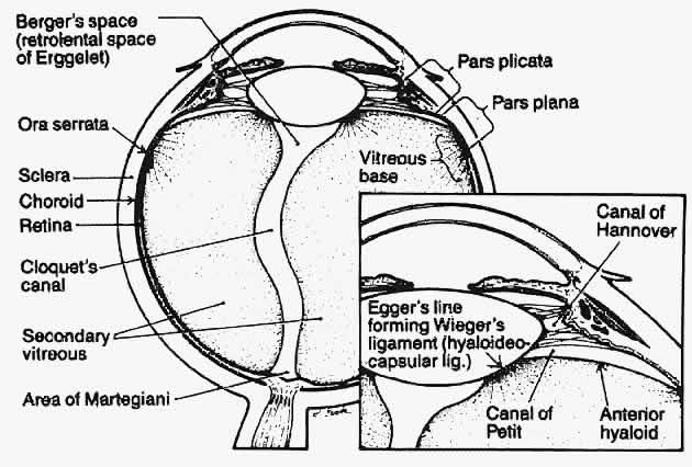
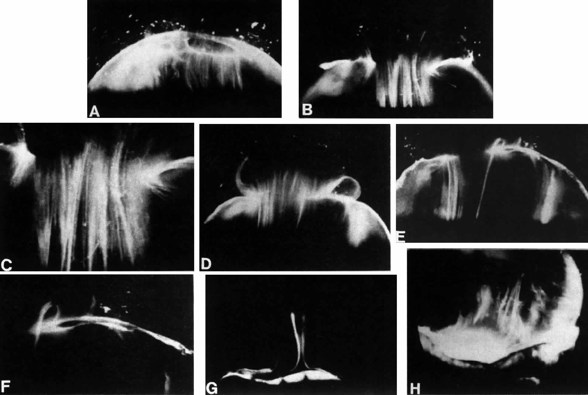
1. Duke-Elder WS: The nature of the vitreous body. Br J Ophthalmol 14(suppl):6, 1930

2. Demours: Observations anatomiques sur la structure cellulaire du corps vitre. In: Memoires de Paris, 1741

3. Zinn: Descriptio anatomica oculi humani. Göttingen

4. Hannover A: Endeckung des baues des Glaskorpers. Muller Arch 00:467, 1845

5. Bowman W: Observations on the structure of the vitreous humour. Dublin Q J Med Sci 6:102, 1848

6. Retzius R: Om membrana limitans retinae interna. Nord Arch 3(2):1, 1871

7. Szent-Gyorgi A: Untersuchungen ueber die Struktur des Glaskoerpers des Menschen. Arch Mikroscop Anat 89:324, 1917

8. Baurmann M: Untersuchungen ueber die Struktur des Glaskoerpers bei Saeugetieren. Graefes Arch Ophthalmol 110:352, 1922

9. Stroemberg: Zur Frage nach dem Bau des Glaskoerpers. Acta Soc Med Suecanae, 57, 1931

10. Redslob E: Le Corps Vitre, pp 174–178. Paris, Masson, 1932

11. Gullstrand A: Die Nernspaltlampe in der ophthalmologischen Praxis. Klin Monatsbl Augenheilkd 50:483, 1912

12. Koeppe L: Clinical observations with the slit lamp. Arch Ophthalmol 90:232, 1917

13. Baurmann M: Ueber die Beziehungen der ultramikroscopischen Glaskorperstruktur zu den Spaltlampenbefunden. Graefes Arch Ophthalmol 117:304, 1926

14. Goedbloed J: Studien am Glaskorper: I. Die Struktur des Glaskorpers. Graefes Arch Ophthalmol p 323, 1934

15. Friedenwald JF, Stiehler RD: Structure of the vitreous. Arch Ophthalmol 14:789, 1935

16. Eisner G: Biomicroscopy of the Peripheral Fundus. New York, Springer-Verlag, 1973

17. Worst JGF: Cisternal systems of the fully developed vitreous body in the young adult. Trans Ophthalmol Soc UK 97:550, 1977

18. Kishi S, Shimizu K: Posterior precortical vitreous pocket. Arch Ophthalmol 108:979, 1990

19. Mann I: The vitreous and suspensory ligament of the lens. In Mann I (ed): The Development of the Human Eye. New York, Grune & Stratton, 1964

20. Sebag J: The Treacher Collins Prize Essay: Lasers in Ophthalmic Diagnosis. Trans Ophthalmol Soc UK 105:607, 1986

21. Packer AJ, Newsome DA: Practical guidelines for posterior segment biomicroscopy. In Newsome DA (ed): Retinal Dystrophies and Degenerations, pp 1–4. New York, Raven Press, 1988

22. Jalkh AE, Trempe CL: Clinical methods of vitreous examination. In Schepens CL, Neetens A (eds): The Vitreous and Vitreoretinal Interface, pp 73–83. New York, Springer-Verlag, 1987

23. Kiryu J, Ogura Y, Shahidi M et al: Enhanced visualization of vitreoretinal interface by laser biomicroscopy. Ophthalmology 100:1040, 1993

24. Charles S: Vitreous Microsurgery, pp 7–8. Baltimore, Williams & Wilkins, 1981

25. Boruchoff SA: Corneo-vitreal contact. Trans Ophthalmol Soc UK 95:417, 1975

26. Lacqua H, Machemer R: Clinico-pathologic correlation in massive preretinal proliferation. Am J Ophthalmol 80:913, 1975

27. Scott JD: Treatment of massive vitreous retraction. Trans Ophthalmol Soc UK 95:429, 1975

28. Awan KJ, Thurmayan M: Changes in the contralateral eye in uncomplicated persistent hyperplastic primary vitreous. Am J Ophthalmol 99:122, 1985

29. El Bayadi G: A new method of slit-lamp micro-ophthalmoscopy. Br J Ophthalmol 37:625, 1953

30. Takahashi M et al: Biomicroscopic evaluation and photography of posterior vitreous detachment. Arch Ophthalmol 98:665, 1980

31. Schepens CL: Retinal Detachment and Allied Diseases. Philadelphia, WB Saunders, 1983

32. Buzney SM, Weiter JJ, Furukawa H et al: Examination of the vitreous: Comparison of biomicroscopy using the Goldmann and El Bayadi-Kajiura lenses. Ophthalmology 92:1745, 1985

33. Goldmann H: Slit-lamp examination of the vitreous and fundus. Br J Ophthalmol 33:242, 1949

34. Jaffe NS: Complications of acute posterior vitreous detachment. Arch Ophthalmol 79:568, 1968

35. Mainster MA, Grossman JL, Erickson PJ, Heacock GL: Retinal laser lenses: Magnification, spot size, and field of view. Br J Ophthalmol 74:177, 1990

36. Fischer YL, Slatter JS, Friedman RA, Yannuzzi LA: Kinetic ultrasound evaluation of the posterior vitreoretinal interface. Ophthalmology 98:1135, 1991

37. Arzabe CW, Akiba J, Jalkh AE et al: Comparative study of vitreoretinal relationships using biomicroscopy and ultrasound. Graefes Arch Clin Exp Ophthalmol 220:66, 1991

38. Morner CT: Untersuchung der Proteinsubstanzen in der lichtbrechenden Medien des Auges. Z Physiol Chem 18:223, 1894

39. Young RA: The ground substance of connective tissue. J Physiol 16:325, 1894

40. Pirie A, Schmidt G, Waters JW: Ox vitreous humor: I. The residual protein. Br J Ophthalmol 32:321, 1948

41. Bembridge BA, Crawford CNC, Pirie A: Phase-contrast microscopy of the animal vitreous body. Br J Ophthalmol 36:131, 1952

42. Matolstsky AG, Gross J, Grignolo A: A study of the fibrous components of the vitreous body with the electron microscope. Proc Soc Exp Biol Med 76:857, 1951

43. Schwarz W: Electron microscopic observations on the human vitreous body. In Smelser GK (ed): Structure of the Eye, pp 283–291. New York, Academic Press, 1961

44. Young RG, Williams HH: Biochemistry of the eye--gelatinous protein of the vitreous body. Arch Ophthalmol 51:593, 1954

45. Swann DA: Chemistry and biology of vitreous body. Int Rev Exp Pathol 22:1, 1980

46. Bishop P, McLeod D, Ayad S: Extraction and characterization of the intact form of bovine vitreous type IX collagen. Biochem Biophys Res Commun 185:392, 1992

47. Snowden JM, Swann DA: Vitreous structure: V. The morphology and thermal stability of vitreous collagen fibers and comparison to articular cartilage (type II) collagen. Invest Ophthalmol Vis Sci 19:610, 1980

48. Hogan MJ: The vitreous: Its structure in relation to the ciliary body and retina. Invest Ophthalmol 2:418, 1963

49. François J, Victoria-Troncoso V, Albarran E: The histochemical structure of the vitreous fibers studied by phase contrast microscopy. Am J Ophthalmol 69:763, 1970

50. Streeten BA: Disorders of the vitreous. In Garner A, Klintworth GK (eds): Pathobiology of Ocular Disease--A Dynamic Approach, part B, chap 49, pp 1381–1419. New York, Marcel Dekker, 1982

51. Balazs EA: Physiology of the vitreous body in retina surgery with special emphasis on reoperations. In Schepens CL (ed): Proceedings of the 11th Conference of the Retina Foundation, pp 29–48. St. Louis, CV Mosby, 1960

52. Balazs EA: The vitreous. Int Ophthalmol Clin 15:53–63, 1973

53. Berman ER, Michaelson IC: The chemical composition of the human vitreous body as related to age and myopia. Exp Eye Res 3:9, 1964

54. Weber H, Landuehr G, Kilp H, Neubauer H: Mechanical properties of the vitreous in pig and human donor eyes. Ophthalmic Res 14:335, 1982

55. Balazs EA, Laurent TC, Laurent UBG et al: Studies on the structure of the vitreous body: VIII. Comparative biochemistry. Arch Biochem Biophys 81:464, 1959

56. Bettelheim FA, Balazs EA: Light-scattering patterns of the vitreous humor. Biochem Biophys Acta 158:309, 1968

57. Birck DE, Zychard EI: Collagen fibrillogenesis in situ: Fibril segments are intermediates in matrix assembly. Proc Natl Acad Sci USA 86:4549, 1989

58. Meyer K, Palmer JW: The polysaccharide of the vitreous humor. J Biol Chem 107:629, 1934

59. Meyer K: Chemical structure of hyaluronic acid. Fed Proc 17:1075, 1958

60. Balazs EA: Molecular morphology of the vitreous body. In Smelser GK (ed): The Structure of the Eye, pp 293–310. New York, Academic Press, 1961

61. Comper WD, Laurent TC: Physiological functions of connective tissue polysaccharides. Physiol Rev 58:255, 1978

62. Balazs EA, Denlinger JL: Aging changes in the vitreous. In: Aging and Human Visual Function, pp 45–57. New York, Alan R Liss, 1982

63. Atkins EDT, Sheehan JK: Structure for hyaluronic acid. Nature (New Biol) 235:253, 1972

64. Atkins EDT, Phelps CF, Sheehan JK: The conformation of the mucopolysaccharides--hyaluronates. Biochem J 128:1255, 1972

65. Chakrabarti B, Park JW: Glycosaminoglycans--Structure and interaction. CRC Crit Rev Biochem 8:255, 1980

66. Christiansson J: Changes in mucopolysaccharides during alloxan diabetes in the rabbit. Acta Ophthalmol 36:141, 1958

67. Xiong H, Cheng HM: Change of vitreous toxicity in “sugar” cataracts. Invest Ophthalmol Vis Sci 29:149, 1988

68. Yamashita H, Hori S: A new model of neovascularization caused by tractional force. Invest Ophthalmol Vis Sci 29:177, 1988

69. Tasman WS: Diabetic vitreous hemorrhage and its relationship to hypoglycemia. Mod Probl Ophthalmol 20:413, 1979

70. Osterlin SE, Balazs EA: Macromolecular composition and fine structure of the vitreous in the owl monkey. Exp Eye Res 7:534, 1968

71. Balazs EA: Structure of vitreous gel. In: Acta XVII Concilium Ophthalmologicum, vol 11, p 1019. 1954

72. Balazs EA: In Acta XVIII Concilium Ophthalmologicum, vol II, p 1296. Brussels, 1958

73. Balazs EA: Die Mikrostruktur und Chimie des Glaskorpers. In Jaeger W (ed): Bericht uber die 68 Zusammen Kunst der Deutschen Ophthalmologischen Besemschaft, Heidelberg, 1967, pp 536–572. Munich, JF Bergmann Verlag, 1968

74. Balazs EA: Functional anatomy of the vitreous. In Duane TD, Jaeger EA (eds): Biomedical Foundations of Ophthalmology, vol 1, chap 17. Philadelphia, JB Lippincott, 1982

75. Ali S, Bettelheim FA: Distribution of freezable and non-freezable water in bovine vitreous. Curr Eye Res 3:1233, 1984

76. Schwarz W: Electron microscopic study on the gel of the central part of the corpus vitreum in the ox. Cell Tissue Res 168:271, 1976

77. Streeten BA: The nature of the ocular zonule. Trans Am Ophthalmol Soc 80:823, 1982

78. Asakura A: Histochemistry of hyaluronic acid of the bovine vitreous body as studied by electron microscopy. Acta Soc Ophthalmol Jpn 89:179, 1985

79. Sebag J: The Vitreous--Structure, Function, Pathobiology, pp 60–61. New York, Springer-Verlag, 1989

80. Scott JE: Proteoglycan: Collagen interactions and corneal ultrastructure. Biochem Soc Trans 19:877, 1991

81. Scott JE: The chemical morphology of the vitreous. Eye 6:553, 1992

82. Sebag J: Ageing of the vitreous. Eye 1:254, 1987

83. Sebag J, Buckingham B, Reiser KA, Charles MA: Biochemical abnormalities in vitreous collagen from patients with proliferative diabetic retinopathy. Arch Ophthalmol 110:1472, 1992

84. Bettelheim FA, Popdimitrova N: Hyaluronic acid--a syneretic glycosaminoglycans. Curr Eye Res 11:411, 1992

85. Hamming NA, Apple DJ, Geiser DK, Vygantas CM: Ultrastructure of hyaloid vasculature in primates. Invest Ophthalmol Vis Sci 16:408, 1977

86. Bloom GD, Balazs EA, Ozanics V: The fine structure of the hyaloid arteriole in bovine vitreous. Exp Eye Res 31:129, 1988

87. Balazs EA, Denlinger JL: The vitreous. In Davson H (ed): The Eye, vol 1A, pp 533–589. London, Academic Press, 1984

88. Sebag J: La structure fibreuse du corps vitre humain. Bull Mem Soc Fr Ophthalmol 96:395, 1985

89. Sebag J, Balazs EA: Human vitreous fibres and vitreoretinal disease. Trans Ophthalmol Soc UK 104:123, 1985

90. Sebag J, Balazs EA: Morphology and ultrastructure of human vitreous fibers. Invest Ophthalmol Vis Sci 30:1867, 1989

91. Sebag J: Age-related differences in the human vitreoretinal interface. Arch Ophthalmol 109:966, 1991

92. Sebag J: Age-related changes in human vitreous structure. Graefes Arch Clin Exp Ophthalmol 225:89, 1987

93. Sebag J: Letter to the editor. Arch Ophthalmol 109:1059, 1991

94. Jongebloed WL, Humalda D, Worst JFG: A SEM correlation of the anatomy of the vitreous body: Making visible the invisible. Doc Ophthalmol 64:117, 1986

95. Jongebloed WL, Worst JFG: The cisternal anatomy of the vitreous body. Doc Ophthalmol 67:183, 1987

96. O'Malley P: The pattern of vitreous syneresis: A study of 800 autopsy eyes. In Irvine AR, O'Malley P (eds): Advances in Vitreous Surgery, pp 17–33. Springfield, IL, Charles C Thomas, 1976

97. Oksala A: Ultrasonic findings in the vitreous body at various ages. Graefes Arch Klin Exp Ophthalmol 207:275, 1978

98. Reeser FH, Aaberg T: Vitreous humor. In Records PE (ed): Physiology of the Human Eye and Visual System, pp 1–31. Hagerstown, MD, Harper & Row, 1979

99. Gartner J: Electron microscopic study on the fibrillar network and fibrocyte-collagen interactions in the vitreous cortex at the ora serrata of human eyes with special regard to the role of disintegrating cells. Exp Eye Res 42:21, 1986

100. Gloor BP, Daicker BC: Pathology of the vitreo-retinal border structures. Trans Ophthalmol Soc UK 95:387, 1975

101. Gartner J: The fine structure of the vitreous base of the human eye and the pathogenesis of pars planitis. Am J Ophthalmol 71:1317, 1971

102. Teng CC, Chi HH: Vitreous changes and the mechanism of retinal detachment. Am J Ophthalmol 44:335, 1957

103. Schepens CL: Personal communication, 1983

104. Gartner J: New research on the aetiology and surgery of retinal detachment. Mod Probl Ophthalmol 15:112, 1975

105. Foos RY: Posterior vitreous detachment. Trans Am Acad Ophthalmol Otolaryngol 76:480, 1972

106. Chaine G, Sebag J, Coscas G: The induction of retinal detachment. Trans Ophthalmol Soc UK 103:480, 1983

107. Best F: Der Glaskoerper bei Augen bewegangen. Klin Monatsbl Augenheilkd 42:538, 1984

108. Leber T, Graefe A, Saemisde ET: Handbuch der Gesamten Augenheilkunde, vol 7. Berlin, Springer, 1916

109. Teng CC, Katzin HM: An anatomic study of the peripheral retina: III. Congenital retinal rosettes. Am J Ophthalmol 36:169, 1953

110. Kanski JJ: Complications of acute posterior vitreous detachment. Am J Ophthalmol 80:44, 1975

111. Schepens CL: Subclinical retinal detachments. Arch Ophthalmol 47:593, 1952

112. Rutnin U, Schepens CL: Fundus appearance in normal eyes: III. Peripheral degeneration. Am J Ophthalmol 64:1040, 1967

113. Byer NE: Lattice degeneration of the retina. Surv Ophthalmol 23:213, 1979

114. Foos RY: Vitreous base, retinal tufts and retinal tears: Pathogenic relationships. In Pruett RC, Regan CDJ (eds): Retina Congress. New York, Appleton-Century-Crofts, 1974

115. Sigelman J: Vitreous base classification of retinal tears: Clinical application. Surv Ophthalmol 25:59, 1980

116. Straatsma BR, Allen RA: Lattice degeneration of the retina. Trans Am Acad Ophthalmol Otolaryngol 66:600, 1962

117. Sato K, Tsunakawa N, Inaba K, Yanagisawa Y: Fluorescein angiography on retinal detachment and lattice degenerations: I. Equatorial degeneration with idiopathic retinal detachment. Acta Soc Ophthalmol Jpn 75:635, 1971

118. Foos RY, Wheeler NC: Vitreoretinal juncture--synchisis senilis and posterior vitreous detachment. Ophthalmology 89:1502, 1982

119. Foos RY, Simons KB: Vitreous in lattice degeneration of retina. Ophthalmology 91:452, 1984

120. Byer NE: Discussion: Vitreous in lattice degeneration of retina. Ophthalmology 91:457, 1984

121. Tolentino FI, Schepens CL, Freeman HM: Vitreoretinal Disorders, p 1976. Philadelphia, WB Saunders, 1967

122. Daicker B: Sind die symptome “Weiss mit Druck” und “Weiss ohne Druck” durch die peripherie netzshautsklerose bedingt? Mod Probl Ophthalmol 15:82, 1975

123. Watzke RC: The ophthalmoscopic sign “white with pressure”: A clinicopathologic correlation. Arch Ophthalmol 66:812, 1961

124. Green WR: Vitreoretinal juncture. In Ryan SJ (ed): Retinal Disease, pp 13–69. St. Louis, CV Mosby, 1989

125. Byer NE: The Peripheral Retina in Profile: A Stereoscopic Atlas. Torrance, CA, Criterion Press, 1982

126. Lindner B: Acute posterior vitreous detachment and its retinal complications. Acta Ophthalmol 87(suppl):1, 1966

127. Tasman WS: Posterior vitreous detachment and peripheral retinal breaks. Trans Am Acad Ophthalmol Otolaryngol 72:217, 1968

128. Novak MA, Welch RB: Complications of acute symptomatic posterior vitreous detachment. Am J Ophthalmol 97:308, 1984

129. Hyams SW, Neumann E: Peripheral retina in myopia with particular reference to retinal breaks. Br J Ophthalmol 53:300, 1969

130. Pickett-Seltner RL, Doughty MJ, Pasternak JJ, Sivak JG: Proteins of the vitreous humor during experimentally induced myopia. Invest Ophthalmol Vis Sci 33:3424, 1992

131. Hyams SW, Neumann E, Friedman Z: Myopia-aphakia: II. Vitreous and peripheral retina. Br J Ophthalmol 59:483, 1975

132. Hovland KR: Vitreous findings in fellow eyes of aphakic retinal detachment. Am J Ophthalmol 86:350, 1978

133. Bradford JD, Wilkinson CP, Fransen SR: Pseudophakic retinal detachments--the relationships between retinal tears and the time following cataract surgery at which they occur. Retina 9:181, 1989

134. Byer NE: Clinical study of retinal breaks. Trans Am Acad Ophthalmol Otolaryngol 71:461, 1967

135. Combs JL, Welch RB: Retinal breaks without detachment: Natural history, management, and long-term follow-up. Trans Am Ophthalmol Soc 80:64, 1982

136. Lindner K: Zur Klinik des Glaskorpers: III. Glaskorper und Netzhautabhebung. Graefes Arch Ophthalmol 137:157, 1937

137. Rosengren B, Osterlin S: Hydrodynamic events in the vitreous space accompanying eye movements: Significance for the pathogenesis of retinal detachment. Ophthalmologica 173:513, 1976

138. Oksala A: Ultrasonic findings in the vitreous space in patients with detachment of the retina. Graefes Arch Klin Exp Ophthalmol 202:197, 1977

139. Duke-Elder WS, MacFaul PA: Concussion changes in the vitreous. In Duke-Elder WS (ed): System of Ophthalmology, vol 14, p 197. London, Henry Kimpton, 1972

140. Tolentino FI, Lee PF, Schepens CL: Biomicroscopic study of the vitreous cavity in diabetic retinopathy. Arch Ophthalmol 75:238, 1966

141. Scheiffarth OF, Kampik A, Gunther H et al: Proteins of the extracellular matrix in vitreoretinal membranes. Graefes Arch Clin Exp Ophthalmol 226:357, 1988

142. Bellhorn MB, Friedman AH, Wise GN, Henkind P: Ultrastructure and clinicopathologic correlation of idiopathic preretinal macular fibrosis. Am J Ophthalmol 79:366, 1975

143. Clarkson JG, Green WR, Massof D: A histopathologic review of 168 cases of preretinal membranes. Am J Ophthalmol 84:1, 1977

144. Wallow IHL, Tso MOM: Proliferation of the RPE over malignant choroidal tumors. Am J Ophthalmol 73:914, 1972

145. Mueller-Jensen J, Machemer R, Azuronia R: Auto-transplantation of retinal pigment epithelium in intravitreal diffusion chamber. Am J Ophthalmol 80:530, 1975

146. Campochiaro PA, Jerdan JA, Glaser BM et al: Vitreous aspirates from patients with PVR stimulate RPE cell migration. Arch Ophthalmol 103:1403, 1985

147. Sebag J, Boulton M, Tang M et al: Surface markers and growth factors in human hyalocytes. Invest Ophthalmol Vis Sci 34:1419, 1993

148. Lutty GA, Merges C, Threlkeld AB et al: Heterogeneity in localization of isoforms of TGF-beta in human retina, vitreous and choroid. Invest Ophthalmol Vis Sci 34:477, 1993

149. Fine BS, Tousimis AJ: The structure of the vitreous body and the suspensory ligaments of the lens. Arch Ophthalmol 65:95, 119, 1961

150. Faulborn J, Bowald S: Combined macroscopic, light microscopic, scanning and transmission electron microscopic investigation of the vitreous body: II. The anterior vitreous cortex. Ophthalmic Res 14:117, 1982

151. Rhodes RH: An ultrastructural study of complex carbohydrates in the posterior chamber and vitreous base of the mouse. Histochem J 17:291, 1985

152. Theopold H, Faulborn J: Scanning electron microscopic aspects of the vitreous body. Mod Probl Ophthalmol 20:92, 1979

153. Jaffe NS: Vitreous traction at the posterior pole of the fundus due to alterations in the vitreous posterior. Trans Am Acad Ophthalmol Otolaryngol 71:642, 1967

154. Jaffe NS: Macular retinopathy after separation of vitreoretinal adherence. Arch Ophthalmol 78:585, 1967

155. Sebag J: Vitreoretinal interface and the role of vitreous in macular disease. In Brancato R, Coscasa G, Lumbrosos B (eds): Proceedings of the Retina Workshop, pp 3–6. Amsterdam, Kugler & Ghedini, 1987

156. Hageman G, Russel G: Enzymatic disinsertion of the primate vitreous body. Exp Eye Res 55:751, 1992

157. Verstraeten TC, Chapman C, Hartzer M et al: Pharmacologic induction of posterior vitreous detachment in the rabbit. Arch Ophthalmol 111:849, 1993

158. Schwalbe G: In Engelmann W (ed): Von Graefe--Saemisch's Handbuch der Gesamnten Augenheilkunde, vol I, p 457. Leipzig, 1874

159. Schwalbe G: Lehrbuch der Anatomie des Auges, p 288. Erlangen, E Besold, 1887

160. Balazs EA, Toth LZ, Eckl EA, Mitchell AP: Studies on the structure of the vitreous body: XII. Cytological and histochemical studies on the cortical tissue layer. Exp Eye Res 3:57, 1964

161. Gloor BP: Cellular proliferation on the vitreous surface after photocoagulation. Graefes Arch Clin Exp Ophthalmol 178:99, 1969

162. Bloom GD, Balazs EA: An electron microscope study of hyalocytes. Exp Eye Res 4:249, 1965

163. Hogan MJ, Alvarado JA, Weddel JE: Histology of the Human Eye: An Atlas and Textbook, p 607. Philadelphia, WB Saunders, 1971

164. Saga T, Tagawa Y, Takeuchi T et al: Electron microscopic study of cells in vitreous of guinea pig. Jpn J Ophthalmol 28:239, 1984

165. Balazs EA: Studies on structure of vitreous body: Absorption of ultraviolet light. Am J Ophthalmol 38:21, 1954

166. Jacobson B, Osterlin S, Balazs EA: A soluble hyaluronic acid synthesizing system from calf vitreous. Proc Fed Am Soc Exp Biol 25:588, 1966

167. Osterlin SE: The synthesis of hyaluronic acid in the vitreous. III. In vivo metabolism in the owl monkey. Exp Eye Res 7:524, 1968

168. Osterlin SE: The synthesis of hyaluronic acid in the vitreous: IV. Regeneration in the owl monkey. Exp Eye Res 8:27, 1969

169. Berman ER, Gambos GM: Studies on the incorporation of U-14 C-glucose into vitreous polymers in vitro and in vivo. Invest Ophthalmol 18:521, 1969

170. Balazs EA, Sundblad L, Toth LZJ: In vitro formation of hyaluronic acid by cells in the vitreous body and by lamb tissue. Abstr Fed Proc 17:184, 1958

171. Bleckmann H: Glycosaminoglycan metabolism of cultured fibroblasts from bovine vitreous. Graefes Arch Clin Exp Ophthalmol 222:90, 1984

172. François J, Victoria-Troncoso V, Maudgal PC: Immunology of the vitreous body. Mod Probl Ophthalmol 16:196, 1976

173. Rhodes RH, Mandelbaum SH, Minckler DS, Cleary PE: Tritiated fucose incorporation in the vitreous body, lens and zonules of the pigmented rabbit. Exp Eye Res 34:921, 1982

174. Jacobson B: Degradation of glycosaminoglycans by extracts of calf vitreous hyalocytes. Exp Eye Res 39:373, 1984

175. Haddad A, Almeida JC, Laicine EM et al: The origin of the intrinsic glycoproteins of the rabbit vitreous body: An immunohistochemical and autoradiographic study. Exp Eye Res 50:555, 1990

176. Haddad A, Laicine EM: Studies on the origin of the glycoproteins of the rabbit vitreous body using a protein synthesis inhibitor and radioactive fucose and amino acids. Germ J Ophthalmol 2:127, 1993

177. Newsome DA, Linsemayer TF, Trelstad RJ: Vitreous body collagen: Evidence for a dual origin from the neural retina and hyalocytes. J Cell Biol 71:59, 1976

178. Ayad S, Weiss JB: A new look at vitreous humour collagen. Biochem J 218:835, 1984

179. Hoffmann K, Baurwieg H, Riese K: Uber Gehalt und Vertailang niederund hoch molekularer Substanzen in Glaskorper: II. Hoch molekulare Substanzen (LDH, MDH, GOT). Graefes Arch Clin Exp Ophthalmol 191:231, 1974

180. Teng CC: An electron microscopic study of cells in the vitreous of the rabbit eye: I. The macrophage. Eye Ear Nose Throat Month 48:91, 1969

181. Freeman MI, Jacobson B, Toth LZ, Balazs EA: Lysosomal enzymes associated with hyalocyte granules: I. Intercellular distribution patterns of enzymes. Exp Eye Res 7:113, 1968

182. Grabner G, Baltz G, Forster O: Macrophage-like properties of human hyalocytes. Invest Ophthalmol Vis Sci 19:333, 1980

183. Forrester JV, Balazs EA: Inhibition of phagocytosis by high molecular weight hyaluronate. Immunology 40:435, 1980

184. Sebag J, Balazs EA, Eakins KE, Kulkarni P: The effect of Na-hyaluronate on prostaglandin synthesis and phagocytosis by mononuclear phagocytes. Invest Ophthalmol Vis Sci 20:33, 1981

185. Balazs EA, Toth LZ, Ozanics V: Cytological studies on the developing vitreous as related to the hyaloid vessel system. Graefes Arch Clin Exp Ophthalmol 213:71, 1980

186. Kefalides NA: The biology and chemistry of basement membranes. In Kefalidese NA (ed): Proceedings of the First International Symposium on the Biology and Chemistry of Basement Membranes, pp 215–228. New York, Academic Press, 1978

187. Goodnight R, Nagy AR, Ryan SJ: Differential distribution of laminin in rabbit and monkey retinas. Invest Ophthalmol Vis Sci 29:203, 1988

188. Yamada E: Some structural features of the fovea centralis in the human retina. Arch Ophthalmol 82:151, 1969

189. Anderson DR: Ultrastructure of the optic nerve head. Arch Ophthalmol 83:63, 1970

190. Uga A: Some structural features of the retinal Muellerian cells in the juxtaoptic nerve region. Exp Eye Res 19:105, 1974

191. Heergaard S, Jensen OA, Prause JU: Structure of the vitread face of the monkey optic disc (Macacca mulatta): SEM on frozen resin-cracked optic nerve heads supplemented by TEM and immunohistochemistry. Graefes Arch Clin Exp Ophthalmol 226:377, 1988

192. Sebag J, Balazs EA: Pathogenesis of C.M.E.: Anatomic consideration of vitreoretinal adhesions. Surv Ophthalmol 28(suppl):493, 1984

193. Zimmerman LE, Straatsma BR: Anatomic relationships of the retina to the vitreous body and to the pigment epithelium. In Schepens CL (ed): Importance of the Vitreous Body in Retina Surgery with Special Emphasis on Regeneration. St. Louis, CV Mosby, 1960

194. Kuwabara T, Cogan DG: Studies of retinal vascular patterns: I. Normal architecture. Arch Ophthalmol 64:904, 1960

195. Pedler C: The inner limiting membrane of the retina. Br J Ophthalmol 45:423, 1961

196. Wolter JR: Pores in the internal limiting membrane of the human retina. Acta Ophthalmol 42:971, 1964

197. Mutlu F, Leopold IH: Structure of the human retinal vascular system. Arch Ophthalmol 71:93, 1964

198. Gartner J: Electron microscopic observations on the ciliozonular border of the human eye with particular reference to the aging changes. Z Anat Entwickl Gesch 131:263, 1970

199. Gersh I, Catchpole HR: The organization of ground substance and basement membrane and its significance in tissue injury, disease and growth. Am J Anat 85:457, 1949

200. Caulfield JB: Medical progress in the application of the electron microscope to renal disease. N Engl J Med 270:183, 1964

201. Michaelson IC: The mode of development of the vascular system of the retina with some observations on its significance for certain retinal diseases. Trans Ophthalmol Soc UK 68:137, 1948

202. Sebag J, McMeel JW: Diabetic Retinopathy: Pathogenesis and role of retina-derived growth factor in angiogenesis. Surv Ophthalmol 30:377, 1986

203. Glaser BM: Extracellular modulating factors and the control of intraocular neovascularization. Arch Ophthalmol 106:603, 1988

204. Jalkh A, Takahashi M, Topilow HW et al: Prognostic value of vitreous findings in diabetic retinopathy. Arch Ophthalmol 100:432, 1982

205. Foos RY, Krieger AE, Forsythe AB et al: Posterior vitreous detachment in diabetic subjects. Ophthalmology 87:122, 1980

206. Tagawa H, McMeel JW, Furukawa H et al: Role of the vitreous in diabetic retinopathy: I. Vitreous changes in diabetic retinopathy and in physiologic aging. Ophthalmology 93:596, 1986

207. Sebag J: Abnormalities of human vitreous structure in diabetes. Graefes Arch Clin Exp Ophthalmol 231:257, 1993

208. Sebag J: Letter to the editor. Ophthalmology 100:1599, 1993

209. Kishi S, Shimizu K: Clinical manifestations of posterior precortical vitreous pocket in proliferative diabetic retinopathy. Ophthalmology 100:225, 1993

210. Chu TG, Green RL, Cano MR et al: Schisis of the posterior vitreous cortex: An ultrasonographic finding in diabetic retinopathy. Invest Ophthalmol Vis Sci 32:1028, 1992

211. Raviola G: The fine structure of the ciliary zonule and ciliary epithelium. Invest Ophthalmol 10:851, 1971

212. Barraquer J: Totale Linsenextraktion nach Aulosung der Zonula durch Chymotrypsin enzymatische zonulyse. Klin Monatsbl Augenheilkd 133:609, 1958

213. Takei Y, Smelsen GK: Electron microscopic studies on zonular fibers: II. Changes of the zonular fibers after the treatment with collagenase, alpha-chymotrypsin and hyaluronidase. Graefes Arch Klin Exp Ophthalmol 194:153, 1975

214. Swann DA, Sotman SS: The chemical composition of bovine vitreous humour collagen fibres. Biochem J 185:545, 1980

215. Streeten BA, Licari PA, Marucci AA et al: Immunohistochemical comparison of ocular zonules and the microfibrils of elastic tissue. Invest Ophthalmol Vis Sci 21:130, 1981

216. Kaczurowski MI: The surface of the vitreous. Am J Ophthalmol 63:419, 1967

217. Hilton GF, Grizzard WS: Pneumatic retinopexy: A two-step outpatient operation without conjunctival incision. Ophthalmology 93:626, 1986

218. Tornambe PE: Pneumatic retinopexy. Surv Ophthalmol 32:270, 1988

219. Sebag J, Tang M: Pneumatic retinopexy using only air. Retina 13:8, 1993

220. Busacca A, Goldmann H, Schiff-Wertheimer S: Biomicroscopie du Corps Vitre et du Fond d'Oeil. Paris, Masson, 1957

221. Rieger M: Uber die Bedeutung der Aderhausveranderungen fur die Entstehung der Glaskorperabhebung. Graefes Arch Klin Ophthalmol 136:119, 1936

222. Smith TJ, Murata Y, Horwitz AL et al: Regulation of glycosaminoglycan synthesis by thyroid hormone in vitro. J Clin Invest 70:1066, 1982

223. Smith TJ: Dexamethasone regulation of glycosaminoglycan synthesis in cultured human skin fibroblasts: Similar effects of glucocorticoid and thyroid hormones. J Clin Invest 74:2157, 1984

224. Likar LH, Likar IN, Robinson RW: Levels of acid mucopolysaccharides of the bovine aorta at different stages of the sexual cycle. J Atheroscl Res 5:388, 1965

225. Sirek OV, Sirek A, Fikar K: The effect of sex hormones on glycosaminoglycan content of canine aorta and coronary arteries. Atherosclerosis 17:227, 1977

226. Larsen JS: The hyaluronic acid in the rabbit vitreous: Variations following hormonal treatment. Arch Ophthalmol 60:815, 1971

227. Larsson L, Osterlin S: Posterior vitreous detachment: A combined clinical and physiochemical study. Graefes Arch Clin Exp Ophthalmol 223:92, 1985

228. Spencer WH: Vitreous. In Spencer WH (ed): Ophthalmic Pathology--An Atlas and Textbook, vol 2, pp 548–588. Philadelphia, WB Saunders, 1985

229. Kuhn W, Hargitay B, Katcholsky M, Eisenberg H: Reversible dilation and contraction by changing the state of ionization of high polymer acid networks. Nature 165:514, 1956

230. Akiba J, Ueono N, Chakrabarti B: Molecular mechanisms of posterior vitreous detachment. Graefes Arch Clin Exp Ophthalmol 231:408, 1993

231. Foos RY, Gloor BP: Vitreoretinal juncture: Healing of experimental wounds. Graefes Arch Klin Exp Ophthalmol 196:213, 1975

232. Jost BF, Hutton WL, Fuller DG et al: Vitrectomy in eyes at risk for macular hole formation. Ophthalmology 97:843, 1990

233. Berman ER, Voaden M: The vitreous body. In Graymore CN (ed): Biochemistry of the Eye, pp 373–471. New York, Academic Press, 1970

234. Osterlin SE: Changes in the vitreous with age. Trans Ophthalmol Soc UK 95:372, 1975

235. Wolf E, Gardiner JS: Studies on the scatter of light in the dioptric media of the eye as a basis of visual glare. Arch Ophthalmol 74:338, 1965

236. Deiter P, Wolf E, Geer S: Glare and the scatter of light in the vitreous: Effect in postoperative retinal detachment patients. Arch Ophthalmol 87:12, 1972

237. Cibis GE, Watzke RC, Chua J: Retinal hemorrhages: Posterior vitreous detachment. Am J Ophthalmol 79:358, 1975

238. Foos RY, Roth AM: Surface structure of the optic nerve head: II. Vitreopapillary attachments and posterior vitreous detachment. Am J Ophthalmol 76:622, 1973

239. Murakami K et al: Vitreous floaters. Ophthalmology 90:1271, 1983

240. Moore RF: Subjective “lightning streak.” Br J Ophthalmol 19:545, 1935

241. Wise GN: Relationship of idiopathic preretinal macular fibrosis to posterior vitreous detachment. Am J Ophthalmol 79:358, 1975

242. Voerhoeff FH: Are Moore's lightning streaks of serious portent. Am J Ophthalmol 41:837, 1956

243. Schachat AP, Sommer A: Macular hemorrhages associated with posterior vitreous detachment. Am J Ophthalmol 102:647, 1986

244. Spencer LM, Foos RY: Paravascular vitreoretinal attachments: Role in retinal tears. Arch Ophthalmol 84:557, 1970

245. Foos RY: Vitreoretinal juncture, epiretinal membranes and vitreous. Invest Ophthalmol Vis Sci 16:416, 1977

246. Kishi S, Demaria C, Shimizu K: Vitreous cortex remmants at the fovea after spontaneous vitreous detachment. Int Ophthalmol Clin 9:253, 1986

247. Gartner J: Photoelastic and ultrasonic studies on the structure and senile changes of the intervertebral disc and of the vitreous body. Mod Probl Ophthalmol 8:136, 1969

248. Smiddy WE, Michels RG, Glaser BM et al: Vitrectomy for macular traction caused by incomplete vitreous separation. Arch Ophthalmol 197:624, 1988

249. Sebag J, de Bustros S, Wendell R: Disorders of the vitreomacular interface. In Margo CE, Hamed LM, Mames RN (eds): Diagnosis Problems in Clinical Ophthalmology, pp 556-563. Philadelphia, WB Saunders, 1994

250. Michels RG: A clinical and histopathologic study of epiretinal membranes affecting the macula and removed by vitreous surgery. Trans Am Ophthalmol Soc 80:580, 1982

251. Smiddy WE, Maguire AM, Green WR et al: Idiopathic epiretinal membranes: Ultrastructural characteristics and clinicopathologic correlation. Ophthalmology 96:811, 1989

252. Kelly NE, Wendell RT: Vitreous surgery for idiopathic macular holes. Arch Ophthalmol 109:654, 1991

253. Glaser BM, Michels RG, Kuppermann BD et al: Transforming growth factor ß2 for the treatment of full-thickness macular holes. Ophthalmology 99:1162, 1992

254. Liggett P, Alfaro DV, Horio B et al: Autologous serum as a tissue adhesive in the treatment of idiopathic macular holes. Ophthalmology 100 (suppl):73, 1993

Back to Top