|
|
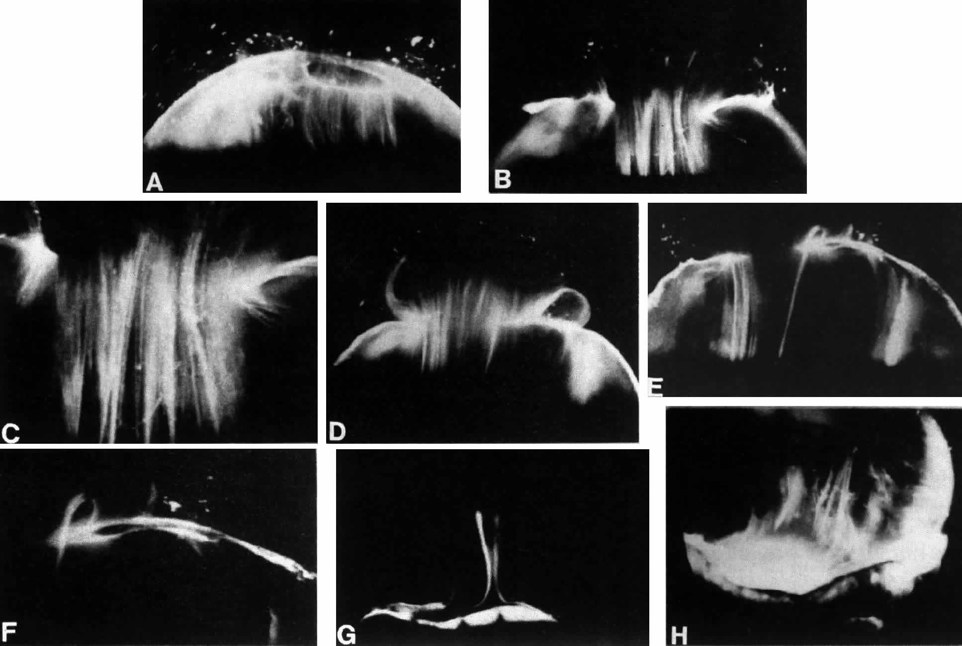
| Fig. 4. Human vitreous structure is visualized by darkfield slit microscopy. All photographs are oriented with the anterior segment below and the posterior pole above. Photographs are sequential, beginning in the upper left hand corner and moving left to right. A. Posterior vitreous in the left eye of a 52-year-old man. The corpus vitreous is enclosed by the vitreous cortex. There is a hole in the prepapillary (small, to the left) vitreous cortex. B. Posterior vitreous in a 57-year-old man. A large bundle of prominent fibers is seen coursing anteroposteriorly and entering the retrohyaloid space through the premacular vitreous cortex. C. Same view as B at higher magnification. D. Posterior vitreous in the right eye of a 53-year-old woman. There is posterior extrusion of vitreous out the prepapillary hole (to the right) and premacular (large extrusion to the left) vitreous cortex. Fibers course anteroposteriorly in the central vitreous and out into the retrocortical (formerly preretinal, before dissection) space. E. Horizontal optical section of the same specimen as D at a different level. A large fiber courses posteriorly from the central vitreous and inserts into the premacular vitreous cortex. F. Same view as E at higher magnification. The large fiber has a curvilinear appearance because of traction by vitreous extruding into the retrocortical space. However, because of its attachment to the posterior vitreous cortex the fiber arcs back to its point of insertion. G. Anterior and central vitreous in a 33-year-old woman. Cloquet's canal is seen forming the retrolental space of Berger. H. Anterior and peripheral vitreous in a 57-year-old man. The specimen is tilted forward to enable visualization of the posterior aspect of the lens and the peripheral anterior vitreous. Behind and to the right of the lens there are fibers coursing anteroposteriorly that insert into the vitreous base. These fibers “splay out” to insert anterior and posterior to the ora serrata. (A, E, and F: Sebag J, Balazs EA: Pathogenesis of cystoid macular edema: An anatomic consideration of vitreoretinal adhesions. Surv Ophthalmol 28[suppl]:493, 1984; B and C: Sebag J, Balazs EA: Morphology and ultrastructure of human vitreous fibers. Invest Ophthalmol Vis Sci 30:187, 1989) |