|
|
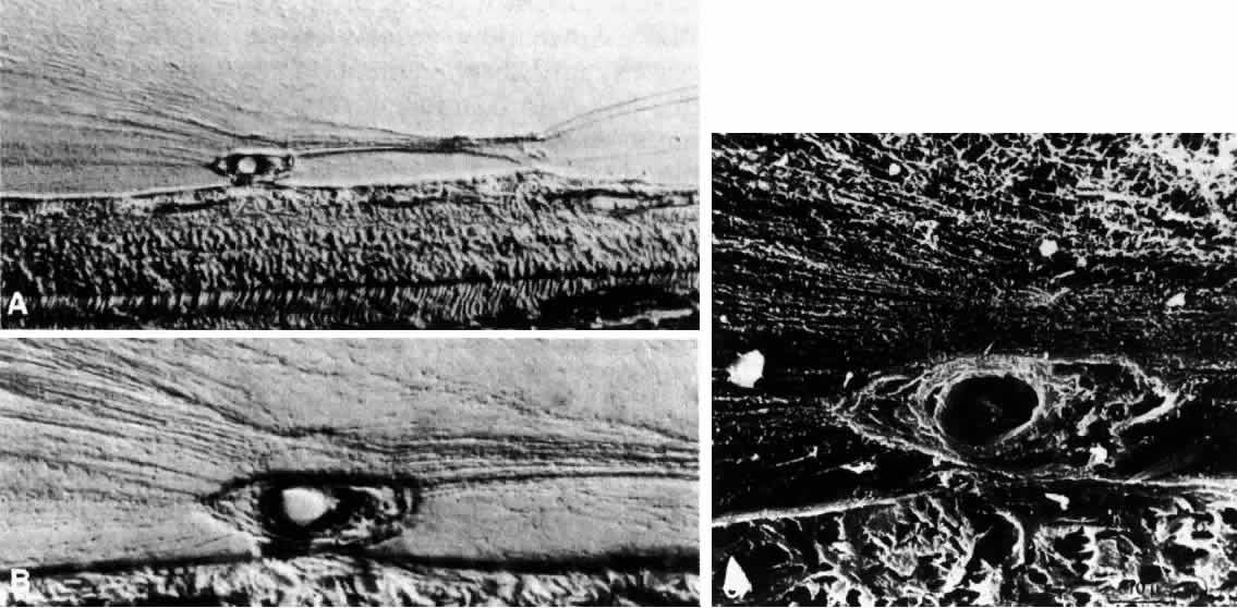
| Fig. 18. A-C. Neovascularization into the posterior vitreous cortex of a patient with proliferative diabetic retinopathy. Neovascularization from the disc and retina involves vascular endothelial cell migration and proliferation onto and into the posterior vitreous cortex. These photomicrographs demonstrate the formation of neovascular complexes arising from the retina (bottom of figures A-C) into the posterior vitreous cortex of a human eye and the insertion of vitreous collagen fibrils onto the new vessels (bar = 10 μm). (Faulborn J, Bowald S: Microproliferations in proliferative diabetic retinopathy and their relation to the vitreous: Corresponding light and electron microscopic studies. Graefes Arch Clin Exp Ophthalmol 223:130, 1985) |