|
|
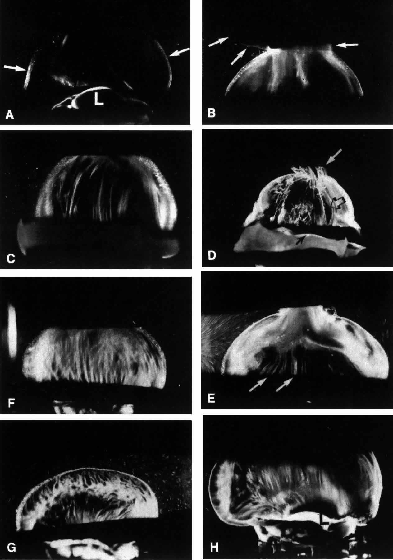
| Fig. 19. Vitreous structure in diabetes. Darkfield slit microscopy of human vitreous morphology at different stages of life. The anterior segment is below and the posterior pole is above in these optical horizontal sections. A. Whole vitreous in a 6-year-old boy, who died from trauma caused by a motor vehicle accident, demonstrates a dense vitreous cortex (arrows) and no fibers within the corpus vitreous. L, lens. B. Whole vitreous in an 11-year-old boy who died as a result of a head injury. Same findings are noted in A, even though vitreous extrudes out of the posterior vitreous cortex (arrows), placing sagittal traction on the central vitreous. C. Whole vitreous of a 56-year-old woman who died of cardiac arrest. Fibers with an anteroposterior orientation are present in the central vitreous. Adjacent to these fibers are areas devoid of structure, filled with liquid vitreous. D. Whole vitreous of an 82-year-old white woman. The corpus vitreous is collapsed (syneresis) and contains aggregated fibers extruding through the posterior vitreous cortex into the retrohyaloid space (white arrow). The central vitreous has lacunae (open black arrow) adjacent to the fibers. The closed black arrows indicate the posterior aspect of the lens. E. Right eye shows extrusion of whole vitreous through the posterior vitreous cortex (top) in a 9-year-old girl with type I diabetes. The subcortical vitreous appears very dense and scatters light intensely. Centrally, there are vitreous fibers (arrows) with an anteroposterior orientation and adjacent areas of liquefaction. F. Central vitreous in the left eye of same patient as in E shows prominent fibers that resemble those seen in nondiabetic adults (compare with C). G. Peripheral vitreous in the left eye of same patient in E and F shows fibers inserting into the vitreous cortex with adjacent pockets of liquid vitreous. H. Anterior vitreous in the left eye of same 9-year-old girl shows fiber insertion into the vitreous base about the lens (L). (Sebag J: Abnormalities of human vitreous structure in diabetes. Graefes Arch Clin Exp Ophthalmol 231:257, 1993) |