|
|
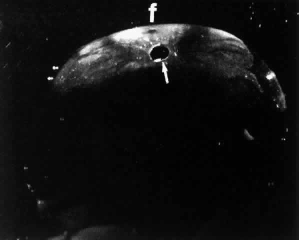
| Fig. 21. Darkfield microscopy of one form of anomalous PVD. Retinal elements may remain attached to the posterior vitreous cortex following PVD. Dissection of the retina off the vitreous cortex in this 14-year-old child resulted in the appearance shown in this photograph. A “cap” of tissue is adherent to the posterior vitreous cortex. A “hole” is present corresponding to the prepapillary region (arrow) and an “imprint” can be seen in the prefoveal region (f). There are linear, branching structures arising from the prepapillary region that likely correspond to “imprints” of the retinal blood vessels. (Sebag J: Age-related differences in the human vitreoretinal interface. Arch Ophthalmol 109:966, 1991. Copyright (χ) 1991, American Medical Association) |