|
|
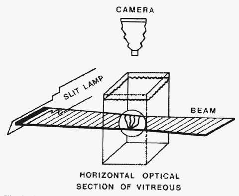
| Fig. 3. Schematic diagram of darkfield illumination system for the study of vitreous structure. The dissected specimen is mounted and immersed in a Lucite chamber containing a physiologic solution (see Fig. 2B). A slit lamp beam is shown from the side through the intact vitreous. The beam enters and exits laterally, avoiding any scattering of light by the structures of the anterior segment. The position of the beam can be raised or lowered, and the thickness of the illuminated portion can be regulated. Observation is performed from above, perpendicular to the plane of illumination. This achieves a 90° illumination/observation angle and thus maximizes the Tyndall effect. (Sebag J: The Vitreous--Structure, Function and Pathobiology. New York, Springer-Verlag, 1989) |