|
|
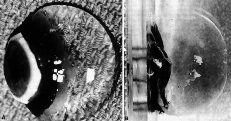
| Fig. 2. Human vitreous dissection. A. Vitreous obtained at autopsy from a 9-month-old child. The sclera, choroid, and retina were dissected off the vitreous, which remains attached to the anterior segment. A band of gray tissue can be seen posterior to the ora serrata. This is neural retina that was firmly adherent to the vitreous base and could not be dissected. Because of the young age of the donor, the corpus vitreous is almost entirely gel. Thus, it is solid and maintains its shape, although situated on a surgical towel exposed to room air. (Sebag J: The Vitreous--Structure, Function and Pathobiology. New York, Springer-Verlag, 1989) B. Human vitreous with the sclera, choroid, and retina dissected away; the corpus vitreous is still attached to the anterior segment. The specimen is mounted on a Lucite frame using sutures through the limbus and then immersed in a Lucite chamber containing an isotonic, physiologic solution. This maintains vitreous turgescence and avoids collapse and artifactual distortion of vitreous structure. (Sebag J, Balazs EA: Pathogenesis of cystoid macular edema: An anatomic consideration of vitreoretinal adhesions. Survey Ophthalmol 28[suppl]:493, 1984) |