Chapter 16
Cataract Extraction in Patients with Glaucoma
RONALD L. FELLMAN and GEORGE L. SPAETH
Main Menu   Table Of Contents

Search

GUIDING PRINCIPLES
CLINICAL ASPECTS OF OPTIC NERVE HEAD DAMAGE
VISUAL FIELD LOSS AND ITS RELATION TO CATARACT–GLAUCOMA SURGERY
ANTERIOR-SEGMENT TISSUE ALTERATIONS SECONDARY TO GLAUCOMATOUS DISEASE REQUIRE ADDITIONAL PLANNING
INTRAOCULAR PRESSURE CHANGES ASSOCIATED WITH LENS EXTRACTION
CASE STUDIES
CASE 1: ANTICIPATED LENS EXTRACTION WITH MILD TO MODERATE DISC DAMAGE AND IOP WITHIN TARGET PRESSURE RANGE
CASE 2: ANTICIPATED LENS EXTRACTION WITH MILD TO MODERATE DISC DAMAGE AND IOP WITHIN 4 MM HG OF TARGET PRESSURE RANGE
CASE 3: ANTICIPATED LENS EXTRACTION WITH MILD TO MODERATE DISC DAMAGE AND IOP FAR OUTSIDE OF TARGET PRESSURE RANGE
CASE 4: ANTICIPATED LENS EXTRACTION WITH ADVANCED DISC DAMAGE AND IOP WITHIN OR OUTSIDE TARGET PRESSURE RANGE
CASE 5: ANTICIPATED LENS EXTRACTION POST SUCCESSFUL TRABECULECTOMY
CASE 6: ANTICIPATED LENS EXTRACTION POST FAILED TRABECULECTOMY WITH ADVANCED DISC DAMAGE AND IOP OUTSIDE TARGET PRESSURE RANGE
PREOPERATIVE ASSESSMENT
SURGICAL TECHNIQUE
EXTRACAPSULAR CATARACT EXTRACTION VIA NUCLEAR EXPRESSION COMBINED WITH GLAUCOMA FILTRATION SURGERY
REFERENCES

This chapter provides a framework for meaningful decision-making regarding patient care in the setting of both cataract and glaucoma. The multitude of technological, procedural, and pharmacologic advances now available to ophthalmologists enables them to provide superior outcomes for their patients afflicted with cataract and glaucoma. The knowledge necessary to accomplish this is straightforward for some cases and exceedingly complex in others. The underlying theme throughout this chapter is that the management approach in the patient who has both glaucoma and cataract must be in many ways totally different from that of a person with cataract uncomplicated by glaucoma. The failure to appreciate this difference may lead to unnecessary morbidity and visual loss.

The trend in conversion from extracapsular cataract extraction (ECCE) to small incision cataract surgery continues to improve visual outcomes. The benefits from small incision cataract surgery for the stable glaucoma patient are extraordinary. The capacity to immediately improve vision with small incision cataract surgery alone, avoid the complications of trabeculectomy, potentially lower intraocular pressure (IOP) over the long-term and spare valuable conjunctiva for future glaucoma surgery is a surgical triumph for these patients. Consequently, a stable glaucoma patient on one or two medications with mild disc damage and symptomatic cataract may no longer require a combined or staged procedure. When filtration surgery is required, the capability to modulate wound healing and alter aqueous flow after trabeculectomy decreases complications and increases efficacy. From a pharmacologic viewpoint, the potent prostaglandin analogues enhance outflow thereby decreasing the need for more complex filtration surgery.

Patients with advanced disc damage, uncontrolled glaucoma at any stage, or complex cataracts present a much more challenging situation. They are far from elementary and present the ophthalmic surgeon and patient with many demanding problems. The knowledge and wisdom necessary to achieve a favorable outcome in these difficult cases requires a fusion of higher level preoperative, intraoperative, and postoperative decision-making skills. This situation is seen most commonly in patients requiring combined procedures who have advanced optic nerve damage complicated by any of the following: systemic diseases, especially diabetes mellitus; secondary glaucomas; anterior segment anomalies; miosis with posterior synechiae; pseudoexfoliation; keratopathy and complex cataracts. Society's perception of cataract surgery as a quick-fix operation with a rapid return of splendid vision is often not true for these patients who invariably perceive they are undergoing “routine” cataract surgery.1 Optic nerve damage or other concomitant eye diseases may limit visual recovery after flawless phacotrabeculectomy leaving a disappointed patient. Moreover, filtration surgery slows improvement in visual acuity.2,3 Informed consent with special emphasis on postoperative complications is especially important in glaucoma patients undergoing high-risk filtration surgery. Surgeons and their staff must spend additional time counseling patients who require filtration surgery due to the myriad of potential complications inherent to this procedure, which are far greater than routine lens extraction.

In years past, patients with both cataract and glaucoma frequently provided overwhelming surgical challenges for the ophthalmologist. The ability to carry out phacoemulsification through a 3.2-mm corneal incision along with inserting a foldable IOL is a vast improvement over 11-mm incisions that were common a decade ago (Fig. 1). The anatomical and inflammatory changes to the eye are less with small incision techniques, improving the likelihood of success with concomitant glaucoma surgery. Pharmacologic inhibition of fibrosis along with postoperative wound revision increases the long-term success rate of filtration surgery when combined with lens extraction. (Fig. 2). The learning curve may be steep at times, but the blending of cataract and glaucoma surgical skills slowly falls into place as the surgeon constantly learns and upgrades his or her technique.

Fig. 1. The anatomic advantage of small incision cataract surgery for the glaucoma patient. A. Long-term bleb function with a large cataract incision is difficult to achieve with either ECCE-trabeculectomy or trabeculectomy followed later by ECCE. This bleb failed to form sufficiently when combined with large incision ECCE. The inflammation, bleeding, and long-term wound healing with stimulation of fibroblasts associated with this technique are more likely to cause bleb failure. In addition, the increased iris manipulation necessary to deliver the nucleus and subsequent iris repair adds to the long-term breakdown of the blood aqueous barrier. B and C. Two-site phacotrabeculectomy has the advantage of small incision cataract surgery combined with separate site trabeculectomy. The incision size is one third the size of the standard ECCE. The inflammation is less severe, and cataract wound healing is confined to the temporal area. Visual rehabilitation with phacoemulsification and foldable IOL is much faster. Phacoemulsification allows successful lens extraction even in the unfriendly environment of a smaller pupil compared with ECCE. The trabeculectomy is performed in an entirely different site, well away from the wound healing associated with temporal phacoemulsification. The likelihood of this filter functioning long-term is greater than with ECCE-trabeculectomy. D. The surgeon also has the option of single-site phacotrabeculectomy with foldable IOL. Both the lens extraction and trabeculectomy are performed through one small 3.5-mm limbal incision.

Fig. 2. Wound modulation and revision. A. MMC decreases the likelihood of bleb failure in high-risk eyes undergoing combined cataract and glaucoma surgery. Overusage of this potent cytotoxic drug should be avoided owing to a myriad of problems related to the formation of an avascular bleb. B. Office-based laser revision of the operative wound allows for increased postoperative flexibility with less complications. The correct number and tension of scleral flap sutures allows the surgeon to strive for a safe immediate postoperative IOP range. When fibrosis occurs at approximately 1 week, suturelysis or release of sutures increases filtration while avoiding unwanted hypotony.

Small incision cataract surgery combined with continuous curvilinear capsulorrhexis (CCC), improved management of miotic pupils, divide-and-conquer nucleofractis phacoemulsification, clear corneal incision, capsular tension rings,4 phaco-chop, improved viscoelastics, piggy-back IOL systems, and foldable intraocular lenses (IOL) greatly improve cataract surgical outcomes. The use of dyes to stain the anterior capsule in patients with an absent red reflex as in a white cataract improves visualization for anterior capsulorrhexis. These techniques, in combination with antimetabolite-assisted guarded filtration surgery (GFS) and postoperative scleral flap suture lysis or releasable sutures, improve results for glaucoma patients with surgical cataracts. The ability to combine these two procedures through one small incision and simultaneously modulate wound healing provides excellent visual results.

Small-incision cataract surgery, however, is not appropriate for every glaucoma patient. The surgeon must be facile and maintain skill in all branches of cataract surgery, including intracapsular cataract extraction (ICCE), manual nuclear expression with extracapsular cataract extraction (ECCE), and phacoemulsification of soft or hard nuclei. A fusion of these skills is necessary because a subluxated rock hard nucleus may require ICCE, the nucleus may be too dense for emulsification requiring manual expression, the cornea may be cloudy preventing phacoemulsification, or the surgeon may find it necessary to convert to ECCE techniques during small-incision surgery.

Nonpenetrating glaucoma surgery is an alternative to trabeculectomy in the management of the patient with combined cataract and glaucoma. These procedures decrease the complications related to trabeculectomy, but fail to reduce IOP as effectively long-term. Nonpenetrating techniques are still in development and there place in the management of the patient with combined cataract and glaucoma continues to evolve.

This chapter provides a framework to help the surgeon caring for a patient with both cataract and glaucoma decide when to perform a cataract extraction alone, a glaucoma procedure alone, or a combined cataract-glaucoma procedure. The case studies found later in this chapter emphasize the order and timing of surgery for patients with both cataract and glaucoma. Furthermore, techniques that are especially appropriate when performing cataract extraction in patients with glaucoma will be presented in detail. The theme running through these decisions is based on the following section on guiding principles and on the concept that any additional surgical step carries with it the increased risk of complication.

Back to Top
GUIDING PRINCIPLES
Glaucoma is a disease in which ocular tissues become damaged by IOP that is higher than the tissues can tolerate. Glaucoma is not a single condition; the word encompasses a variety of entities of different pathogenesis and different intensity. Common to all the glaucomas is the end organ damage of an optic neuropathy with characteristic structural changes to the optic disc with associated visual dysfunction.

Knowledge of the basic principles related to the effects and side effects of cataract extraction in patients with glaucoma is essential if the surgeon is to choose a course that is most appropriate for each individual case. Some of these principles are known by all, and many are established but not well appreciated. Some of the principles that follow are neither well known nor well established but are based on our personal experience or the experiences of those whose clinical judgment appears to us to be sound. Principles not given a supporting reference usually fall into this category. An evidence-based approach to the literature is emphasized where appropriate and recently thoroughly reviewed.5,6

Back to Top
CLINICAL ASPECTS OF OPTIC NERVE HEAD DAMAGE
Short-term, moderate elevation of IOP is not likely to affect a healthy optic nerve.

Optic nerves vary in their ability to resist the damaging effects of IOP.7–15 Normal optic nerves of healthy adults are not usually damaged by IOPs below the diastolic ophthalmic artery pressure (around 30 to 35 mm Hg) unless the pressure persists for a minimum of 2 to 3 months. The exception is patients with sickle-cell disease.16–18 In addition, healthy optic nerves in adults can withstand short-term (perhaps up to 2 weeks) elevations of IOP below the systolic level of ophthalmic artery blood pressure (average, 70 mm Hg) without sustaining apparent damage (with the exception of sickle-cell abnormalities). IOPs greater than systolic retinal artery blood pressure damage all optic nerves permanently when such an elevation persists for more than a few minutes.

Glaucomatous optic nerves are more likely to be damaged by elevated IOP.

There are a variety of factors associated with IOP induced visual field loss; most notable is ocular perfusion pressure.19 Optic discs already compromised by glaucomatous disease or other optic neuropathies are at greater risk for further damage and can be permanently damaged by increases in IOP of relatively small magnitude and short duration. Precise data are lacking, but pressure elevations as small as a 50% increase (i.e., 20 to 30 mm Hg) for 1 month can be expected to cause a permanent worsening of optic nerve function in patients with serious disc disease prior to the pressure elevation.

Optic discs that are badly damaged by glaucoma (“sick discs”) can be further damaged permanently by pressure elevations as short as: 1 day or less when the pressure elevation is in the range of 50 mm Hg or more; 1 day when IOP is 30 to 50 mm Hg; or several days when the IOP spike is in the range of the diastolic ophthalmic artery blood pressure.

The visually damaging pressure level of IOP can be estimated.

The pressure at which a patient develops a hemorrhage of the optic disc or at which progressive disc damage or visual field loss occurs before cataract extraction gives a rough estimate of the level of IOP the optic nerve is able to tolerate. This pressure provides a baseline to use for predicting future damage. For example, a patient whose IOPs are fairly consistent at about 15 mm Hg and who develops an optic disc hemorrhage with pressures in that range is very likely to be at risk for progressive optic disc deterioration with IOPs above 15 mm Hg. In contrast, a patient who develops a disc hemorrhage when IOPs are averaging around 25 mm Hg probably has an optic disc that is more resistant to the damaging effects of IOP. This patient may do well with a long-term IOP of 15 to 20 mm Hg. These factors help the comprehensive ophthalmologist decide how high and how long a postoperative IOP spike is tolerable.

Pseudopits of the optic nerve are a sign of a pressure-sensitive optic nerve.

Optic discs in which there is an acquired pit of the optic nerve are probably damaged more rapidly by pressure elevations than are optic discs without pits of the optic nerve.20,21

Back to Top
VISUAL FIELD LOSS AND ITS RELATION TO CATARACT–GLAUCOMA SURGERY
Visual field defects of a diffuse type are generally characteristic of patients whose optic nerves are relatively resistant to the damaging effects of IOP.22–24 Dense paracentral defects, however, are characteristic of patients whose optic discs are more sensitive to the damaging effects of IOP.25,26

FACTORS AFFECTING THE LIKELIHOOD OF VISUAL FIELD DAMAGE

Patients with visual field loss that extends into fixation are more likely to have their visual acuity damaged by postoperative pressure elevations than are patients whose field defects spare the area of fixation. Appropriate preoperative planning, such as combined or staged surgery to eliminate damaging postoperative pressure elevations is essential for this group of patients. In addition, patients with preoperative visual field defects that split into fixation may experience a sudden decrease in acuity associated with an otherwise uncomplicated intraocular surgery. This phenomenon of wipeout occurs in 1% to 5% of cases.27,28 “Wipeout” also appears to be related to severe postoperative hypotony and is more common in patients whose postoperative IOPs are less than 5 mm Hg. Hypotony maculopathy is a well-recognized cause of visual loss following filtration surgery. Every conceivable effort should be made to avoid long-term maculopathy and keratopathy due to excessive filtration. In spite of a surgeon's best effort, this complication will always be an issue with filtration surgery.

THE EFFECT OF CATARACT ON AUTOMATED PERIMETRY

Nonglaucomatous patients with progressive nuclear sclerosis develop a generalized depression in their field. This is detected during automated perimetry on the Humphrey Field Analyzer (HFA) and is expressed with software analysis on the glaucoma hemifield test as a generalized depression. This generalized depression is also reported as an abnormal global index expressed as decreased mean deviation. Glaucoma patients develop both generalized or localized visual field defects. Nuclear sclerosis causes a worsening of both pre-existing generalized and localized glaucomatous field defects. A cataract by itself does not produce a dense scotoma.29 Worsening of a scotoma without an increase in generalized depression of the rest of the field is not due to a nuclear cataract. The best method of determining whether field progression is due to cataract or glaucoma is to dilate the pupil and compare the optic disc to an earlier less-cataractous state. A thorough understanding of normal and abnormal visual field indices related to automated perimetry is essential in order to reliably interpret automated visual fields.30

Following uncomplicated cataract extraction in glaucoma patients, there is an overall improvement in mean deviation,31,32 and foveal threshold33 however, dense paracentral scotomas may appear deeper as noted by a worsening of the corrected pattern standard deviation, (CPSD).34 This worsening of CPSD appears to be less with phacotrabeculectomy augmented with Mitomycin C.35 From a perimetric psychophysical viewpoint,36 the statistical worsening of the CPSD is likely due to raising the height of the island of vision while the floor of the scotoma remains the same.

Back to Top
ANTERIOR-SEGMENT TISSUE ALTERATIONS SECONDARY TO GLAUCOMATOUS DISEASE REQUIRE ADDITIONAL PLANNING
The anatomic and physiologic alterations of glaucomatous eyes are protean.

Recognition of these alterations facilitates treatment of the disease process. A severe attack of angle-closure glaucoma causes corneal endothelial damage, cataract, trabecular damage, posterior synechiae, and iris necrosis. These tissue alterations will alter the surgeon's approach to cataract surgery. Corneal decompensation after otherwise uncomplicated cataract extraction is common in patients having sustained a severe attack of acute angle-closure glaucoma.37 Surgeons contemplating cataract extraction in these eyes naturally desire to avoid as much corneal trauma as possible. Glaucoma patients with concomitant corneal disease such as iridocorneal endothelial syndrome (ICE) should be informed of likely corneal decompensation associated with glaucoma or cataract surgery.

The atonic pupil that follows a severe angle-closure attack is fixed and dilated. This pupil alteration may cause the surgeon to choose an IOL with a larger optic to prevent glare and monocular diplopia associated with the larger pupil. Patients should be instructed that visual aberrations associated with abnormal pupils from glaucomatous disease might alter visual performance post cataract surgery. Eyes with short axial lengths typically have a very shallow anterior chamber. This makes it even more difficult for the surgeon to work, especially when introducing any instrument into the eye. In addition, eyes with unusual axial lengths require more sophisticated IOL calculation formulas; otherwise, undesirable postoperative hyperopia occurs.

The iris of glaucoma patients is usually abnormal.

Widespread glaucomatous iris abnormalities include abnormal blood vessel permeability, flaccid iris tone, rigid or atrophic muscles, and friability. The iris of patients with the exfoliation syndrome is extremely tender and easily torn. Pigment dispersion, breakdown of the blood–aqueous barrier, and hyphema at the time of surgery and postoperatively are common in these eyes. The lens capsule and zonules tend to be more fragile in patients with glaucoma than in those without glaucoma.38,39 The problem is most severe in those with the most advanced glaucoma who have been treated most intensively. Surgery is extremely complicated in the exfoliation syndrome because of zonular dehiscence, loose zonules, sticky cortex, and altered blood–aqueous barrier. The lenses of patients with long-standing glaucoma and advanced cataracts are often partially “loose” even though they do not appear frankly dislocated. Long-term parasympathomimetic drug therapy produces leaky iris vessels with breakdown of the blood–aqueous barrier, posterior synechiae, peripheral anterior synechiae, and rigid miotic pupils. Inability to sufficiently dilate the pupil is a leading cause of vitreous loss at the time of cataract surgery. The final outcome of cataract extraction is worsened by preoperative long-term use of para sympathomimetic drugs because the pupil does not dilate well, the iris is more likely to bleed when traumatized, breakdown of the blood–aqueous barrier is excessive,40 and there is an increased likelihood of vitreoretinal interface problems.

The conjunctiva of patients with glaucoma is often abnormal.

The tissues of patients with glaucoma who have been treated medically for glaucoma are not as healthy as the tissues of patients who do not have glaucoma.41 The conjunctiva undergoes several tissue alterations due to topical antiglaucoma therapy. These tissue alterations may lead to higher intraoperative complications and long-term filtration failure.

Back to Top
INTRAOCULAR PRESSURE CHANGES ASSOCIATED WITH LENS EXTRACTION
IOP rises during the first 24 hours following cataract extraction.

Cataract extraction with complete wound closure is associated in all cases with a postoperative rise in IOP. The greater the inflammation and the more seriously damaged the outflow channels, the greater the intensity and rise of IOP after cataract extraction. Viscoelastic substance,42 severity of pre-existing glaucomatous trabecular damage, suture deformation with trabecular collapse, cortical and pigmentary debris, blood components, breakdown of the blood–aqueous barrier, and altered prostaglandin metabolism all factor into the decrease in outflow facility. The severity, height, and duration of IOP rise are worse if the surgery is complicated by loss of vitreous. The highest IOP after either ICCE or ECCE appears to occur around 6 hours after surgery.43–45 As many as 15% of nonglaucomatous patients undergoing uncomplicated ECCE with posterior chamber intraocular lens (PCIOL) develop pressure spikes greater than 40 mm Hg within 24 hours of surgery without prophylactic pressure-lowering drugs.46 On the first postoperative day, as many as 55% of patients post-ECCE with a PCIOL develop IOPs in excess of 25 mm Hg.47 This occurs even when viscoelastic material is removed at the time of surgery but without the advantage of pressure-reducing drugs used at the conclusion of surgery.

Even in normal eyes, mean IOP rises approximately 30% on the first day after uncomplicated phacoemulsification with 6% of patients experiencing IOPs greater than 30 mm Hg.48 IOP elevations during the first postoperative day are common following uncomplicated phacoemulsification with either scleral or clear corneal incisions.49 After phacoemulsification and foldable IOL implantation, the immediate postoperative IOP increase is higher in eyes undergoing sclerocorneal incisions compared with clear corneal incisions.50

Pressure elevation in glaucomatous eyes after cataract extraction is much worse and of longer duration than in nonglaucomatous eyes.

Postoperative pressure spikes may lead to blindness and visual field loss in glaucomatous eyes.51 These pressure elevations are a particular problem in patients unable to tolerate carbonic anhydrase inhibitors (CAIs) or topical beta-adrenergic blocking agents. Several measures can be taken to control and monitor immediate postoperative IOP rises in all eyes undergoing cataract extraction:

  1. Avoid excessive intraocular trauma.
  2. IOP response varies with viscoelastic,52,53 which should be thoroughly removed.
  3. Inject intracameral carbachol or acetylcholine to enhance outflow (avoid in uveitis).54
  4. Perform peripheral iridectomy in patients with diabetes mellitus, high hyperopia, rubeosis iridis, intraoperative vitreous loss, and uveitis in order to avoid pupillary block glaucoma.
  5. IOP check 30 minutes after cataract extraction is insufficient to detect elevated IOP55
  6. Topical CAIs reduce postoperative IOP better than prostaglandin analogues,56 and prostaglandin analogues lower IOP better than placebo57
  7. Oral CAIs are effective in blunting postoperative IOP spikes58
  8. Prostaglandin analogues are not as effective in the immediate postoperative period as combination fixed aqueous suppressants59
  9. Use topical parasympathomimetic drugs such as 2 % pilocarpine if inflammation is not excessive.
  10. Use nonselective alpha agonist short-term, pre and postoperatively, 60,61 less effective after phacoemulsification62
  11. Sole use of perioperative alpha 2 agonists is ineffective in preventing short- term IOP spikes63,64
  12. Consider serial anterior chamber paracentesis when pressure is dangerously high.65

Postoperatively, patients with badly damaged optic nerves may lose considerable visual acuity or visual field during the above pressure-lowering efforts. Patients with mild to moderate glaucoma damage generally tolerate the above-mentioned procedures without significant visual loss. Patients with badly damaged optic nerves require additional planning in order to prevent loss of vision. This may require treatment by combining cataract and glaucoma surgery, or staging the procedures with filtration surgery followed later by cataract extraction. Patients with advanced nerve damage require more frequent postoperative visits starting as early as 4 to 6 hours after surgery in order to detect the first potentially damaging postoperative IOP spike. If the surgeon is not comfortable with a combined procedure and the IOP is dangerously elevated, a filtration procedure alone may be preferable to stabilize the glaucoma, followed later by lens extraction.

Argon laser trabeculoplasty (ALT) tends to be less effective in aphakic or pseudophakic patients than in phakic patients.

Use laser trabeculoplasty before cataract extraction to gain better control of glaucoma in preparation for cataract surgery. If indicated, ALT is more effective when performed before lens extraction; in addition, postoperative pressure spikes are dampened in these eyes67–68 Following effective ALT, decrease glaucoma medications in anticipation of cataract extraction, allowing room to resume drugs after surgery if IOP is problematic. ALT is effective in approximately two thirds of patients with open angles post-failed filtration surgery.69 Selective laser trabeculoplasty (SLT) appears similar to ALT in reducing IOP before lens extraction while maintaining IOP control in the postoperative period.

Cataract extraction alone, after the initial pressure spike, often leads to long-term lowering of IOP, most notably after clear corneal phacoemulsification.

This lowering of IOP occurs with both ICCE and ECCE surgery. The magnitude of the pressure lowering appears to be increased in patients having PCIOL implantation at the time of the cataract extraction.70–73 The reduction in IOP associated with phacoemulsification is greater than ECCE. Stable, medically controlled glaucoma patients who undergo uncomplicated clear corneal phacoemulsification show a significant reduction in the number of postoperative glaucoma medications at one year, particularly in patients with pseudoexfoliation syndrome.74,75 Even nonglaucomatous patients with normal IOP before uncomplicated clear corneal phacoemulsification experience reduced postoperative IOP.76 A similar IOP lowering effect is seen at 5 years with phacoemulsification through a scleral approach.77 The mechanism of IOP lowering after phacoemulsification is unclear. However, patients with a reduced preoperative facility of outflow show a significant improvement in outflow facility following phacoemulsification at one year.78 Long-term IOP control after phacoemulsification with a foldable IOL is slightly better with a clear corneal approach compared to sclerocorneal.79

IOP CHANGES ASSOCIATED WITH COMBINED CATARACT AND GLAUCOMA SURGERY

Immediate IOP control after combined cataract-glaucoma surgery is better than cataract extraction alone.

Immediate IOP control after combined procedures is especially beneficial in patients with advanced disc damage who are already on maximal medical therapy. Phacotrabeculectomy with eventual bleb failure has a similar long-term pressure-lowering efficacy of phacoemulsification alone. However, the combined procedure that eventually fails is not a complete failure if the optic nerve damage from the immediate spike in IOP is prevented.80 Combining cataract extraction with any of a variety of methods that allows for increased outflow effectively minimizes the pressure spike that occurs after cataract surgery (Table 1). Phacotrabeculectomy protects against early postoperative elevations in IOP compared with phacoemulsification alone. This difference in IOP is detectable at 4 hours after surgery with 5% of phacotrabeculectomy eyes experiencing IOP greater than 30 mm Hg compared with 23% with phacoemulsification alone.81 If the IOP is elevated a few days after surgery, release of scleral flap sutures enhances filtration, and the timing of this is critical to long-term bleb survival.82


Table 1. Patients Who Are Candidates for Combined Cataract and Glaucoma Procedures Include Symptomatic Cataract and Any of the Following:

  1. Progressive glaucomatous optic nerve head cupping
  2. Uncontrolled IOP on maximal medical therapy
  3. Advanced nerve damage on two or more antiglaucoma medications
  4. Intolerance to multiple glaucoma medications
  5. Failed trabeculectomy with uncontrolled IOP
  6. Progressive glaucomatous visual field progression
  7. Anticipated optic disc damage as a result of excessive postoperative IOP
  8. Lack of compliance with anticipated glaucomatous visual loss


Long-term IOP control after combined cataract-glaucoma surgery is better than cataract extraction alone.

Combining a cataract extraction with a filtration procedure without using an antimetabolite, results in a long-term lowering of IOP slightly greater than that caused by cataract extraction alone.83–87 Long-term IOP control after phacotrabeculectomy is better than with ECCE-trabeculectomy, with fewer medications needed in the phacotrabeculectomy group.88

5-Fluorouracil (5-FU) does not improve the IOP-lowering effect of combined cataract and glaucoma surgery.

Adjunctive use of 5-FU at the time of cataract extraction has, in the dosage and methods so far used, not appreciably improved the long-term pressure lowering associated with ECCE combined with a guarded filtration procedure.89–91 There also appears to be limited pressure-lowering benefit from postoperative 5-FU injections for combined same-site phacotrabeculectomy.92–94 There is limited data on the efficacy of intraoperative 5-FU for combined phacotrabeculectomy.

Mitomycin-C (MMC) does improve the IOP-lowering effect of combined cataract and glaucoma surgery.

Antimetabolite usage is worth the long-term risks of blebitis and hypotony in patients who are likely to go blind without its application. However, patients without advanced disc damage who can tolerate one or two postoperative glaucoma medications may not need an antimetabolite and certainly should not be exposed to high concentrations of MMC.

Combined same-site phacotrabeculectomy with MMC lowers IOP more effectively with fewer postoperative medications and larger filtration blebs than without MMC.95–97 MMC also improves the success rate in blacks, eyes of patients on two or more glaucoma medications, eyes with IOP 20 mm Hg or higher, and prior failed trabeculectomy.98,99 Antimetabolite use in combined procedures is associated with a lower postoperative IOP with reduced need for long-term glaucoma medications.100 MMC appears to have a beneficial effect on long-term filtration surgery and on combined surgery without the corneal toxicity of 5-FU.101 However, long-term bleb morphology with MMC is different from that with 5-FU. Excessive concentrations or durations of MMC cause progressive conjunctival necrosis with bleb leaks, hypotony, and possible endophthalmitis. Antimetabolite usage has decreased with trabeculectomy surgery and is reserved for high-risk patients likely to scar down without its usage.

COMBINED CATARACT AND GLAUCOMA SURGERY

Phacotrabeculectomy is more effective than ECCE-trabeculectomy with or without postoperative 5-FU injections.102–105 The smaller incision associated with phacoemulsification likely leads to less intraocular inflammation and wound healing, allowing better bleb formation. Phacotrabeculectomy with intraoperative or postoperative 5-FU significantly lowers IOP but not as successfully as 5-FU trabeculectomy alone.106 This is probably related to the prolonged anterior chamber flare following phacoemulsification compared with trabeculectomy with peripheral iridectomy.107 Combined same-site phacoemulsification, posterior chamber IOL, and trabeculectomy without antimetabolite significantly lowers IOP.108–111 Results of same-site phacotrabeculectomy appear similar with either a 3.5-mm incision with a foldable IOL or a 5.2-mm incision with a rigid single piece PMMA lens.112,113

Phacotrabeculectomy with intraoperative 5-FU is as efficacious as a 5-FU trabeculectomy followed by phacoemulsification.114 Visual acuity and complications of combined phacotrabeculectomy are comparable to a two-staged approach with the obvious benefit of earlier visual rehabilitation.115 Foldable silicone lenses are associated with an increase in postoperative inflammation compared to PMMA lenses.116

SINGLE-SITE VERSUS TWO-SITE COMBINED CATARACT AND GLAUCOMA SURGERY

Combined ECCE-trabeculectomy techniques now yield to smaller incision combined phacotrabeculectomy. The methods for combining phacotrabeculectomy vary, depending on physician training, preference and patient anatomy. For example, physicians with two-site experience will have an easier access to the surgical site in a patient with enophthalmos and a prominent brow who requires a combined procedure. A temporal approach for the lens extraction is much easier in this particular case. IOP control following one-site versus two-site phacotrabeculectomy with MMC is similar but with a trend for less postoperative glaucoma medications,117 improved IOP control,118 less induced astigmatism,119 and better bleb formation in the two-site group.120

CATARACT EXTRACTION CAUSES PARTIAL OR COMPLETE FAILURE OF EXISTING FILTERING BLEBS

Cataract extraction by any technique performed in a patient with a pre-existing filter will have an effect on the previous filtering bleb.121–124 Bleb failure is more likely with ECCE compared with small-incision phacoemulsification.125 However, even patients undergoing topical anesthesia with clear corneal phacoemulsification and foldable IOL may experience bleb failure. One of three patients with a functioning filter and preoperative mean IOP of 12 mm Hg without antiglaucoma medications experiences bleb failure after lens extraction (Table 2). These patients require long-term drug therapy or bleb needling to control IOP. 126 Additional incisional glaucoma surgery may eventually be required in up to 10% of patients.127 Intraoperative iris manipulation may cause significant breakdown of the blood–aqueous barrier, resulting in inflammation that causes bleb failure. Even after uncomplicated clear corneal phacoemulsification, IOP may increase an average of 2 to 3 mm Hg due to bleb fibrosis.128 Approximately 20% of filtered patients require a long-term increase in glaucoma medications following uncomplicated clear corneal phacoemulsification with a foldable copolymer acrylic IOL129 (Fig. 3). In situations in which the bleb is not working at all, the eye will have a postoperative pressure spike that mimics that in the patient not having had a prior filtering procedure. In situations in which the bleb is marginal, the pressure spikes tend to be lower, and the final postoperative IOP tends to be around 50% higher than it was preoperatively. These patients require combined procedures in order to reestablish long-term filtration. In situations in which the bleb is very thin, polycystic, and associated with an IOP around 5 to 8 mm Hg on no antiglaucoma therapy; uncomplicated cataract extraction will have a minimal effect on the level of IOP. Patients with functioning glaucoma drainage implants usually have minimal long-term changes in IOP after uncomplicated cataract extraction.130


Table 2. Risk Factors for bleb failure following lens extraction

Preoperative

    1.  Uveitic glaucoma
    2.  Neovascular glaucoma
    3.  Period less than 6 months between trabeculectomy and lensextraction
    4.  Preoperative IOP greater than 10 mm Hg
    5.  Age of 50 years or younger

Intraoperative

    6.  Large cataract incision
    7.  Failure to adequately close wound
    8. Excessive iris manipulation
    9.  Vitreous loss
   10.  Inadequate fixation or positioning of IOL

Postoperative

   11.  Iridocapsular adhesions secondary to can-opener capsulotomy
   12.  Postoperative hyphema
   13.  Failure to adequately use postoperative corticosteroids


Fig. 3. Partial bleb failure following clear corneal phacoemulsification with foldable IOL. A. Preoperative bleb appearance prior to temporal lens extraction. Preoperative IOP was 12 mm Hg on no antiglaucoma medications. Time from 5-FU trabeculectomy surgery to lens extraction was one year. B. Bleb appearance 2 months after clear corneal cataract surgery with topical anesthesia. Following lens extraction, increased vascularity was noted along with decreased size of the filtering bleb. IOP increased to 20 mm Hg as early as 2 weeks after surgery, necessitating topical antiglaucoma therapy. C. High magnification view of bleb before lens extraction demonstrates diffuse pale bleb. D. High magnification view of bleb 2 months after surgery. There are vessels surrounding the nasal side of the bleb and the overall bleb size is smaller.

Tissue plasminogen activator (TPA), a protease with fibrinolytic-thrombolytic activity, may be helpful in dissolving either fibrin or blood clots that are obstructing bleb flow. TPA may be injected into the anterior chamber to dissolve fibrin clots that impede flow into the trabeculectomy site.131 The application of TPA may salvage a failing bleb after cataract surgery but sometimes leads to hypotony or hyphema.

Gonioscopy is essential in determining the etiology of bleb failure post cataract extraction. If a gossamer fibrin membrane forms over the internal trabeculectomy stoma post lens extraction, use the YAG laser to disrupt the membrane and re-establish flow. Needle the bleb when filtration becomes delimited by fibrosis. There are a variety of bleb needling techniques that are all designed to increase external filtration into and around the bleb.132, 133

Bleb failure after cataract extraction is actually beneficial in the setting of preoperative hypotony maculopathy.134,135 The surgeon may elect to use postoperative corticosteroids sparingly in order to encourage inflammation and wound healing. However, elderly patients with long-standing pale avascular blebs and reduced aqueous production are less likely to experience long-term IOP elevation.

PHACOTRABECULOTOMY FOR UNCONTROLLED GLAUCOMA AND SYMPTOMATIC CATARACT

Patients who are at high risk for complications related to trabeculectomy are candidates for trabeculotomy. The magnitude of IOP reduction with trabeculotomy is significantly less than with trabeculectomy. Patients with a history of bleb related endophthalmitis, profound hypotony from trabeculectomy, symptomatic filtering blebs or cicatricial ocular surface disease are candidates for trabeculotomy. Trabeculotomy with careful scleral and conjunctival wound closure in adults is not associated with postoperative hypotony. Postoperative hyphema is common but generally clears within a week and a pressure spike on postoperative day 1 is common. The technique varies depending on surgeon preference.136 The results with either a one- or two-site technique are similar.137 Postoperative IOP control with phacotrabeculotomy is superior to phacoemulsification alone138 and appears to be most effective in patients older than age 70.139

PHACOVISCOCANALOSTOMY AND PHACO/DEEP SCLERECTOMY FOR UNCONTROLLED GLAUCOMA AND SYMPTOMATIC CATARACT

Nonpenetrating procedures as they exist today generally decrease postoperative complications but do not reduce IOP as successfully as trabeculectomy.140 The success rate also appears to be highly dependent on race, length of topical antiglaucoma therapy, and prior ocular surgery.141,142 Deep sclerectomy combined with phacoemulsification results in an IOP reduction similar to phacotrabeculectomy at one year with comparable visual outcome.143 Phacoviscocanalostomy lowers IOP by approximately 33%144 through either a one- or two-site approach145 (Fig. 4). Nonpenetrating procedures are in evolution, and their place in long-term glaucoma care is still unclear.


Fig. 4. Viscocanalostomy with deep sclerectomy and phacoemulsification. Nonpenetrating filtration procedures (NPFS) may be combined with phacoemulsification. Patients with mild disc damage and a history of limited topical drug therapy are the best candidates. Patients who require an IOP in the low teens are not good candidates for NPFS. By definition, NPFS is designed to lower IOP without penetrating into the anterior chamber, thereby avoiding the complications associated with trabeculectomy. Viscocanalostomy is intended to allow aqueous to percolate through a trabeculodescemetic membrane into a subscleral cavern created by the deep sclerectomy. The aqueous diffuses from the cavern into the dilated ostia of Schlemm's canal and into the episcleral venous plexus. A. Fashion a uniform 300-micron superficial scleral flap 1 mm into clear cornea. B. Construct a second 600-micron deep flap that facilitates the unroofing of Schlemm's canal, seen as the darker area. C. Use viscoelastic to dilate the ostia of Schlemm's canal. The major problem with viscocanalostomy is the eventual closure of the ostium decreasing flow to the episcleral plexus. D. Dissect the deep flap anteriorly into clear cornea creating the trabeculodescemetic membrane. This membrane is clearly seen between the scleral spur and the bend of the deep flap. The integrity of this membrane ensures the nonpenetrating portion of the surgery. Another problem with NPFS is the eventual fibrosis of this initially transparent membrane requiring goniopuncture. E. Deep sclerectomy gets its name from removal of the deep flap. Removal of this flap creates the potential subscleral space for accumulation of aqueous before it enters Schlemm's canal and exits the episcleral venous plexus. After removal of the deep flap, the superficial flap is sutured into place and conjunctiva closed. Approximately half of these procedures develop a shallow bleb.

CATARACT EXTRACTION ALONE REDUCES IOP IN MOST EYES WITH ANGLE-CLOSURE GLAUCOMA

Uncomplicated cataract extraction substantially reduces IOP, along with the number of postoperative glaucoma medications in eyes with angle-closure glaucoma.146,147 When preoperative gonioscopy reveals PAS, along with adjacent areas of appositional closure, lens extraction alone in select cases may be a reasonable alternative to filtration surgery.148,149 Phacomorphic angle-closure disease due to enlargement of the lens with progressive angle crowding is eliminated following lens extraction. The width and depth of the anterior chamber angle in eyes with angle-closure glaucoma increases significantly after cataract extraction with IOL implantation and becomes similar to open-angle glaucoma and normal eyes.150,151 (Fig. 5). Combining phacoemulsification, IOL implantation, and limited goniosynechialysis is effective in the treatment of cataract and chronic angle-closure glaucoma.152 Phacoemulsification with implantation of a foldable IOL is more effective in reducing IOP and improving visual acuity than surgical peripheral iridectomy in eyes with acute angle-closure glaucoma.153

Fig. 5. Anterior chamber angle changes associated with lens extraction and PCIOL This 65-year-old Vietnamese woman has a long-standing history of chronic angle-closure glaucoma treated with laser peripheral iridectomy. The optic nerve demonstrated mild glaucomatous damage and IOP was moderately controlled on two antiglaucoma medications. The cataract was removed through temporal clear corneal phacoemulsification with foldable acrylic IOL. A. Symptomatic cataract in narrow-angle glaucoma eye with patent iridectomy. B. Intraoperative goniophotograph showing crowding of angle with increasing narrowness due to phacomorphic component. C. Intraoperative photograph showing temporal clear corneal approach with IOL in the capsular bag. D. Intraoperative goniophotograph demonstrating deepening of chamber angle following lens extraction. Proposed theories for IOP reduction following lens extraction with complete wound closure:

  1. Anterior chamber deepening with improved access to trabecular meshwork
  2. Increase in traction on the trabecular meshwork
  3. Improved outflow facility mediated by an increase in prostaglandin release
  4. Reduction in aqueous humor production
  5. Atrophy of ciliary body processes
  6. Goniosynechialysis due to intraoperative over deepening of AC with viscoelastic
  7. Relief of undiagnosed pupil block

SUCCESSFUL TRABECULECTOMY SIGNIFICANTLY DECREASES AXIAL LENGTH CAUSING INACCURATE IOL CALCULATIONS

Surgeons contemplating “routine” cataract extraction in patients with pre-existing successful trabeculectomy surgery must convey their concerns regarding postoperative refractive surprises. Axial length decreases an average of 0.4 mm after trabeculectomy, with some eyes decreasing as much as 2.8 mm.154 After lens extraction the axial length increases toward the eyes' original shape, thereby inducing myopia. There is a significant association between decrease in axial length and use of antimetabolites.155 Because the incidence of cataract after filtering surgery is increased,156 surgeons benefit from obtaining preoperative biometry for future comparisons.

ANTERIOR CHAMBER LENSES ARE LESS PREFERABLE THAN POSTERIOR CHAMBER LENSES IN PATIENTS WITH GLAUCOMA

First-generation anterior chamber lenses contacted uveal tissue and caused inflammation, glaucoma, and hyphema. Superior haptic design and AC stability are worthy improvements in these lenses. Owing to the greater inflammatory response and potential for suprachoroidal hemorrhage associated with suturing in a posterior chamber lens, it is not known whether a sutured-in posterior chamber lens or a new-generation anterior chamber lens is preferable in cases in which a bag fixated posterior chamber IOL cannot be used. Placement of the PCIOL in the bag is preferable to sulcus placement because the lens tends to be more posterior and thereby causes less inflammation. Posterior chamber lenses designed for bag fixation should not be placed in the sulcus owing to the increased likelihood of iris chaffing, pigment release, inflammation, and worsening of glaucoma.

EYES WITH GLAUCOMA ARE PREDISPOSED TO SUPRACHOROIDAL HEMORRHAGE, BOTH INTRAOPERATIVELY AND POSTOPERATIVELY

Suprachoroidal hemorrhage is a devastating, painful complication of ocular surgery. It may occur intraoperatively157 or postoperatively. The major predisposing factor is always hypotony. This occurs most frequently in glaucoma patients with aphakic vitrectomized eyes undergoing filtration surgery.158–165 Suprachoroidal hemorrhage is also more likely to occur in myopic, hypertensive patients with long-standing glaucoma with high IOPs at the time of cataract extraction. Visual loss may be severe or mild depending on immediate extrusion of uvea and retina, length of time for wound closure, and subsequent retinal detachment. Should a suprachoroidal effusion or hemorrhage occur during small-incision cataract surgery, the self-sealing anatomy of the wound and the controlled irrigation pressure system allow for a rapid watertight wound closure, preventing expulsion of intraocular contents.

Identification of patients at high risk for suprachoroidal hemorrhage may considerably alter the surgeon's thought processes. The following factors are considered:

  1. Aggressive preoperative IOP control
  2. Small-incision surgery safer than large incision
  3. Preplace sutures
  4. Minimize surgical time
  5. Avoid collapse of the globe during surgery
  6. Prophylactic sclerostomies (Sturge-Weber and nanophthalmos)
  7. Discontinuation of anticoagulants at appropriate time
  8. Superb control of blood pressure before, during, and after surgery
  9. Prevention of postoperative hypotony by placing extra sutures in the trabeculectomy flap at the time of filtration surgery
  10. Avoid excessive concentration or duration of antimetabolites
  11. Protect the blood–aqueous barrier by preventing excessive inflammation with generous postoperative topical and, if necessary, oral corticosteroids
  12. Recognition of postoperative choroidal effusions and vigorous treatment of same, knowing that these choroidal effusions are often the triggering event for hemorrhage and may require drainage in high-risk eyes
  13. Counseling of patients not to do those things that mimic a Valsalva maneuver
  14. Use stool softeners, encouraging liquid intake
  15. Use bandage contact lenses to tamponade the bleb and prevent hypotony
  16. Return to surgery to place additional scleral flap sutures if postoperative IOP is lower than anticipated
  17. Injection of viscoelastic into anterior chamber
  18. In very high risk eyes, injection of gas bubble into the vitreous cavity
  19. Do not release or laser sutures prematurely, test first with slight focal pressure on the wound

Postoperative suprachoroidal hemorrhage is most likely to occur during the first 2 weeks after trabeculectomy, especially following premature suture lysis. Commonly, a choroidal effusion develops as a result of hypotony. As the choroidal effusion increases in size, stretching of the long ciliary artery in the suprachoroidal space occurs. This vessel may break at a critical point, precipitating this sight-threatening event.166 Surgeons should never be timid in returning to the operating room to drain a choroidal effusion, especially in high-risk monocular patients.

THE EFFECT OF GLAUCOMA SURGERY ON VISUAL ACUITY VARIES DEPENDING ON THE PREOPERATIVE STATUS OF THE PATIENT AND THE TECHNIQUE OF GLAUCOMA SURGERY

Patients with small pupils and axial cataracts frequently have significant improvement in visual acuity after glaucoma surgery, especially when accompanied by sector iridectomy. On the other hand, a persistent shallow anterior chamber after glaucoma surgery is associated with development or worsening of pre-existing cataract, especially if accompanied by excessive inflammation. If the anterior chamber is totally flat (lens-corneal touch), development of cataract is rapid and severe. In addition, a flat chamber leads to peripheral anterior synechiae, iridocorneal adhesions, blockage of the trabeculectomy stoma, and corneal decompensation.

A shallow anterior chamber after cataract-glaucoma surgery requires explanation and documentation. Grade the chamber depth at each visit to facilitate determination of change.167 A shallow chamber should be a warning sign of excessive outflow, decreased aqueous inflow, abnormal diversion of aqueous humor168 or pupillary block.169 The management of a shallow chamber is highly dependent on its cause. The most common cause of postoperative shallow chamber is excessive outflow through the trabeculectomy flap or a conjunctival wound leak. Fornix-based conjunctival incisions are more likely to leak than limbus-based incisions, especially with concomitant antimetabolite therapy. Flat anterior chamber after glaucoma filtration surgery is more likely in patients with preoperative hyperopia, shallow anterior chambers, and loose lenses. These same patients are more prone to develop aqueous misdirection syndrome.

The principles just enumerated are not agreed upon by all surgeons. However, most of them appear to be relatively well established. By taking them into account when planning how to proceed in a particular case, the surgeon will find that the most appropriate methodology for that particular case will usually become apparent. It is critical to recall that glaucoma is a protean disease and that the most appropriate procedure is determined by the specific characteristics of the patient in question.

Back to Top
CASE STUDIES
Clinical examples often help clarify the thought processes outlined earlier in the guiding principles. The concept of target pressure implies a range necessary to prevent further glaucomatous damage. This is based on the height of the preoperative IOP, state of the optic nerve, and general health of the patient. These values are not written in stone but serve as a guideline when faced with a patient who has progressive disease in spite of maximal tolerated medical therapy. In general, patients with progressive glaucomatous damage with preoperative IOPs in the high teens to low 20s require postoperative IOPs in the mid teens (14–17 mm Hg). Patients with progressive glaucomatous damage with preoperative IOPs in the high teens require IOPs in the low to mid teens (12–15 mm Hg) and patients worsening with IOPs in the low teens may require single digit IOPs to prevent further glaucomatous damage.170 Complications are obviously more likely to occur when the surgeon must try to reduce IOP into single digits. The risk to the patient may be immediate and apparent as when IOP plummets after suture lysis and vision dramatically decreases. The benefits to the patient at this time seem obscure, but the well-educated patient understands that without trying to lower IOP into a safer range,171 visual loss will progress in a relentless fashion.
Back to Top
CASE 1: ANTICIPATED LENS EXTRACTION WITH MILD TO MODERATE DISC DAMAGE AND IOP WITHIN TARGET PRESSURE RANGE
A patient with long-standing, moderately well-controlled primary open-angle or chronic angle-closure glaucoma tolerating one or two antiglaucoma medications develops nuclear sclerotic cataracts. Visual field loss and optic nerve damage are minimal to moderate; the neuroretinal rim is intact for 360 degrees, and there are no sulfa allergies. Temporal clear corneal cataract surgery with foldable IOL implantation is optimal for this case. The conjunctiva and superior sclera are spared for future glaucoma surgery. Administration of an oral carbonic anhydrase inhibitor blunts the IOP spike associated with lens extraction. Long-term IOP control may improve with fewer glaucoma medications. Considering that the intraoperative and postoperative complications of cataract surgery alone are less than those with a combined procedure, this stable patient is better served with cataract surgery alone. Surgeons who are not comfortable with clear corneal cataract surgery can use either a temporal or superior temporal scleral tunnel approach, avoiding the superior nasal area for future filtration. The patient should understand beforehand that trabeculectomy might be necessary at any time if excessive IOP threatens the health of the optic nerve.
Back to Top
CASE 2: ANTICIPATED LENS EXTRACTION WITH MILD TO MODERATE DISC DAMAGE AND IOP WITHIN 4 MM HG OF TARGET PRESSURE RANGE
The patient's clinical history is the same as the case given earlier, but IOP is elevated approximately 4 mm Hg above target range. The simplest approach is to accomplish laser trabeculoplasty before cataract surgery. If the patient responds to laser trabeculoplasty, preoperative antiglaucoma medications may be decreased, further preparing the eye for cataract surgery. Temporal clear corneal cataract surgery with foldable IOL implantation along with perioperative administration of oral CAIs is again optimal for this case. If the patient has already failed ALT, SLT may reduce IOP before lens extraction.
Back to Top
CASE 3: ANTICIPATED LENS EXTRACTION WITH MILD TO MODERATE DISC DAMAGE AND IOP FAR OUTSIDE OF TARGET PRESSURE RANGE
Open-angle glaucoma patients with symptomatic cataract, mild to moderate disc damage and significantly uncontrolled IOP require phacotrabeculectomy. The spike in IOP associated with lens extraction may damage eyes with uncontrolled preoperative IOP. A combined procedure will protect the optic nerve and associated ocular structures from severe IOP elevation. Some surgeons believe that a filtering procedure should be performed first, before the lens extraction. However, proponents of this approach are rapidly decreasing due to the effectiveness of phacotrabeculectomy.
Back to Top
CASE 4: ANTICIPATED LENS EXTRACTION WITH ADVANCED DISC DAMAGE AND IOP WITHIN OR OUTSIDE TARGET PRESSURE RANGE
As stated earlier in this chapter, badly damaged optic nerves cannot tolerate pressure spikes. The majority of patients with advanced disc damage and cataract require a combined procedure in order to suppress the anticipated marked rise in IOP that may permanently snuff out vision. The additional risk of filtration surgery is justified in these difficult cases. If the cataract is not mature or the risk of lens extraction very high, stage visual rehabilitation with filtration surgery followed months later by cataract extraction.
Back to Top
CASE 5: ANTICIPATED LENS EXTRACTION POST SUCCESSFUL TRABECULECTOMY
A patient with long-standing primary open-angle glaucoma with a successful glaucoma filtration procedure and IOP of 10 mm Hg on no antiglaucoma medications develops a significant, moderately dense nuclear sclerotic cataract. This patient is best served by small incision cataract extraction with foldable IOL implantation. Less desirable options include ECCE with IOL implant via clear cornea, temporal limbus, or inferior approach. Astigmatism, wound instability, prolonged visual recovery, visual fluctuation, exposed sutures, and bleb failure are the major drawbacks to large-incision lens extraction. Even after the surgeon has mastered large-incision clear corneal cataract extraction, long-term wound shift and astigmatism are a chronic problem (Fig. 6). Inferior cataract extraction is difficult for those not experienced with it. With the advent of small-incision phacoemulsification surgery, the surgeon is now able to use a limbus approach adjacent to the bleb or a clear corneal temporal approach. Postoperative inflammation is less with the smaller incision, and filtering blebs are more likely to survive. Patients with IOP greater than 10 mm Hg after a filtering procedure are more likely to experience permanent IOP elevation after phacoemulsification. Patients should understand that IOP elevation post cataract extraction is always an issue, even with a functioning filter.

Fig. 6. ECCE following trabeculectomy. The larger corneal incision associated with ECCE leads to a host of problems that are typically less severe with small incision cataract surgery. With improved phacoemulsification techniques, this type of incision is infrequently encountered. This bleb failed following ECCE, and topical antiglaucoma therapy was restarted. Long-term wound drift; astigmatism, and corneal decompensation are additional visual factors. However, when the nucleus is brunescent and phacoemulsification risky, surgeons with the skill to convert to ECCE through a clear corneal incision are at a significant advantage. This 11-mm free hand incision takes considerable practice and skill to master.

Back to Top
CASE 6: ANTICIPATED LENS EXTRACTION POST FAILED TRABECULECTOMY WITH ADVANCED DISC DAMAGE AND IOP OUTSIDE TARGET PRESSURE RANGE
The inflammation associated with lens extraction at any site usually causes complete failure of a marginal functioning bleb. In addition, pressure reduction by medical means is usually minimal in these recalcitrant cases, and combined surgery is indicated. In the past, these cases were approached with large-incision ECCE, clear corneal cataract incision, and bleb revision. Drawbacks included those mentioned earlier for large incision clear corneal cataract extraction, and bleb revision is often associated with conjunctival buttonholes, wound leaks, subconjunctival hematoma, destruction of friable sclera, and associated hypotony. With the advent of modern-day cataract surgery, a phacotrabeculectomy is often possible adjacent to the failed filter (Fig. 7). The ability to combine cataract extraction with implant and filtration surgery all through the same small incision has greatly improved outcomes for patients with marginal preoperative filters. There are several other viable options in this case. If the surgeon elects to remove the cataract through a temporal clear corneal incision, the bleb may be revised or a new adjacent filter fashioned. As mentioned earlier, revising a failed filter is technically challenging. If the surgeon believes it is not feasible to revise the filter or fashion a new one, a glaucoma drainage implant is a reasonable option combined with temporal lens extraction.172

Fig. 7. Phacotrabeculectomy adjacent to a failed filter in cataractous eye. The ability to combine small-incision cataract extraction with trabeculectomy all through the same incision adjacent to the failed filter allows the surgeon to work in a familiar superior area. Avoiding incisions into the existing bleb decreases conjunctival buttonholes, hypotony, operating room time, and subconjunctival bleeding. A. Appearance of failed bleb with exposure of superior temporal quadrant gained with a corneal traction suture. B. Prepare a limbus-based conjunctival flap and a scleral flap. C. This bleb is at high risk to fail again justifying the need for MMC, 0.2 mg/cc applied on a pledget for 4 minutes. D. Insert the keratome and perform phacotrabeculectomy in the usual fashion.

Back to Top
PREOPERATIVE ASSESSMENT
The preoperative assessment of a glaucoma patient under consideration for cataract extraction includes those evaluations used for routine cataract surgery in addition to those for glaucoma surgery. For patients on pilocarpine or other miotic medications, determine the state of visual function with a dilated pupil. Patients who cannot function satisfactorily with a small pupil, such as those with axial cataracts, may perform in an entirely satisfactory manner when the pupil is larger. This is important information to know, because it may be possible to defer cataract extraction in these patients and carry out filtration surgery alone. Miotics are no longer necessary after trabeculectomy, and visual function may be satisfactory for years. At the time of surgery, the surgeon positions the filter slightly nasally for future superior temporal phacoemulsification. Surgeons who are comfortable with clear corneal phacoemulsification can position the filter closer to 12 o'clock and remove the cataract through a temporal approach in the future. Before surgery, inform the patient that their cataract might worsen after filtration surgery, especially if the anterior chamber shallows or if inflammation is severe.173

An assessment of the patient's longevity and a determination of the rate at which glaucoma is progressing helps the physician and the informed patient determine the best procedure to improve vision and stabilize the glaucoma. Patient expectations are typically high before surgery, but glaucomatous damage may dampen these emotions. The surgeon should explain, if possible with the aid of the patient's visual field before surgery, that vision will improve in some areas but will not likely return in regions of the field that have dense scotomas.

The immediate visual needs of the patient, condition of the optic nerve, number and tolerance of antiglaucoma medications, prior surgery, and severity of cataract are important factors in deciding on cataract extraction alone, glaucoma surgery alone, staged surgery, or combined surgery. Patients with life-threatening systemic conditions may only need lens extraction because the risks of filtration surgery may be unjustified granted their tenuous health status. These patients may be candidates for less aggressive pressure lowering procedures at the time of lens extraction such as trabeculotomy or nonpenetrating glaucoma surgery.

During preoperative slit-lamp biomicroscopy, the surgeon evaluates the condition of the conjunctiva and decides on a fornix-based or a limbus-based conjunctival approach.174–177 There are definite advantages and disadvantages of each approach (Table 3). With proper wound construction, there appears to be very little difference in long-term IOP control between the two incisional groups. However, the long-term bleb appearance varies considerably between the two incisional groups,178 with a limbus-based conjunctival flap more likely to develop a cystic bleb especially if an antimetabolite is used179 (Fig. 8).


Table 3. Rationale for Limbus-based versus Fornix-based Conjunctival Flaps


 Limbus-basedFornix-basedReason
Conjunctival Scarring PreferredIf conjunctival adhesions are present, dissecting through them is much easier and safer with a fornix-based approach.
Enophthalmos/prominent brow PreferredBetter access to dissect both conjunctival and scleral flap
Lack of trained assistant PreferredRetracting conjunctival flap very difficult without trained assistant, not necessary when fornix-based
Combined procedure PreferredConjunctival flap less likely to interfere with visualization during phacoemulsification
Lack of experience withfornix-based closurePreferred Postoperative wound leaks are more common with fornix-based approach due to difficulty of closure
Antimetabolite usagePreferred if surgeon not adept at fornix closurePreferred if surgeon highly skilled at wound closure at limbusLimbus-based conjunctival flap requires incision through conjunctival vessels that are needed to prevent long-term bleb necrosis, thus increasing the likelihood of developing a pale avascular bleb
Prevention of postoperative foreign body sensation/short termPreferred Wound closure is far away from the limbus and sutures less likely to be symptomatic
Further glaucoma surgery PreferredLess resultant conjunctival scarring from fornix-based approach allowing either fornix or limbus based approach for second procedure
Desire mature asymptomatic bleb with antimetabolite usage PreferredFornix-based conjunctival incisions are not associated with as much scarring, allowing the bleb to spread out and be shallower and diffuse, less cystic, and less avascular.
Desire mature asymptomatic bleb without use of antimetaboliteSurgeon preferenceSurgeon preferenceBlebs are more likely to be asymptomatic without antimetabolite


Fig. 8. Bleb appearance after limbus-based versus fornix-based conjunctival flaps. Even though the IOP is thought to be equivalent between limbus and fornix-based conjunctival flaps, the final bleb appearance varies considerably. A. During a limbus-based approach, an incision through conjunctiva 10 mm posterior to limbus will sever through multiple arterial vessels, increasing the likelihood of an avascular bleb. B. The tissues are dissected down to the sclera further cutting feeder vessels from Tenon's capsule. C. The wound is closed inciting a cascade of wound healing events that may ultimately lead to scarring producing a barrier to aqueous flow. D. This leads to walling off of a bleb that has lost some of its overlying vascularity (pale cystic avascular bleb). E. During a fornix-based conjunctival approach, the incision is made at the limbus and tissues undermined. F. The incision is closed at the limbus; no conjunctival vessels are severed over the bleb area. G. This fosters the formation of a shallow diffuse pale bleb with a normal vessel pattern.

Careful attention to detail includes degree of pupillary dilation and nuclear sclerosis, along with examination for guttata, phacodonesis, posterior synechiae, and the exfoliation syndrome. The detection of PAS by routine gonioscopy often guides the surgeon to the appropriate limbal site for wound entry. The degree of nuclear sclerosis and level of surgical skill will help the surgeon decide on manual expression of the nucleus or phacoemulsification. If a filtering bleb is present and functioning well, a temporal clear corneal lens extraction with foldable IOL is preferable. If the bleb is marginal and not likely to survive cataract extraction, a combined procedure is warranted. For example, if the marginal existing bleb is located off center, then a combined phacotrabeculectomy may be accomplished in the adjacent quadrant (Fig. 7).

Evaluation of the posterior pole may be very difficult because of extreme miosis and lens opacification. In addition, a preoperative potential acuity meter (PAM) test may be misleading and inaccurate because of scotomas close to fixation. To achieve maximal pupillary dilation and preserve the blood–aqueous barrier, discontinue miotic drugs several days before surgery. Medical clearance before surgery, along with discontinuation of anticoagulants for an appropriate period, is especially important when incising conjunctival and scleral tissues.

PERIOPERATIVE MANAGEMENT OF THE ANTICOAGULATED PATIENT

Perioperative management has become an important issue in light of the increasingly widespread use of anticoagulants that are prescribed, consumed over the counter, or used as alternative medicinals. The ophthalmologist must weigh the potential risk of a life-threatening event due to increased thrombosis related to discontinuing anticoagulants against the potential for visual loss due to intraoperative or postoperative bleeding. Consultation from the patient's primary care physician (PCP) and other appropriate health care professionals to aid in the determination of the risks associated with discontinuing anticoagulants is wise and essential. Patients who are taking anticoagulants require special preoperative planning concerning cataract and glaucoma surgery (Fig. 9):

Fig. 9. Perioperative assessment of anticoagulants. Anticoagulant usage is widespread and patients do not always volunteer the information that they are taking these drugs; thus, a thorough history is mandatory. These powerful agents significantly increase subconjunctival, anterior chamber, and suprachoroidal space bleeding associated with incisional glaucoma surgery. An increasingly common problem is the combined use of ASA and vitamin E; together these drugs severely inhibit platelet function preventing formation of the platelet plug. The following guidelines are useful to decrease bleeding associated with prophylactic antithrombotic drug therapy:
DrugMechanismWhen to discontinue before surgery
Aspirininhibits platelet aggregation2 weeks before surgery
Vitamin Einterferes with platelet adhesiveness2 weeks before surgery
Plavixinhibits platelet aggregation2 weeks before surgery
Ticlidinhibits platelet aggregation2 weeks before surgery
Aggrenoxinhibits platelet aggregation2 weeks before surgery
Coumadininhibits coagulation factors3 to 5 days before surgery
Persantineinhibits platelet adhesionnot necessary

Plavix = clopidogrel, Ticlid = Ticlopidine, Aggrenox = ASA and dipyridamole, Coumadin = warfarin, Persantine = dipyridamole.


  1. Patients requiring cataract surgery who are medically unable to discontinue their anticoagulant are optimal candidates for topical clear corneal phacoemulsification with foldable IOL. The use of preservative free topical and intracameral lidocaine (Xylocaine) eliminates the need for peribulbar injection reducing the risk of peribulbar hemorrhage. Additionally, a clear corneal incision obviates the need for a subconjunctival or scleral incision, again reducing the likelihood of a subconjunctival hemorrhage.
  2. Obviously, all anticoagulated patients who must maintain their anticoagulants are not candidates for topical anesthesia. In this instance, use a short-acting low-molecular-weight heparin in place of the long-acting anticoagulant during the perioperative period to provide protection again thrombosis until the longer acting anticoagulant can be restarted. These short acting drugs allow a window of opportunity to carry out surgery and at the same time protect the patient against thrombosis. This technique is useful for patients requiring a combined procedure receiving a peribulbar block.
  3. Stable anticoagulated patients at moderately low risk for thromboembolic recurrence may hold their anticoagulant before surgery and then restart as soon as possible after surgery. In these cases, on approval from the patient's primary care physician, discontinue the anticoagulant at the appropriate time interval prior to surgery and restart as soon as possible. This approach is practical for either cataract surgery alone or combined procedures. Patients with life-threatening thrombotic potentials such as high risk for stroke, mechanical heart valve, atrial fibrillation with transient ischemic attack (TIA), are not candidates for this approach and are better served with short acting low-molecular-weight heparin coverage during the perioperative period.

The topical anesthesia approach is much more difficult in patients requiring combined cataract and glaucoma surgery. Manipulation of the conjunctiva and scleral cautery are more painful without a peribulbar anesthetic. The skill necessary to do these procedures under topical anesthesia is considerable. Patients requiring phacotrabeculectomy who must be anticoagulated at all times require coverage with short acting low-molecular-weight heparin in the perioperative period coordinated by the patients PCP.

Back to Top
SURGICAL TECHNIQUE
As exemplified earlier in this chapter, cataract extraction in patients with glaucoma is a highly individual matter, requiring almost totally different approaches in different patients. What follows is a description of techniques that are appropriate for some individuals, specifically patients with significant cupping and visual field loss, elevated IOPs despite maximal medical therapy, and vision that has been reduced to a level interfering with the patient's quality of life because of lens opacification.
Back to Top
EXTRACAPSULAR CATARACT EXTRACTION VIA NUCLEAR EXPRESSION COMBINED WITH GLAUCOMA FILTRATION SURGERY
All patients are not candidates for phacoemulsification; thus, ophthalmic surgeons must be prepared to carry out ECCE with trabeculectomy. Perform a peritomy that extends over the superior 180 degrees of the limbus and develop a loose fornix-based flap that can later be pulled over the limbus without traction. Apply the antimetabolite before scleral incisions. A MMC-soaked sponge cut to cover the proposed trabeculectomy flap is used. Dosages remain controversial, but 0.2 mg/mL of MMC for 2 to 4 minutes seems to be safe for most patients undergoing combined procedures. Care should be taken to avoid spread of the chemical onto the edges of the conjunctiva where wound healing is critical. The size of the pledget should cover a generous area, approximately 8 by 5 mm with the hope of encouraging a diffuse nonlocalized pale bleb that is not avascular.

Cauterize all bleeders with wet-field cautery in an area approximately 4 mm × 4 mm at the 12-o'clock position. Remove the corneal epithelium adjacent to the exposed limbus for a width of around 0.5 to 1 mm with a 69 Beaver blade. Scraping the epithelium from the limbus ensures better wound healing at the limbus with less likelihood of an incisional leak. Create a temporal paracentesis site for future injection of balanced salt solution.

Develop a 3 to 3.5 mm wide scleral flap starting approximately 3 mm from the limbus at the 12-o'clock position. One should fashion the scleral flap approximately one half to two thirds thickness and the shape of the flap is of little significance. Extend the wound at two thirds depth on the scleral side of the limbus on both sides of the trabeculectomy flap to a chord length of approximately 10 mm. Place sutures 4 mm apart and free the loops; the two ends are located so as to allow easy identification in case of immediate need.

METHODS OF PUPIL ENLARGEMENT

Enter the anterior chamber through the clear corneal extension of the scleral bed with a super-sharp blade and inject viscoelastic material to facilitate chamber maintenance. If needed, instill epinephrine into the chamber under the iris before viscoelastic injection. The surgeon evaluates the pupil at this time and decides on the method of pupil enlargement if indicated. Enlargement of the pupil is most often necessary in patients on long-term miotic therapy or a history of uveitis. Posterior synechiae are broken with push-pull instruments and viscoelastic is reinserted. If the pupil can be enlarged to 7 mm with this technique, then one may safely continue surgery.

However, often the pupil is still small, and it is not safe to perform a capsulotomy without good visualization. At this juncture, it is helpful to enlarge the pupil by creating a peripheral iridectomy with radial iridotomy extension to the sphincter. Grasp the iris with 0.12 forceps and excise tissue with the DeWecker scissors through the slit like wound in the scleral bed and then reinsert viscoelastic on both sides of the iris to position the iris in midchamber. The radial iridotomy is made by inserting capsulotomy scissors into the wound on both sides of the iris through the iridectomy and cutting all the way to the sphincter. This opens the pupil superiorly, but multiple small inferior sphincterotomies may still be necessary for adequate visualization of the anterior capsule. This technique is particularly suited for nucleus expression because the superior iris is no longer an impediment to extracting the superior pole of the nucleus.

Iris clips are not as helpful for manual expression of the nucleus as they are for phacoemulsification because the superior iris still remains an impediment for manipulating the superior pole of the nucleus.

CAPSULOTOMY

Once the pupil is adequately enlarged, the surgeon may safely proceed to capsulotomy. A can-opener capsulotomy with two superior relaxing slits is made with a bent 27-gauge cystotome. The slits are beneficial for safe delivery of large dense nuclei. Invariably, there are extensions of the anterior capsulotomy, but the relaxing incisions, combined with good nuclear delivery, allow for posterior capsule integrity. It is possible to deliver the nucleus through a continuous curvilinear capsulorhexis opening, but this is not a consistent maneuver with a dense large nucleus. However, some surgeons prefer to prolapse the nucleus into the anterior chamber and divide the nucleus with phacosection techniques.

NUCLEAR EXPRESSION

Proper expression of the nucleus is the most difficult part of the operation. Separate the nucleus from the epinucleus by hydrodissection with a blunt 27-gauge cannula. Complete hydrodissection in all four quadrants until a fluid wave is seen separating the nucleus from the epinucleus and cortex. Often, the inferior pole of the nucleus will lift up slightly, indicating a thorough hydrodissection plane. One should then rock the nucleus slightly and thoroughly rotate it with a bent cystotome. With the nucleus totally free from epinucleus attachments, tilt the nucleus so the superior pole is positioned above the anterior capsule. If the superior pole is difficult to elevate at this time, it can be rectified at the next surgical step.

Extend the preplaced cataract incision on both sides of the trabeculectomy site by inserting scissors into the scleral bed incision site, with care taken to avoid the iris. Viscoelastic may be necessary to push the iris posteriorly; otherwise, a large iridodialysis may inadvertently be created.

If the superior pole of the nucleus is not well positioned for easy delivery, inject a small volume of viscoelastic gently around the side of the nucleus, and the pole will elevate into the anterior chamber. Raise the irrigation bottle and have the assistant elevate the cornea, using a toothed forceps. Place a lens loop against the superior incision while holding back the tissues to provide a support for the superior zonules. In most cases, the nucleus will deliver itself without any additional work, especially if one uses an irrigating lens loop. If the nucleus does not move, use a Kelman-McPherson forceps or similar instrument to gently skewer the superior pole and rotate the nucleus out of the eye. No counter pressure is applied at the inferior portion of the globe at any stage of nuclear delivery.

Tie the preplaced sutures and add 10-0 nylon sutures as necessary to close the cataract wound. Insufflate the globe to facilitate proper suture tension in order to avoid excessive astigmatism. The corneal block should not be removed at this juncture in order to avoid excessive flow through this area during irrigation and aspiration of cortex. Typically, two additional temporary sutures close the trabeculectomy flap allowing irrigation and aspiration. Once cortical cleanup is complete, remove the temporary sutures and insert a posterior chamber IOL into the capsular bag.

METHOD OF RESTORING A ROUND PUPIL

After the scleral block has been removed, inspect the anterior chamber to make sure it contains sufficient viscoelastic material for safe iris manipulation. Often, the iris is turned on itself and requires correct positioning before suturing. Place a Kelman or similar forceps through the 12-o'clock incision, and grasp the edge of the iris at the pupillary margin. After the iris is carefully withdrawn through the open incision, pass a 10-0 prolene suture on a small needle through the margin of the pupil (not farther up in the iridectomy). There is a tendency not to place the suture all the way at the edge of the pupillary margin. With the Kelman forceps, grasp the iris on the other side of the iridectomy at the pupillary margin, retracting the iris into the incision. Place the needle through the iris at the pupillary margin and tie the suture. After several throws, trim the suture immediately adjacent to the knot. Reposit the iris by gentle manipulation and irrigation with intracameral acetylcholine. Close the trabeculectomy flap with an adequate number of sutures to prevent excessive outflow, but at the same time, allowing enough flow to prevent postoperative pressure spikes. Remove all viscoelastic from the anterior chamber and evaluate flow through the flap with Week cell sponges. If no flow is present, sutures are adjusted or removed. If excessive flow is present, sutures are added to the flap. Once the flow is adjusted, bring Tenon's capsule forward and suture to bare sclera on either side of the trabeculectomy site. Hood the conjunctival flap tightly over the limbal incision and suture on either side of the limbus in a watertight fashion. An alternate method is a running mattress suture closure explained in the next section. Reinsufflate the anterior chamber with balanced salt solution and check the bleb for leaks. Use a tapered needle to close limbal leaks.

SINGLE-SITE PHACOTRABECULECTOMY

Limbus vs fornix based conjunctival flap

In most patients, the surgeon is able to remove the nucleus through a small incision technique and be comfortable with both one- and two- site approaches. The technique for trabeculectomy varies slightly depending on training, exposure, conjunctival scarring and risk factors for filtration failure.180 Preoperatively, the pupil is maximally dilated and fluoroquinolone antibiotic drops instilled. A Honan balloon to soften the globe is not used in patients with fragile optic nerves. Insert a Barraquer wire lid speculum after adequate peribulbar anesthesia, akinesia, prep, and drape. If at all possible, make the decision to create a limbus-based or fornix-based conjunctival flap preoperatively, preferably during visualization of limbus anatomy during biomicroscopy. If scarring is present, a fornix-based flap is preferred. Because of the increase in sutureless clear corneal cataract extractions, surgeons are less familiar with conjunctival incisions and their adequate closure. More often, a limbus-based conjunctival flap is prepared, because ophthalmologists are familiar with its placement and closure associated with trabeculectomy. Surgeons familiar with both limbus and fornix techniques enjoy the versatility to tailor the incision to the needs of the patient (Figs. 10 and 11). Inspect the sclera, apply light cautery and antimetabolite as indicated (Fig. 12).

Fig. 10. Exposure and limbus-based conjunctival flap preparation. When preparing a limbus-based conjunctival flap, place an 8-0 Vicryl corneal traction suture 1 mm from the limbus at the 12-o'clock position at two thirds corneal thickness. Infraduct the globe, and insufflate the conjunctiva with balanced salt solution through a 30-gauge needle 10 mm. from the limbus. This allows easier dissection of the conjunctiva from the episclera. Insert sharp Wescott scissors into the conjunctival needle tract, and extend the conjunctival incision. The flap is dissected down to the limbus in a buttonhole-free manner with blunt Wescott scissors. Some surgeons prefer a superior rectus bridle suture; the technique used to rotate the globe is dependent on physician training, comfort level and type of flap.

Fig. 11. Fornix-based conjunctival flap preparation. A. Exposure was excellent, and a traction suture was not necessary. Grasp the conjunctiva gently with smooth forceps next to the limbus, and incise with sharp Wescott scissors. Extend the incision 6 mm, staying as close to the cornea as possible. B. Use blunt Wescott scissors to tunnel under Tenon's capsule, creating a potential space for aqueous flow. A relaxing incision (not shown) at one end of the peritomy may be necessary if limbal exposure is difficult. This is particularly helpful in eyes with limbal scarring, as is commonly seen following a scleral buckle. C. Scrape the limbus with a 69 Beaver blade to remove epithelium in order to encourage rapid wound healing at the limbus.

Fig. 12. Scleral preparation and application of antimetabolite. A. Hemostasis is critical in order to obtain proper visualization of limbal structures. Use wet-field cautery sparingly to avoid scleral shrinkage and excessive astigmatism. B. and C. Application of an antimetabolite is a highly individual manner. Most surgeons use an antimetabolite in high-risk cases. MMC is typically applied before any incision is made on the sclera. Apply MMC 0.2 mg/mL soaked onto a Merocel 8 mm × 6 mm sponge (see also Fig. 2A) to the sclera for 3 to 4 minutes, with the conjunctiva and Tenon's capsule draped over the sponge. Obviously, the dose, duration, and location of the sponge will vary on a case-by-case basis. D. Remove the sponge, dry the area, and then irrigate vigorously with balanced salt solution.

Preparation of the Scleral Tunnel

Creation of the scleral tunnel is a crucial step in successful wound construction. If the resultant scleral flap is too thin and a tear occurs during closure, excessive filtration with all the risks of hypotony will occur. When the scleral flap is too thick due to a deep plane, premature entry into the anterior chamber with subsequent intraoperative iris prolapse and bleeding occurs. During surgery, this results in a shallow anterior chamber with excessive iris and corneal trauma. Typically, a 50% to 75% depth flap seems to be safest for development of the flap and scleral tunnel. Additionally, if the scleral tunnel incision is extended too far into clear cornea, corneal striae inhibit adequate visualization during phacoemulsification.

After achieving approximately 75% scleral depth, extend the scratch incision for 3 mm and use a crescent blade to tunnel 1 mm into clear cornea (Fig. 13). During development of the tunnel, keep the underside of the crescent blade flat against the curvature of the eye to establish the proper plane. Direct a 3-mm keratome to the end of the scleral pocket and with a dimpling-down maneuver enter the anterior chamber. This completes a triplanar incision, a critical prerequisite for small-incision wound stability.

Fig. 13. Formation of the scleral tunnel. A. and B Use a razor blade or super sharp to start the scleral tunnel 2 to 3 mm posterior to the limbus with a length of 3 mm. The initial depth should be 50% to 75% scleral thickness to achieve the proper plane for tunneling. The sides of the flap are not fashioned at this point until the decision is made on the type of IOL. C and D. Create the scleral tunnel with a crescent blade. The plane of the incision should be kept constant to ensure proper entry through the limbus into clear cornea. The crescent blade should be barely visible through the scleral flap as the surgeon advances the blade 1 mm into clear cornea. As the limbus is approached, reflect the conjunctiva posteriorly if limbus-based, allowing adequate viewing for advancement of the crescent blade into clear cornea. After the angled crescent blade is advanced 1 mm into clear cornea, it is removed. E. and F. A 3-mm keratome follows the same plane of the scleral tunnel to its end, where a dimpling-down of the corneal tissues, followed by anterior chamber entry, ensures a corneal valve effect and triplanar incision. Immediately after removal of the keratome, instill epinephrine underneath the iris to enhance pupillary dilation if necessary. Inject viscoelastic to maintain anterior chamber depth.

Pupil management

Inadequate visualization of the anterior capsule due to miosis may lead to a series of surgical mishaps. Inadequate dilation of the pupil is a common problem for all cataract surgeons and occurs most frequently in glaucoma patients. In these patients, the pupils are not only small but also bound down to the lens with pigment and fibrous tissue adherent to the anterior capsule. As these adhesions are lysed, pigment and blood may be liberated into the anterior chamber, further interfering with the surgeon's view.

If the pupil is less than 4 mm after preoperative and intraoperative pharmacologic means, the surgeon must be prepared to use ancillary surgical methods of pupil enlargement (Fig. 14). The methods of pupil enlargement associated with phacoemulsification are different than those required for ECCE. Both are thoroughly covered in this chapter because miosis is still a common problem encountered when performing cataract surgery in glaucoma patients. The most popular methods of pupil enlargement associated with phacoemulsification are two-handed pupil stretching techniques,181 pupil expanders,182 and iris hooks,183,184 (Table 4) Microsphincterotomies created by mechanical iris stretching are preferable to incisional sphincterotomies185 made with scissors. If the sphincter is completely transected by scissors or if stretching is too vigorous, the pupil will remain flaccid postoperatively and an atonic pupil develops.186 If a can-opener capsulorhexis is used, pupil capture is more common as the IOL migrates and the cut end of the capsule adheres to the iris. Iridocapsular adhesions serve as a bridge for the migration of macrophages onto the surface of the IOL preventing optimal vision. Properly performed sphincterotomies along with a complete capsulorrhexis allow excellent postoperative pupil function as long as postoperative inflammation is minimal and adhesions do not occur.


Table 4. Methods of Intraoperative Pupil Enlargement

  1. Multiple tiny radial sphincterotomies
  2. Peripheral iridectomy with radial extension through sphincter
  3. Self-retaining iris retractors
  4. Pupil expanders
  5. Two-instrument pupillary stretch technique
  6. Removal of pupillary membrane
  7. Viscoelastic
  8. Epinephrine
  9. Preservative-free 1% Lidocaine (Xylocaine)


Fig. 14. Management of intraoperative miosis.One of the most common explanations for a surgical complication related to lens extraction is insufficient pupillary dilatation and the failure to adequately deal with it (Table 4). A. Multiple minisphincterotomies are made with capsulotomy or iris scissors, with only part of the sphincter being incised. However this requires a fairly large scleral opening and controlling the extent of the radial iridotomy is difficult. These maneuvers are difficult in a narrow-angled eye. B. Self-retaining iris clips require four paracentesis tracts and additional time to insert. Pupil function postoperatively is usually excellent. The hooks can be easily placed through a paracentesis track made with a super sharp. The track should start at the edge of the limbus and be directed toward the center of the pupil so as to avoid the hooks being too anterior resulting in upward tenting of the iris when retracted. If the track is too oblique, aiming at the root of the iris, it is impossible to engage the hook onto the edge of the pupil. The surgeon must carefully retract these hooks when finished or occasionally the hook will tear the anterior capsule. C. Appearance of pupil before stretching technique. D. A two-instrument pupil stretching technique is efficient, simple and allows adequate visualization for most patients. Occasionally, a pupillary membrane must be lifted off the capsule with a viscoelastic and then removed before engaging the pupillary border. Use two Kuglen hooks, one with a straight handle, the other angled. Insert the straight hook first and engage the iris at 12 o'clock and engage the iris at 6 o'clock with the angled hook. A slow steady push toward the respective limbus will stretch the pupil. The pupil is stretched approximately 80% of the translimbal distance. E. If the pupil is not large enough after the first stretch, cross the instruments at the wound margin and engage the pupil border in the iris plane at 3 and 9 o'clock; repeat the same motion. This creates multiple tiny sphincterotomies leaving a functional 3.5-mm pupil. If the pupil is extremely miotic, fibrotic and bound down, only stretch the pupil 60% of the translimbal distance, otherwise an atonic pupil is more likely to occur. F. The pupil is now adequately dilated by mechanical means to safely proceed with surgery.

Phacoemulsification

Once the continuous curvilinear pupil is enlarged to a minimum of 6 mm, perform a capsulorrhexis, followed by hydrodissection (Fig. 15). The size of the capsulorrhexis varies depending on the type and size of the IOL. The use of dyes to stain the anterior capsule significantly helps visualize the poorly seen anterior capsule facilitating completion of the capsulorrhexis.187,188 Hydrodissection allows separation of the cortex from the capsule allowing rotation of the nucleus. These maneuvers are essential for easy nucleus rotation and are especially critical for patients with weak or fragile zonules. Make a paracentesis site in the temporal cornea.

Fig. 15. Continuous curvilinear capsulorrhexis (CCC), hydrodissection, and paracentesis. A. After achieving adequate pupillary dilation, continuous curvilinear capsulorrhexis (CCC) is most commonly performed with either a bent cystotome and Utrata forceps, or sometimes a combination of both. If the anterior chamber starts to shallow during this maneuver, the surgeon should pause and insert more viscoelastic to prevent the capsular tear from extending into the periphery. CCC is most difficult in patients with very dense nuclei and minimal red reflex, or with miosis. B. After CCC, the surgeon gently lifts the anterior capsule with a 27-gauge cannula and simultaneously directs fluid between the posterior capsule and cortex. Repeat this hydrodissection maneuver in all quadrants to separate cortex from the posterior capsule. Depress the anterior surface of the lens with the shaft of the cannula so that the nucleus does not move too far anteriorly. This practice ensures that the fluid injected separates the cortex from the anterior capsule. C. A paracentesis is essential in order to have a portal for a two-handed nuclear disassembly and lastly to inject balanced salt solution to gauge flow through the scleral flap.

At this point, incise only one side of the scleral tunnel to create a partial scleral flap. This allows phacoemulsification without creating striae in the cornea that inhibit visualization but prevents excessive outflow. With a 30-degree tip phacoemulsification handpiece, emulsify and crack the nucleus with a two-handed divide-and-conquer technique. Each quadrant is fragmented with the emulsifier and removed on pulse mode, leaving the epinucleus, which is removed with aspiration and low-power ultrasound (Fig. 16). If the cataract is very dense, use a chopping technique or a Kelman tip. Surgeons comfortable with nuclear cracking techniques have an advantage working with very dense nuclei. Employ the irrigation and aspiration handpiece to remove residual cortex, and clean the posterior capsule with low vacuum. A U-shaped cannula is often very helpful for removing residual subincisional cortex. After irrigation, aspiration, and capsule vacuum, inject viscoelastic into the anterior chamber and expand the capsular bag.

Fig. 16. Divide-and-conquer phacoemulsification. A. Divide-and-conquer techniques are particularly suited for glaucoma patients with moderately hard nuclei and small pupils. The small incision prevents wide fluctuations in chamber depth and maintains normal IOP. Perform deep sculpting with moderate phaco power and minimal vacuum settings at 90-degree angles until the nucleus can be divided into four pieces. Modify the instrument settings to encourage capture of the pieces into the phaco port at the same time protecting the cornea. Increase the vacuum and aspiration, decrease the phaco power, and activate the pulse and burst modes. Emulsify the nucleus with a divide-and-conquer approach in the middle of the pupil slightly below the pupillary plane. Remove the epinucleus and cortex and polish the capsule.

Trabeculectomy

Construct the scleral flap, enlarge the wound and insert the IOL (Fig. 17). Instill miochol into the anterior chamber to achieve miosis. Remove the trabecular block and accomplish the peripheral iridectomy (Fig. 18). Some surgeons feel iridectomy may not be necessary;189 however, patients with increased inflammation due to intraoperative complications, uveitis, diabetes, neovascularization, or at increased risk for pupil block require iridectomy.

Fig. 17. IOL insertion and formation of scleral flap. A. Expand the capsular bag with viscoelastic before insertion of the foldable IOL. B. Extend both sides of the tunnel with scleral scissors in a radial fashion to the limbus. C. Widen the incision with a keratome to 3.5 mm. D. Insert the IOL through an injector system.

Fig. 18. Removal of trabecular block and peripheral iridectomy. A. Before removing the block, preplace four 10-0 nylon scleral flap sutures. With a Kelly or Crozafon-DeLaage punch, remove a block of trabecular tissue. B. With the scleral flap retracted over the cornea, place the punch at the anterior extent of the tunnel and excise tissue as needed to create the sclerostomy. C. The punch is used two to three times in order to remove an adequate block of tissue. Do not excise tissue posterior to the scleral spur or ciliary body bleeding occurs. D. A peripheral iridectomy ensures that the iris will not block the opening of the trabeculectomy. A peripheral iridectomy may be unnecessary in select cases.

There is a difference in wound architecture required for insertion of a rigid IOL and closure of a limbus-based conjunctival flap (Fig. 19).

Fig. 19. Trapezoidal flap and insertion of rigid PMMA IOL in place of a foldable IOL. If the surgeon elects to insert a rigid IOL instead of a foldable one, a different course of action is required. Construct the scleral flap in a trapezoidal fashion to allow for a larger wound for IOL insertion. Mark 5.5 mm at the limbus with a caliper and use a razor blade to connect the posterior lateral edge of the scratch incision to the limbal mark on both sides. Dissect the flap with a razor blade to the limbus and extend the wound with a 5.1-mm keratome. Insert a 6 mm one-piece polymethylmethacrylate (PMMA) IOL into the capsular bag and rotated as needed. Instill a short-acting miotic into the anterior chamber. The assistant retracts the scleral flap exposing the scleral bed. Grasp the anterior edge of the bed with a 0.12-mm forceps, and with the other hand incise with a super-sharp blade the tissues 1 mm from the anterior lip, parallel to the lip of the wound. Insert a Kelly punch into this slit and remove a block of trabecular tissue, followed by a peripheral iridectomy. This leaves a corneal bridge that prevents long-term sliding of the wound. Close the scleral flap, remove viscoelastic, and tie the preplaced sutures while estimating flow. In this example of a limbus-based conjunctival flap, the globe is rotated inferiorly with the corneal traction suture, and the wound is closed in two layers with absorbable sutures on a vascular needle. The double-layer closure prevents wound leaks that are more likely to occur with antimetabolite usage. Insufflate the chamber with balanced salt solution through the paracentesis track and check the bleb for leaks. Remove the traction suture, inject subconjunctival antibiotic and corticosteroid and patch and shield the eye.

Close the scleral flap and titrate flow (Fig. 20). There are a variety of techniques to gauge flow through the flap. The punch technique is simple especially when used in conjunction with a scleral tunnel. Extend the punch to the end of the tunnel and excise tissue. This functions similar to a valve effect and only one or two sutures are needed to close the flap. Some surgeons prefer an adjustable releasable suture for this step. It is slightly more difficult to titrate flow with this technique.190 If both sides of the scleral flap are opened and a block removed, the valve-like effect is lost and more sutures are necessary to close the flap. This requires more effort and increased surgical time but increases the ability to titrate flow postoperatively.

Fig. 20. Scleral flap closure. Tie the preplaced scleral flap sutures with just the right tension to allow some flow through the flap. With a fornix-based conjunctival flap as seen here, the most desirable place for exit of fluid is through the posterior region of the flap. Direct the fluid posteriorly to decrease the likelihood of an early leak at the limbus.

CONJUNCTIVAL CLOSURE

Adequate closure of a fornix-based conjunctival flap ensures there is no postoperative wound leak. There are a variety of techniques that have been described. Clearly, electrocoaptation is not desirable for a watertight closure and is avoided. The usual method of pulling a fornix-based flap onto the cornea under tension and held in place by two lateral sutures is simple but is more likely to leak with antimetabolite usage. An MMC compatible suture technique for fornix-based conjunctival flaps is well described and used by the author. 191,192 This technique is time consuming and requires a considerable learning curve but is well worth it (Fig. 21). Close the wound in a watertight fashion; inject subconjunctival antibiotic and corticosteroid. Patch and shield the eye in the usual fashion.

Fig. 21. Fornix-based conjunctival closure. This is the author's preferred conjunctival closure for all fornix-based antimetabolite filtration procedures including combined procedures. The recurrent theme behind this closure is a conjunctivopexy approach. The conjunctiva must be tacked down to the limbus in an absolutely watertight fashion. The conjunctiva is brought forward onto the previously denuded limbus and sutured meticulously into place. This is a much more complicated time consuming closure than the usual hood technique. The wound is always closed from the surgeons' right to left for a right-handed surgeon. A. Use a 9-0 nylon Ethicon 2890 suture. First anchor the suture horizontally to epicornea beyond the conjunctival incision and trim the knot. B. Direct the needle across the limbus by first obtaining a limbal bite then impaling the underside of the conjunctiva and exit through the surface of the conjunctiva 1 mm from its edge. C. Redirect the needle back through the conjunctiva approximately 2 mm further to the left. This creates a suture loop over the conjunctiva. D. Tack down this loop of conjunctiva to the limbus by reintroducing the suture horizontally along the limbus 1 mm back toward the anchor bite. Follow the arc of the needle and exit the limbus. Pull to the left and tighten the suture while holding the edge of the conjunctiva encompassed by the conjunctival loop toward the cornea. This ensures the overlying suture loop is tacked directly down to the limbus. E. Direct the needle across the limbus again and pierce the underside of the conjunctiva approximately 1 mm back toward the anchor suture. Again, tack the conjunctiva down to the limbus in the same fashion by repeating steps C and D. F. It is especially important to tighten the suture loop-by-loop to make sure that the conjunctival edge (G) stays properly positioned beneath the loop. This constitutes a running vertical mattress suture and is repeated (H- J) until the wound is closed. K. Make a final epicorneal bite at the end of the incision, tie the suture to itself, and trim the knot. L. Final appearance of leak-free wound. In summary, this technique uses two ways to tack the conjunctiva down to the denuded limbus: First, directly as the suture loop compresses the conjunctiva down onto the limbus as in figure G much like a horizontal mattress suture. Second, the gap in the conjunctiva between the suture loops is normally the weak point in the wound. However with this particular wound closure, the conjunctiva between the tacked loops is stretched over the limbus due to the two adjacent conjunctival sutures (medial and lateral) that pull in opposite directions. This direct and indirect methodology ensures the best possible leak-free closure.

POSTOPERATIVE CARE

The status of the optic nerve is the major guiding factor in determining the level of postoperative IOP control. If the optic nerve is fragile and the surgeon is unsure of immediate postoperative filtration, use aqueous suppressants such as CAIs to protect the nerve until the first postoperative day when the IOP is monitored. Some patients are evaluated the same day of surgery. It is more desirable to start with an IOP in the teens or higher compared with subnormal IOP because some patients experience aqueous shutdown. With antimetabolites, it is preferable to control IOP medically for a few days before scleral flap suture lysis to avoid hypotony. The timing of suture lysis is critical and factors associated with this should be thoroughly reviewed before lysis. However, if the IOP is elevated, apply focal pressure193 to the filtration site or start medical therapy. Immediate suture lysis is dangerous194 because the wound hasn't adequately healed, subconjunctival resistance has not developed, wound leaks are more common, choroidal effusions may develop, and the blood–aqueous barrier breaks down; all of these factors may lead to a suprachoroidal hemorrhage.

Administer topical corticosteroids in the immediate postoperative to inhibit fibrosis and slowly taper over several weeks.195 The tendency is to discontinue these drugs too early. If it appears that the bleb is failing, 5-FU may be given postoperatively in the usual dosage, but one must watch carefully for corneal toxicity.

The perfect stereotypical operation for combined cataract–glaucoma surgery does not exist because each case demands a slightly different surgical approach. The above-mentioned principles help guide the surgeon to the most rational surgical approach for his or her patient. A recurrent theme is to perform the simplest and safest procedure with the least risk in your hands that will restore and maintain visual acuity and field.

Back to Top
REFERENCES

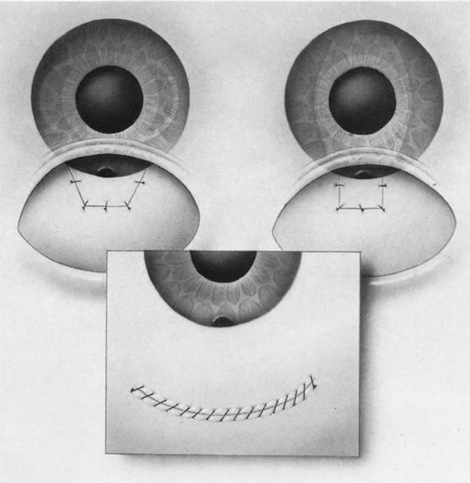
1. Vanbuskirk EM. The technology dilemma. J Glaucoma 5:77, 1996

2. Cunliffe IA, Dapling RB, Longstaff S: A prospective study examining the changes in factors that affect visual acuity following trabeculectomy. Eye 6:618, 1992

3. Dietze PJ, Oram O, Kohnen T et al: Visual function following trabeculectomy: effect on corneal topography and contrast sensitivity. J Glaucoma 6:90, 1997

4. Menapace R, Findl O, Georgopoulos M: The capsular tension ring:designs, applications, and techniques. J Cataract Refract Surg, 26:898, 2000

5. Friedman DS, Jampel HD, Lubomski LH et al: Surgical strategies for coexisting glaucoma and cataract. Ophthalmology 109:1902, 2002

6. Jampel HD, Friedman DS, Lubomski LH et al: Effect of technique on intraocular pressure after combined cataract and glaucoma surgery. Ophthalmology, 109:2215, 2002

7. Chandler PA, Grant WM: General considerations. In Lectures on Glaucoma, pp 13–16. Philadelphia, Lea & Febiger, 1965

8. Drance SM: Doyne Memorial Lecture: Correlation of optic nerve and visual field defects in simple glaucoma. Trans Ophthalmol Soc UK 95:288, 1975

9. Quigley HA, Anderson DR: The dynamics and location of axonal transport blockage by acute intraocular pressure elevation in primate optic nerves. Invest Ophthalmol Vis Sci 15:606, 1976

10. Quigley HA, Addick EM, Green WR et al: Optic nerve damage in human glaucoma: II. The site of injury and susceptibility to damage. Arch Ophthalmol 99:635, 1981

11. Quigley HA: The future. In Spaeth GL, Varma R, Parker K (eds): The Optic Nerve in Glaucoma, p 347. Philadelphia, JB Lippincott, 1993

12. Quigley HA, Dunkelberger GR, Sanchez RM: Chronic experimental glaucoma causes selectively greater loss of larger optic nerve fibers. Invest Ophthalmol Vis Sci 27:42, 1986

13. Spaeth GL: Appearance of the optic disc in glaucoma: A pathogenic classification. In New Orleans Academy of Ophthalmology: A Symposium on Glaucoma, p 114. St. Louis, CV Mosby, 1981

14. Caprioli J: Correlation between optic disc appearance and type of glaucoma. In Spaeth GL, Varma R, Parke K (eds): The Optic Nerve in Glaucoma, p 91. Philadelphia, JB Lippincott, 1993

15. Spaeth GL: Development of glaucomatous changes of the optic nerve. In Spaeth GL, Varma R, Parker K (eds): The Optic Nerve in Glaucoma, pp 63-81. Philadelphia, JB Lippincott, 1993

16. Crouch ER Jr, Frenkel M: Aminocaproic acid in the treatment of traumatic hyphema. Am J Ophthalmol 81:355,1976

17. Goldberg MF: Sickle erythrocytes, hyphema and secondary glaucoma. I. Diagnosis and treatment of sickled erythrocytes in human hyphemas. Trans Am Ophthalmol Soc 76:781, 1978

18. Goldberg MF, Tso MOM: Sickled erythrocytes, hyphema, and secondary glaucoma: VII. The passage of sickled erythrocytes out of the anterior chamber of the human and monkey eye: Light and electron microscopic studies. Ophthalmic Surg 10:89, 1979

19. Trible JR, Anderson DR: Factors associated with intraocular pressure-induced acute visual field depression. Arch Ophthalmolo, 115:1523-27, 1997

20. Javitt JC, Spaeth GL, Katz LJ et al: Acquired pits of the optic nerve. Increased prevalence in patients with low-tension glaucoma. Ophthalmology 97(8): 1038, 1990

21. Samuelson TW, Spaeth GL: Focal and diffuse glaucomatous visual field defects: Their relationship to intraocular pressure. Ophthalmic Surg 24(8):519, 1993

22. Niesel P, Flammer J: Correlations between intraocular pressure, visual field and visual acuity, based on 11 years of observations of treated chronic glaucomas. Int Ophthalmol 3(1):331, 1980

23. King D, Drance MD, Douglas G et al: Comparison of visual field defects in normal-tension glaucoma and high-tension glaucoma. Am J Ophthalmol 101:204, 1986

24. Kurimoto Y, Kobayashi H, Sakaue H et al: Correlation between the optic disc shape and the visual field in glaucoma patients: Objective and quantitative analysis using Heidelberg retinal tomograph and Humphrey field analyzer. Invest Ophthalmol Vis Sci 35(4): 1345, 1994

25. Caprioli J, Spaeth GL: Comparison of visual field defects in the low-tension glaucomas with those in the high-tension glaucomas. Am J Ophthalmol 97:730, 1984

26. Caprioli J, Sears M, Miller JM: Patterns of early visual field loss in open-angle glaucoma. Am J Ophthalmol 103:512, 1987

27. Costa VP, Smith M, Spaeth GL et al: Loss of visual acuity after trabeculectomy. Ophthalmology 100:599, 1993

28. Kolker A: Visual prognosis in advanced glaucoma: A comparison of medical and surgical therapy for retention of vision in 101 eyes with advanced glaucoma. Trans Am Ophthalmol Soc 75:539, 1977

29. Hayashi K, Hayashi H, Nakao F, et al: Influence of cataract surgery on automated perimetry in patients with glaucoma. Am J Ophthalmol, 132:41, 2001

30. Fellman RL: Visual Fields. ed. Gross, RL, Clinical Glaucoma Management: Critical signs in diagnosis and Therapy, pp. 414-473, W.B. Saunders Co, Philadelphia, 2001

31. Kim YY, Kim JS, Shin DH et al: Effect of cataract extraction on blue-on-yellow visual field. Am J Ophthalmol 132:217, 2001

32. The AGIS Investigators: The advanced glaucoma intervention study,6: effect of cataract on visual field and visual acuity. Arch Ophthalmol 118:1639, 2000

33. Chen PP, Budenz DL. The effects of cataract extraction on the visual field of eyes with chronic open-angle glaucoma. Am J Ophthalmol, 125:325-33, 1998

34. Smith SD, Katz J, Quigley HA: Effect of cataract extraction on the results of automated perimetry in glaucoma. Arch Ophthalmol 115:1515, 1997

35. Shin DH, Islander NG, Ahee JA et al: Long-term filtration and visual field outcomes after primary glaucoma triple procedure with and without Mitomycin-C. Ophthalmology 109:1607, 2002

36. Lynn JR, Fellman RL, Starita RJ: Principles of perimetry. In Ritch R, Shields MB, Krupin R (eds): The Glaucomas, p 511 Mosby, St Louis, 1996

37. Bigar F, Witmer R: Corneal endothelial changes in primary acute angle-closure glaucoma. Ophthalmology 89:596,1982

38. Ritch R, Schlotzer-Schrehardt U: Exfoliation syndrome. Surv Ophthalmol 45:265-315, 2001

39. Naumann GO, Schlotzer-Schrehardt U, Kuchle M: Pseudoexfoliation syndrome for the comprehensive ophthalmologist. Intraocualr and systemic manifestations. Ophthalmology 105:951, 1998

40. Chen HSL, Steinmann WC, Spaeth GL: The effect of chronic miotic therapy on the results of posterior chamber intraocular lens implantation and trabeculectomy in patients with glaucoma. Ophthalmic Surg 20(11):784, 1989

41. Sherwood MB, Grierson I, Miller L et al: Long-term morphologic effects of antiglaucoma drugs on the conjunctiva and Tenon's capsule in glaucomatous patients. Ophthalmology 96:327, 1989

42. Mastropasqua L, Carpineto P, Ciancaglini et al: Intraocular pressure changes after phacoemulsifiocation and foldable silicone lens implantation using Healon GV. Ophthalmologica, 212:315-21, 1998

43. Rich WJ, Radtke ND, Cohan BE: Early ocular hypertension after cataract extraction. Br J Ophthalmol 58:725, 1974

44. Krupin T, Feitl ME Bishop KI,: Postoperative intraocular pressure rise in open-angle glaucoma patients after cataract or combined cataract-filtration surgery. Ophthalmology 96:579, 1989

45. Spaeth GL: The management of patients with cataract and glaucoma. Ophthalmic Surg 11:780, 1980

46. West J, Burke J, Cunliffe I et al: Prevention of acute postoperative pressure rises in glaucoma patients undergoing cataract extraction with posterior chamber lens implant. Br J Ophthalmol 76:534, 1992

47. Ruiz RS, Rhem MN, Prager TC: Effects of carbachol and acetylcholine on intraocular pressure after cataract extraction. Am J Ophthalmol 197:7, 1989

48. Pohjalainen T, Vesti E, Uusitalo RJ, Laatikainen L: Intyraocular pressure after phacoemulsification and intraocular lens implantation in nonglaucomatous eyes with and without exfoliation. J Cataract Refract Surg, Mar 27(3):426-31, 2001

49. Pohjalainen T, Vesti E, Uusitalo RJ, Laatikainen L: Phacoemulsification and intraocular lens implantation in eyes with open-angle glaucoma. Acta Ophthalmol Scand Jun 79(3):313-6, 2001

50. Schwenn O, Dick HB, Krummenauer F, et al Intraocular pressure after small incision cataract surgery: temporal sclerocorneal versus clear corneal incision. J Cataract Refract Surg Mar:27:421, 2001

51. Savage JA, Thomas JV, Belcher CD et al: Extracapsular cataract extraction and posterior chamber intraocular lens implantation in glaucomatous eyes. Ophthalmology 92:1506, 1988

52. Schwenn O, Dick HB, Krummenauer F et al: Healon5 versus Viscoat during cataract surgery: intraocular pressure, laser flare and corneal changes. Graefes Arch Clin Exp Ophthalmol 238:861, 2000

53. Arshinoff SA, Albiani DA, Taylor-Laporte J: Intraocular pressure after bilateral cataract surgery using Healon, Hedalon5 and Healon GV. J Cataract Refract Surg, 28:617, 2002

54. Ruiz RS, Rhem MN, Prager TC: Effects of carbachol and acetylcholine on intraocular pressure after cataract extraction. Am J Ophthalmol 197:7, 1989

55. Shingleton BJ, Wadhwani RA, O'Donoghue MW et al:Evaluation of intraocular pressure in the immediate period after phacoemulsification. J Cataract Refract Surg 27:524, 2001

56. Rainer G, Menapace R, Schmetterer K, Findl O, et al:Effect of dorzolamide and latanoprost on intraocular pressure after small incision cataract surgery. J Cataract Refract Surg 25:1624, 1999

57. Scherer WJ, Mielke DL, Tidwell PE, Hauber FA: Effect of latanoprost on intraocular pressure following cataract extraction. J Cataract refract Surg, 24:964, 1998

58. Byrd S, Singh K: Medical control of intraocular pressure after cataract surgery. J Cataract Refract Surg 24:1493, 1998

59. Rainer G, Menapace R, Findl O et al: Intraindividual comparison of the effects of a fixed dorzolamide-timolol combination and latanoprost on intraocular pressure after small incision cataract surgery. J Cataract Refract Surg 27:706, 2001

60. Robin AL: Short-term effects of unilateral 1% apraclonidine therapy. Arch Ophthalmol 106:912, 1988

61. Sterk CC,Renzenbrink-Bubberman AC, van Best JA: The effect of 1% aoraclonidine on intraocular pressure after cataract surgery. Ophthalmic Surg Lasers 29:472, 1998

62. Kasetti SR, Shrivatsa PD, Subramaniam W et al: Preventing intraocular pressure increase after phacoemulsification and the role of perioperative apraclonidine. J Cataract Refract Surg 28:2177, 2002

63. Whitehouse G: Brimonidine and postoperative pressure spikes in cataract surgery. Clin Experiment Ophthalmol 289:364, 2000

64. Rainer G, Menapace R, Findl O, et al: Effect of topical brimonidine on intraocular pressure after small incision cataract surgery. J Cataract Refract Surg 27:1227, 2001

65. Carnahan MC, Platt LW: Serial paracentesis in the management of acute elevations of intraocular pressure. Ophthalmology 109:1604, 2002

66. Brooks AMV, Gillies WE: Do any factors predict a favourable response to laser trabeculoplasty? Aust J Ophthalmol 12:149, 1984

67. Galin MA, Obstbaum SA, Asano Y et al: Laser trabeculoplasty and cataract surgery. Trans Ophthalmol Soc UK 104(1):72, 1985

68. Savage JA, Thomas JV, Belcher CD 3rd , Simmons RJ: Extracapsular cataract extraction and posterior chamber lens implantation in glaucomatous eyes. Ophthalmology 92:1506, 1985

69. Fellman RL, Starita RJ, Spaeth GL, et al: Argon laser trabeculoplasty following failed trabeculectomy. Ophthalmic Surg 15:195, 1984

70. Laatikainen L: Late results of surgery on eyes with primary glaucoma and cataract. Acta Ophthalmol 49:281, 1971

71. Steuhl KP, Marahrens P, Frohn C et al: Intraocular pressure and anterior chamber depth before and after extracap-sular cataract extraction with posterior chamber lens implantation. Ophthalmic Surg 23:233, 1992

72. Dimitrov PN, Mukesh BN, Taylor HR, McCarty CA:Intraocular pressure before and after cataract surgery in participants of the Melbourne Visual Impairment Project. Clin Experiemtn Ophthalmol 29:128, 2001

73. Hansen MH, Gyldenkerne GJ, Otland NW et al: Intraocular pressure seven years after extracapsular cataract extraction and sulcus implantation of a posterior chamber intraocular lens. J Cataract Refract Surg 21:676, 1995

74. Shingleton, BJ, Gamell LS, O'Donoghue MW et al: Long-term changes in intraocular pressure after clear corneal phacoemulsification: Normal patients versus glaucoma suspect and glaucoma patients. J Cataract Refract Surg 25:887,1999

75. Merkur A, Damji KF, Mintsioulisw G, Hodge WG: Intraocular pressure decrease after phacoemulsification in patients with pseudoexfoliation syndrome. J Cataract and Refractive Surg 27:528, 2001

76. Wirbelauer C, Anders N, Pham DT et al: Intraocular pressure in nonglaucomatous eyes with pseudoexfoliation syndrome after cataract surgery. Ophthalmic Surg Lasers 29:466, 1998

77. Jahn CE: Reduced intraocular pressure after phacoemulsification and posterior chamber intraocular lens implantation. J Cataract Refract Surg 23:1260, 1997

78. Meyer MA, Savitt ML, Kopitas E: The effect of phacoemulsification on aqueous outflow facility. Ophthalmology 104:1221, 1997

79. Schwenn O, Dick HB, Krummenauer F et al: Intraocular pressure after small incsion cataract surgery:temporal sclerocorneal versus clear corneal incision. J Cataract Refract Surg 27:421, 2001

80. Krupin T, Feitl ME Bishop KI,: Postoperative intraocular pressure rise in open-angle glaucoma patients after cataract or combined cataract-filtration surgery. Ophthalmology 96:579, 1989

81. Hopkins JJ, Apel A, Trope GE, Rootman DS: Early intraocular pressure after phacoemulsification combined with trabeculectomy. Ophthalmic Surg Laser 29:273, 1998

82. Porges Y, Ophir A: Surgical outcome after early intraocular pressure elevation following combined cataract extraction and trabeculectomy. Ophthalmic Surg Lasers 30:727, 1999

83. Bigger JF, Becker B: Cataracts and primary open-angle glaucoma: The effect of uncomplicated cataract extraction on glaucoma control. Am Acad Ophthalmol Otol 75:260, 1971

84. Laatikainen L: Late results of surgery on eyes with primary glaucoma and cataract. Acta Ophthalmol 49:281, 1971

85. Steuhl KP, Marahrens P, Frohn C et al: Intraocular pressure and anterior chamber depth before and after extracapsular cataract extraction with posterior chamber lens implantation. Ophthalmic Surg 23:233, 1992

86. Bobrow JC: Prospective intrapatient comparison of extracapsular cataract extraction and lens implantation with and without trabeculectomy. Am J Ophthalmol 129:291, 2000

87. Anders N, Pham T, Holschbach A, Wollensak J: Combined phacoemuslfication and filtering surgery with the “no-stitch” technique. Arch Ophthalmol 115:1245, 1997

88. Kosmin AS, Wishart PK, Ridges PJ: Long-term intraocular pressure control after cataract extraction with trabeculectomy:phacoemulsification versus extracapsular technique. J Cataract Refract Surg 24:249, 1998

89. Cohen JS: Combined cataract implant and filtering surgery with 5-fluorouracil. Ophthalmic Surg 21:181, 1990

90. Johnstone MA, Ziel CJ: Cataract surgery in the presence of a filtering bleb with postoperative 5-fluorouracil (5-FU). Invest Ophthalmol Vis Sci 33:948, 1992

91. Wong PC, Ruderman JM, Krupin T et al: 5-Fluorouracil (5-FU) after primary combined filtration surgery. Am J Ophthalmol 117:149, 1994

92. Budenz DL, Pyfer M, Singh K et al: Comparison of phacotrabeculectomy with 5-fluorouracil, Mitomycin-C, and without antifibrotic agents. Ophthalmic Surg Lasers 30:367, 1999

93. O'Grady JM, Juzych MS, Shin DH et al: Trabeculectomy, phacoemulsification and posterior chamber lens implantation with and without 5-fluorouracil. Am J Ophthalmol 116:594, 1993

94. Hurvitz, LM: 5-fluorouracil supplemented phacoemulsification, posterior chamber lens implantation, and trabeculectomy. Ophthalmic Surg 24:674, 1993

95. Cohen JS, Greff LJ, Novack GD, Wind BE: A placebo-controlled, double masked evaluation of Mitomycin C in combined glaucoma and cataract procedures. Ophthalmology 103:1934, 1996

96. Carlson DW, Alward WL, Barad JP et al: A randomized study of Mitomycin augmentation in combined phacoemulsification and trabeculectomy. Ophthalmology 104:719, 1997

97. Casson RJ, Salmon JF: Combined surgery in the treatment of patients with cataract and primary open-angle glaucoma. J Cataract Refract Surg 27:1854, 2001

98. Shin DH, Ren J, Juzych MS et al: Primary glaucoma triple procedure in patients with primary open angle glaucoma: The effect of Mitomycin C in patients with and without prognostic factors for filtration failure. Am J Ophthalmol 125:346, 1998

99. Shin DH, Kim YY, Sheth N et al: The role of adjuctive Mitomycin C in seconday glaucoma triple procedure as compared to primary glaucoma triple procedure. Ophthalmology 105:740, 1998

100. Rockwood EJ, Larive B, Hahn J: Outcomes of combined cataract extraction, lens implantation and trabeculectomy surgeries. Am J Ophthalmol 130:704, 2000

101. Belyea DA, Dan JA, Lieberman MF, Stamper RL: Midterm follow-up results of combined phacoemulsification, lens implantation, and mitomycin-C trabeculectomy procedure. J Glaucoma 6:90, 1997

102. Yamagami S, Hamada N, Araie M, Shirato S: Risk factors for unsatisfactory intraocular pressure control in combined trabeculectomy and cataract surgery. Ophthalmic Surg Lasers 28:476, 1997

103. Tezel G, Kolker AE, Kass MA, Wax MB: Comparative results of combined procedures for glaucoma and cataract: I. Extracapsular cataract extraction versus phacoemulsification and foldable versus rigid intraocular lenses. Ophthalmic Surg Lasers 28:539, 1997

104. Chia WLA, Goldberg I: Comparison of extracapsular and phacoemulsification cataract extraction techniques when combined with intraocular lens placement and trabeculectomy:short term results. Aust N Z J Ophthalmol 26:19, 1998

105. Shingleton BJ, Jacobson LM, Kuperwaser MC: Comparison of combined cataract and glaucoma surgery using planned extracapsular and phacoemulsification techniques. Ophthalmology Surg lasers 26:414, 1995

106. Singh RP, Goldberg I, Mohsin M: The efficacy and safety of intraoperative and/or postoperative 5-fluorouracil in trabeculectomy and phacotrabeculectomy. Clin Exp Ophthalmol 29:296, 2001

107. Siriwardena D, Kotecha A, Minassian D et al: Anterior chamber flare after trabeculectomy and after phacoemulsification. Br J Ophthalmol 84:1056, 2000

108. Beckers HJ, DeKroon KE, Nuijts RM, Webers CA: Phacotrabeculectomy. Doc Ophthalmol 100:43, 2000

109. Menapace R, Wedrich A, Muhlbauer-Ries et al: Phacotrabeculectomy with a small-optic PMMA implant:two-year functional and m porphological results. Ophthalmologica 212:322, 1998

110. Caprossi A, Casprini F, Topsi GM, Balestrazzi A: Long-term results of combined 1-way phacoemulsification, intraocular lens implantation and trabeculectomy. J Cataract Refract Surg 25:1641, 1999

111. Mamalis N, Lohner S, Rand AN, Crandall AS: Combined phacoemulsification, intraocular lens implantation, and trabeculectomy. J Cataract Refract Surg 22:467, 1996

112. Vyas AV, Bacon PJ, Percival SP: Phacotrabeculectomy: comparison of results from 3.5 and 5.2 mm incisions. Ophthalmic Surg Lasers 29:227, 1998.

113. Kosmin AS, Wishart PK, Ridges PJ: Silicone versus polymethacrylate lenses in combined phacoemulsification and trabeculectomy. J Cataract Refract Surg 23:97, 1997

114. Donoso R, Rodriguez A: Combined versus sequential phacotrabeculectomy with intraoperative 5-fluorouracil. J Cataract Refract Surg 26:71, 2000

115. El-Sayyad FF, Helal MH, Khalil MM et al: Phacotrabeculectomy versus two-stage operation: a matched study. Ophthalmic Surg Lasers 30:260, 1999

116. Braga-Mele R, Cohen S, Rootman DS: Foldable silicone versus polymethylmethcrylate intraocular lenses in combined phacoemulsification and trabeculectomy. J Cataract Refract Surg 26:1517, 2000

117. Wyse T, Meyer M, Ruderman JM, Krupin T, et al: Combined trabeculectomy and phacoemulsification: a one-site vs a two-site approach. Am J Ophthalmol 125:334, 1998

118. Borggrefe J, Lieb W, Grehn F: A prospective randomized comparison of two techniques of combined cataract-glaucoma surgery. Graefes Arch Clin Exp Ophthalmol 237:887, 1999

119. Rossetti L, Bucci L, Miglior S et al: Temporal corneal phacoemulsification combined with separate incision superior trabeculectomy vs standard phacotrabeculectomy. A comparative study. Acta Ophthalmol Scand Suppl 224:39, 1997

120. el Sayyad F, Helal M, el-Maghraby et al: One-site verus 2-site phacotrabeculectomy: a randomized study. J Cataract Refract Surg 24:77, 1999

121. Spaeth GL: Glaucoma surgery. In Principles & Practice of Ophthalmic Surgery, p 319. Philadelphia, WB Saunders, 1990

122. Kass MA: Cataract extraction in an eye with a filtering bleb. Ophthalmology 89:871, 1988

123. Mietz H, Andresen A, Welsandt G, Krieglstein GK: Effect of cataract surgery on intraocular pressure in eyes with previous trabeculectomy. Graefes Arch Clin Exp Ophthalmol 239:763, 2001

124. Halikiopoulos D, Moster MR, Azuara-Blanco A et al: The outcome of the functioning filter after subsequent cataract extraction. Ophthalmic Surg Lasers 32:108, 2001

125. Manoj B, Chako D, Khan MY: Effect of extracapsular extraction and phacoemulsification performed after trabeculectomy on intraocular pressure. J Cataract Refract Surg 26:75, 2000

126. Rebolleda G, Munoz-Negrete FJ: Phacoemulsificatgion in eyes with functioning filtering blebs: a prospective study. Ophthalmology 109:2248, 2002

127. Chen PP, Weaver YF, Budenz DL et al: Trabeculectomy function after cataract extraction. Ophthalmology 105:1928, 1998

128. Wygnanski-Jaffe T, Barak A, Melamed S, Glovinsky Y: Intraocular pressure increments after cataract extraction in glaucomatous eyes with functioning filtering blerbs. Ophthalmic Surg Lasers 28:657, 1997

129. Crichton ACS, Kirker AW: Intraocular pressure and medication control after clear corneal phacoemulsification and Acrysof posterior chamber intraocular lens implantation in patients with filtering blebs. J Glaucoma 10:38, 2001

130. Bhattacharyya CA, WuDunn D, Lakhani V et al: Cataract surgery after tube shunts. J Glaucoma 9:453, 2000

131. Smith MF, Doyle JW: Use of tissue plasminogen activator to revive blebs following intraocular surgery. Arch Ophthalmol 119:809, 2001

132. Mardelli PG, Lederer CM, Murray PL et al: Slit lamp needle revision of failed filtering blebs using Mitomycin C. Ophthalmology 103:1946, 1996

133. Hawkins AS, Glanagan JK, Brown SVL: Predictors for success of needle revision of failing filtration blebs. Ophthalmology 109:783, 2002

134. Sibayan SA, Igarashi S, Kasahara N et al: Cataract extraction as a means of treating postfiltration hypotony maculopathy. Ophthalmic Surg Lasers 28:241, 1997

135. Doyle JW, Smith MF: Effect of phacoemulsification surgery on hypotony following trabeculectomy surgery. Arch Ophthalmol 118:763, 2000

136. Fellman RL: Trabeculotomy. In George L Spaeth (ed): Ophthalmic Surgery: Principles and Practice, 3rd ed, p XX. Philadelphia, WB Saunders, 2003

137. Tanihara H, Honjo M, Inatani M et al: Trabeculotomy combined with phacoemulsification and implantation of an intraocular lens for the treatment of primary open angle glaucoma and coexisting cataract. Ophthalmic Surg Lasers 28:810, 1997

138. Tanito M, Ohira A, Chihara E: Surgical outcome of combined trabeculotomy and cataract surgery. J Glaucoma 10:302, 2001

139. Tanito M, Ohira A, Chihara E: Factors leading to reduced intraocular pressure after combined trabeculotomy and cataract surgery. J Glaucoma 11:3, 2002

140. Luke C, Dietlein TS, Jacobi PC et al: A prospective randomized trial of viscocanalostomy versus trabeculectomy in open angle glaucoma: a one year follow-up study. J Glaucoma 11:294, 2002

141. Stegmann R, Pienaar A, Miller D: Viscocanalostomy for open-angle glaucoma in black African patients. J Cataract Refgract Surg 25:316, 1999

142. El Sayyad F, Helal M, El-Kholify H et al: Nonpenetrating deep sclerectomy versus trabeculectomy in bilateral primary open angle glaucoma. Ophthalmology 107:1671, 2000

143. Gianoli F, Schyder CC, Bovey E, Mermoud A: combined surgery for cataract and glaucoma:phacoemulsification and deep sclerectomy compared with phacoemulsification and trabeculectomy. J Cataract Refract Surg 25:340, 1999

144. Wishart MS, Shergill T, Porooshani H: Viscocanalostomy and phacoviscocanalostomy: long-term results. J Cataract Refract Surg 28:745, 2002

145. Gimbel HV, Penno EE, Ferensowicz M: Combined cataract surgery, intraocular lens implantation, and viscocanalostomy. J Cataract Refract Surg 25:1370, 1999

146. Hayashi K, Hayashi H, Nakao F, Hayashi F: Effect of cataract surgery on intraocular pressure control in glaucoma patients. J Cataract Refract Surg 27:1779, 2001

147. Yang CH, Hung PT: Intraocular lens position and anterior chamber angle changes after cataract extraction in eyes with primary angle-closure glaucoma. J Cataract Refract Surg 23:1109, 1997.

148. Gunning FP, Greve EL: Lens extraction for uncontrolled angle-closure glaucoma: Long term follow-up. J Cataract Refract Surg 24:1347, 1998

149. Acton J, Salmon JF, Scholtz R: Extracapsular cataract extraction with posterior chamber lens implantation in primary angle-closure glaucoma. J Cataract Refract Surg 23:930, 1997

150. Hayashi K, Hayashi H, Nakao F et al: Changes in anterior chamber angle width and depth after IOL implantation in eyes with glaucoma. Ophthalmology 107:698, 2000

151. Fellman RL: Comment of: Changes in anterior chamber angle width and depth after IOL implantation in eyes with glaucoma. Evidence-based Eye Care 2:53, 2001

152. Lai JS, Tham CC, Lam DS: The efficacy and safety of combined phacoemulsification, intraocular lens implantation, and limited goniosynechialysis, followed by diode laser peripheral iridoplasty, in the treatment of cataract and chronic angle closure glaucoma. J Glaucoma 10:309, 2001

153. Jacobi PC, Dietlein TS, Luke C et al: Primary phacoemulsification and intraocular lens implantation for acute angle closure glaucoma. Ophthalmology 109:1597, 2002

154. Cashwell LF, Martin CA: Axial length decreases accompanying successful glaucoma filtration surgery. Ophthalmology 106:2307, 1999

155. Kook MS, Kim HB, Lee SU: Short-term effect of Mitomycin-C augmented trabeculectomy on acial length and corenal astigmatism. J Cataract Refract Surg 27:518, 2001

156. The advanced glaucoma intervention study group, The advanced glaucoma intervention study, 8: risk of cataract formation after trabeculectomy. Arch Ophthalmol 119:1771, 2001

157. Iaccarino G, Rosa N, Romano M et al: Expulsive hemorrhage before phacoemulsification. J Cataract Refract Surg 28:1074, 2002

158. Cantor LB, Katz LJ, Spaeth GL: Complications of surgery in glaucoma: Suprachoroidal expulsive hemorrhage in glaucoma patients undergoing intraocular surgery. Ophthalmology 92:1266, 1985

159. Ruderman JM, Harbin TS Jr, Campbell DG: Postoperative suprachoroidal hemorrhage following filtration procedures. Arch Ophthalmol 104:201, 1986

160. Givens K, Shields MB: Suprachoroidal hemorrhage after glaucoma filtering surgery. Am J Ophthalmol 103:689, 1987

161. Jaynes JH, Payne JW, Green WR: Clinicopathologic study of eyes obtained postmortem from a patient 6 and 2 years after operative choroidal hemorrhage. Ophthalmic Surg 18:667, 1987

162. Welch JC, Spaeth GL, Benson WE: Massive suprachoroidal hemorrhage: Follow-up and outcome of 30 cases. Ophthalmology 95:1202, 1988

163. Speaker MG, Guerriero PN, Met JA et al: A case-control study of risk factors for intraoperative suprachoroidal expulsive hemorrhage. Ophthalmology 98:202, 1990

164. Canning CR, Lavin M, McCartney ACE et al: Delayed suprachoroidal hemorrhage after glaucoma operations. Eye 3:327, 1989

165. Spaeth GL, Baez KA: Long-term prognosis of eyes having had operative suprachoroidal expulsive hemorrhage. Ger J Ophthalmol 3:159, 1994

166. Bellows AR, Chylack LT, Huthchinson BT: Choroidal detachment:clinical manifestations, therapy and mechanism of formation. Ophthalmology 88:1107, 1981.

167. Herndon LW: Shallow or flat anterior chamber. In Spaeth GL (ed): Ophthalmic surgery: Principles and Practice, 3rd ed. Philadelphia, WB Saunders, 2003

168. Fellman RL, Budenz D: Malignant glaucoma. J Glaucoma 8:149, 1999

169. Fellman RL: Flat chamber following glaucoma filtering procedures. In Spaeth GL, Katz LJ (eds): Current Therapy in Ophthalmic Surgery, p 330. Hamilton, Ontario, BC Decker, 1989

170. Lynn JR, Swanson WH, Fellman RL, Starita RJ.: Does glaucomatous visual filed loss continue despite surgically subnormal IOP? Perimetry Update 1992.93, p 129. Proceedings of the Xth International Perimetric Society Meeting, Kugler Publications, Amsterdam / New York

171. The advanced glaucoma intervention study (AGIS): 7. The relationship between control of intraocular pressure and visual field deterioration. The AGIS Investigators, Am J Ophthalmol, 130:429, 2000

172. Hoffman KB, Feldman RM, Budenz DL et al: Combined cataract extraction and Baerveldt glaucoma drainage implant: indications and outcomes. Ophthalmology 109:1916, 2002

173. The advanced glaucoma intervention study, 8: risk of cataract formation after trabeculectomy. Arch Ophthalmol 119:1771, 2001

174. Kozobolis VP, Siganos CS, Christodoulakis EV et al: Two-site phacotrabeculectomy with intraoperative mitomycin-C:Fornix-versus limbus-based conjunctival opening in fellow eyes. J Cataract Refract Surg 28:1758, 2002

175. Shingleton BJ, Chaudhry IM, O'Donoghue MW et al: Phacotrabeculectomy: limbus-based versus fornix-based conjunctival flaps in fellow eyes. Ophthalmology 106:1152, 1999

176. Berestka JS, Brown SV: Limbus-versus fornix-based conjunctival flaps in combined phacoemulsification and Mitomycin-C trabeculectomy surgery. Ophthalmology 104:187, 1997

177. Lemon LC, Shin DH, Kim C, et al: Limbus-based vs fornix-based conjunctival flap in combined glaucoma and cataract surgery with adjunctive Mitomycin-C. Am J Ophthalmol 125:340, 1998

178. Agbeja AM, Dutton GN: Conjunctival incisions for trabeculectomy and their relationship to the type of bleb formation—a preliminary study. Eye 1:738, 1987

179. el Sayyad F, el-Rashood A, Helal M et al: Fornix-based versus limbal-based conjunctival flaps in initial trabeculectomy with postoperative 5-fluorouracil: four-year follow up findings. J Glaucoma 8:124, 1999

180. Fellman RL: Trabeculectomy. London, Mosby International Ltd., 1999

181. Dinsmore SC, Modified stretch technique for small pupil phacoemulsification with topical 
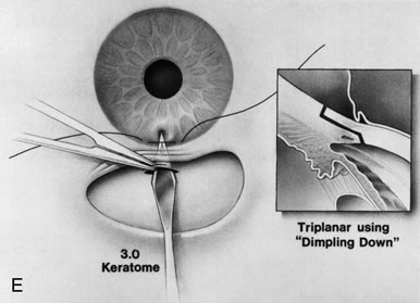
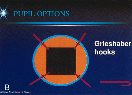
anesthesia. J Cataract Refract Surg 22:27, 1996

182. Graether JM. Graether pupil expander for managing the small pupil during surgery. J Cataract Refract Surg 22:530, 1996.

183. Bacskulin A, Kundt G, Guthoff R: Efficiency of pupillary stretching in cataract surgery. Eur J Ophthalmol 8:230, 1998.

184. Birchall W, Spencer AF: Misalignment of flexible iris hook retractors for small pupil cataract surgery:effects on pupil circumference. J Cataract Refract Surg 27:20, 2001

185. Fine IH: Pupilloplasty for small pupil phacoemulsification. J Cataract Refract Surg 20:192, 1994

186. Yuguchi T, Oshika T, Sawaguchi S et al: Pupillary functions after cataract surgery using flexible iris retractor in patients with small pupil. Jpn J Ophthalmol 43:20, 1999

187. Horiguchi M, Miyake K, Ohta I, Ito Y: Staining of the lens capsule for circular continuous capsulorhexis in eyes with white cataract. Arch Ophthalmol 116:535, 1998

188. Jacob S, Agarwal AA, Agarwal AA et al: Trypan blue as an adjunct for safe phacoemulsification in eyes with white cataract. J Cataract Refract Surg 28:1819, 2002

189. Shingleton BJ, Chaudhry IM, O'Donoghue MW: Phacotrabeculectomy: peripheral iridectomy or no peripheral iridectomy? J Cataract Refract Surg 28:998, 2002

190. Crestani A, DeNatale R, Steindler P: Phacotrabeculectomy with or without punch: preliminary results comparing two techniques. Ophthalmologica 211:72, 1997

191. Wise JB: Mitomycin-C compatible suture technique for fornix-based conjunctival flaps in glaucoma filtration surgery. Arch Ophthalmol 111:992, 1993

192. Philip WC, Yeung BYM, Yick DWF et al: Fornix-based trabeculectomy with Wise's suture technique in Chinese patients. Ophthalmology, 107:2310–2313,2000

193. Traverso CE, Greenridge KC, Spaeth GL et al: Focal pressure:a new method to encourage filtration after trabeculectomy. Ophthalmol Surg, 15:62, 1984

194. Spaeth GL: Laser Suture Lysis. In Spaeth GL (ed): Ophthalmic Surgery: Principles and Practice, 3RD ed. Philadelphia, WB Saunders, 2003

195. Starita, RJ, Fellman RL, Spaeth GL et al: Short and long term effects of postoperative corticosteroids on trabeculectomy. Ophthalmology 92:938, 1985

Back to Top