|
|
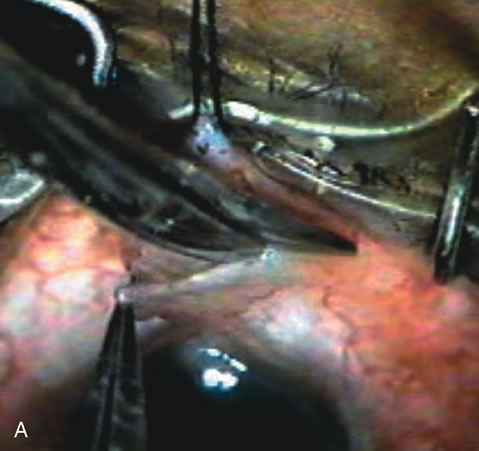
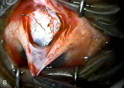
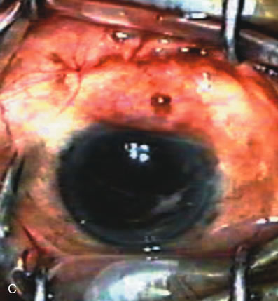
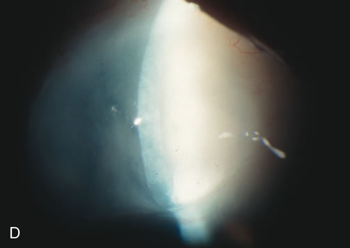
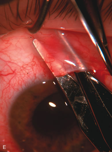
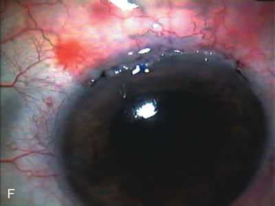
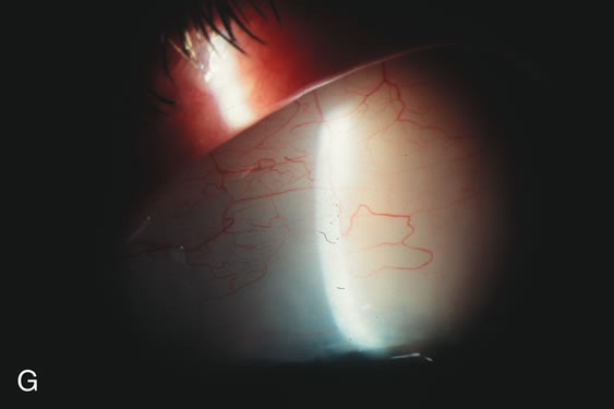
| Fig. 8. Bleb appearance after limbus-based versus fornix-based conjunctival flaps. Even though the IOP is thought to be equivalent between limbus and fornix-based conjunctival flaps, the final bleb appearance varies considerably. A. During a limbus-based approach, an incision through conjunctiva 10 mm posterior to limbus will sever through multiple arterial vessels, increasing the likelihood of an avascular bleb. B. The tissues are dissected down to the sclera further cutting feeder vessels from Tenon's capsule. C. The wound is closed inciting a cascade of wound healing events that may ultimately lead to scarring producing a barrier to aqueous flow. D. This leads to walling off of a bleb that has lost some of its overlying vascularity (pale cystic avascular bleb). E. During a fornix-based conjunctival approach, the incision is made at the limbus and tissues undermined. F. The incision is closed at the limbus; no conjunctival vessels are severed over the bleb area. G. This fosters the formation of a shallow diffuse pale bleb with a normal vessel pattern. |