Chapter 36
The Phakomatoses
DAVID SAMI, ANTHONY VIVIAN, DAVID TAYLOR and DAWN SAUNDERS
Main Menu   Table Of Contents

Search

A PERSPECTIVE
NEUROFIBROMATOSES
TUBEROUS SCLEROSIS COMPLEX
VON HIPPEL-LINDAU SYNDROME
STURGE-WEBER SYNDROME AND OTHER VASCULAR DISORDERS
REFERENCES

A PERSPECTIVE
In 1932, Van der Hoeve suggested that the syndromes of Bourneville, Recklinghausen, and von Hippel-Lindau should be grouped as phakomatoses (“mother-spot”), having in common hereditary multisystem tumors with risk of malignant transformation.1 The spectrum was extended in 1937 to include Sturge-Weber syndrome.2 How is it that a condition with no clear familial pattern or risk of malignant transformation was included as a phakomatosis? Its inclusion was an error based on a single case in which retinoblastoma developed in a patient with Sturge-Weber syndrome.3

Later on, to accommodate Sturge-Weber syndrome, Hogan and Zimmerman reinterpreted the phakomatoses as conditions of multisystem hamartoses, irrespective of malignancy potential.4 Duke-Elder protested by excluding Sturge-Weber from his discussion of phakomatoses.5,6 Over time it seems that the cutaneous lesions, as a marker of multisystem disease, have gained prominence. This may explain inclusion of the phakomatoses as part of the “neurocutaneous disorders,”7–10 even though cutaneous findings are not part of the diagnostic criteria of von Hippel-Lindau syndrome. Given multiple definitions to choose from, the phakomatoses have become a loose category, expanding as a convenient way to group syndromes.5,7,11

We believe that the term phakomatosis should be restricted to the original three syndromes as molecular genetics support Van der Hoeve's observation: neurofibromatosis, tuberous sclerosis complex, and Von Hippel-Lindau syndromes are disorders of tumor suppressor genes,12–15 a common link explaining the familial pattern (autosomal dominant), variable expressivity, multisystem tumors, and risk of malignant transformation. Thus we will restrict the phakomatoses to the original three syndromes:

  1. The neurofibromatoses (NF1 and NF2)
  2. Tuberous sclerosis complex
  3. Von Hippel-Lindau syndrome

Each section is divided into: epidemiology; systemic features (cutaneous, neurologic, skeletal/visceral); ophthalmic features; diagnostic criteria; genetics and screening. Sturge-Weber and related syndromes will be discussed separately.

Back to Top
NEUROFIBROMATOSES
Although up to 10 subtypes have been described,16,17 currently the commonly used clinical subdivision is based on the National Institutes of Health (NIH) consensus classification.18 This classification recognizes two major forms of neurofibromatosis: types 1 and 2. The other types were not formally classified. Both major subtypes are disorders of tumor suppressor genes (Table 1).12,14 Knudsen's two-hit hypothesis states that the statistical odds of developing an inactivating mutation in both alleles of a tumor suppressor gene, in the absence of an underlying disorder of DNA repair, is very low unless there is a germ-line mutation in one of the alleles. The odds of developing “loss of heterozygosity” through a new mutation is doubled, making the individual prone to developing tumors in his/her lifetime.19

TABLE 1. Neurofibromatosis Classification/Subtypes


Major type Subtype
Neurofibromatosis type 1 (NF1)von Recklinghausen's disease
 Peripheral neurofibromatosis
 Multiple neurofibromatosis
Neurofibromatosis type 2 (NF2) 
 Bilateral acoustic neurofibromatosis
 Central neurofibromatosis22
Rare forms of NeurofibromatosisMosaic/segmental20,22
 Familial spinal neurofibromatosis
 Familial intestinal neurofibromatosis
 Schwannomatosis
 Autosomal dominant café au lait spots
 Autosomal dominant neurofibromas
 Watson syndrome (pulmonary stenosis, café au lait patches)
 Noonan/neurofibromatosis syndrome22


NEUROFIBROMATOSIS TYPE 1

Epidemiology

Neurofibromatosis type 1 (NF1) accounts for 90% of the neurofibromatoses and shows autosomal dominant inheritance. Its prevalence is 1/3500 to 1/5000.20,21 (Prevalence is an estimate of the frequency of a condition in the general population; incidence is an estimate of the number of new cases over a given period of time, typically 1 year.) Although the penetrance of NF1 is close to 100% by age 5 years, its expressivity even within the same family may be highly variable. (Penetrance is the proportion of individuals with the defective gene who show clinical evidence of the disorder; expressivity is the degree of variability in clinical findings among individuals with similar mutations.) The NF1 gene has a high mutation rate, with up to half of newly diagnosed cases representing de novo mutations.22

Systemic Features

NF1 can involve any organ system.

CUTANEOUS FINDINGS.

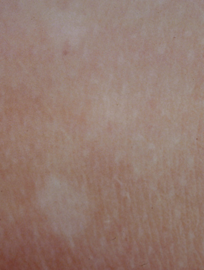
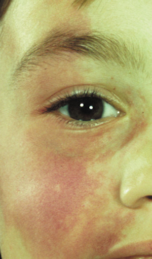
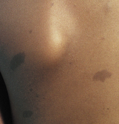
  1. Café au lait spots: Evenly pigmented flat brown macules that generally appear in the first year of life, are present in 95% of adults with NF1, and tend to fade in the elderly.23 One or two café au lait spots are not uncommon in normal patients.24 Overall, 75% of children with six or more café au lait spots develop other clinical findings of NF1 by 5 years of age (Fig. 1).24 A regular border differentiates café au lait spots in NF1 from the café au lait spots of McCune Albright syndrome (fibrous dysplasia and precocious puberty). Café au lait spots may be an isolated dominant condition in rare instances.25
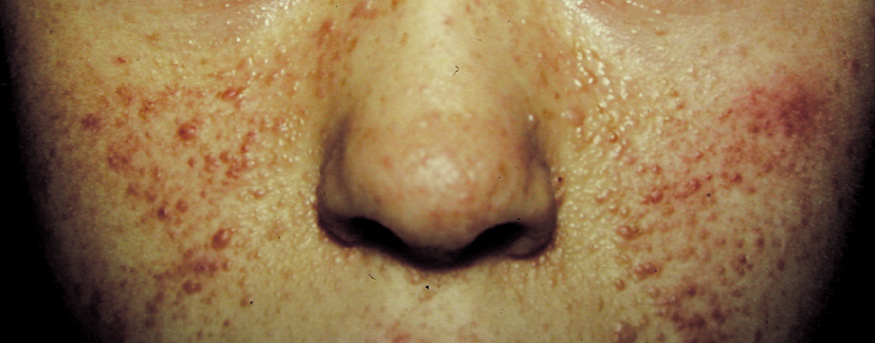
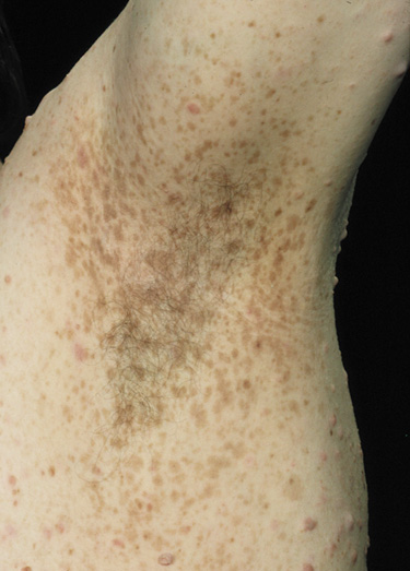
  2. Freckling in non–sun-exposed areas: the axilla, inguinal region, and the submammary folds (typically after age 20 years). Freckling may not have developed by the time café au lait spots appear (first year of life). Eighty percent to 90% of NFI patients show axillary or inguinal freckling by 5 years (Fig. 2).25,26
  3. The skin overlying dermal neurofibromas and plexiform neurofibromas is often abnormal and will be discussed with nerve-associated tumors below.

Fig. 1. Neurofibromatosis type 1: café au lait spots. These are well demarcated, evenly pigmented brown macules (flat). They generally appear in the first year of life.

Fig. 2. Neurofibromatosis type 1: axillary freckling. By 5 years of age, 80% to90% show axillary or inguinal freckling.25,26

NEUROLOGIC FINDINGS.

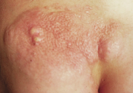
  1. Cutaneous neurofibromas are benign tumors associated with peripheral nerves. There is evidence for a single-cell origin despite multiple cell types within the tumors, although the cell of origin is unclear.27,28 Neurofibromas are typically soft and mobile on palpation. The overlying skin may be dimpled and have a violacious color due to dilated capillaries. Neurofibromas often enlarge and increase in number during puberty or pregnancy. In all, 95% of adults over age 30 with NF1 have neurofibromas, compared with less than half of adolescents and rarely in infants. Neurofibromas occur predominantly on the trunk—only 20% of patients have lesions on the head or neck. Less common subcutaneous peripheral neurofibromas are palpable as firm tumors along the trunks of peripheral nerves.22,25,29
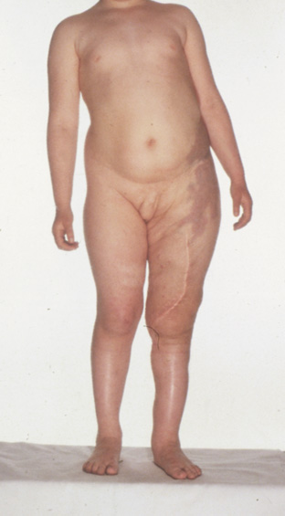
  2. Plexiform neurofibromas are subcutaneous tumors with ill-defined borders. The overlying the skin is often hypertrophic, hyperpigmented, and may have excessive hair growth. They affect 25% to 30% of individuals with NF1, most commonly on the trunk, less commonly on the limbs, head, and neck. They may carry about a 5% lifetime risk for malignant transformation (neurofibrosarcoma).25,29,30 Plexiform neurofibromas are a surgical challenge. They do not respect tissue planes, making complete removal difficult. They regrow when not fully excised and are not radiosensitive.25
  3. Brain tumors. Gliomas will be discussed below. Other brain tumors occur infrequently (<1%) in NF1. With the exception of brainstem astrocytomas and optic gliomas, the outcome of brain tumors in children affected with NF1 is similar to children without NF1.25 Brainstem and optic gliomas show slower progression in NF1.31 Vestibular schwannomas and spinal meningiomas, known complications of NF2, are probably no more common in NF1 patients than in the general population.22,25
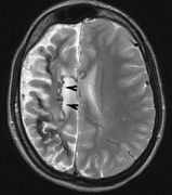
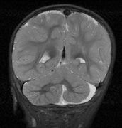
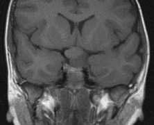
  4. Cognitive impairment. Thirty percent to 60% of children with NF1 have learning difficulties, which are typically mild and nonprogressive. These include visual-spatial problem-solving difficulty, language disorders, and attention deficit disorder. The reason for this is not clear but may be related 
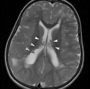
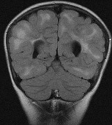
to unidentified bright objects (UBOs) seen on T2-weighted brain magnetic resonance imaging (MRI) scans. These hyperintensities are common in young patients with NF1 but decrease with advancing age. The histopathologic correlate of UBOs is unclear. It has been postulated that the prevalence of learning difficulties in children with NF1 may be related to heterozygosity of the NF1 gene (i.e., the gene may have additional functions that affect cognition when the full complement of its gene product is not expressed in the central nervous system).25,32–34

SKELETAL/VISCERAL FINDINGS.

  1. Bone dysplasia25
    • Scoliosis affects a significant number of NF1 patients (10% to 15%) and may be severe enough to require surgical correction
    • Thinning of long bones, especially medial bowing of the tibia, may predispose to fractures and pseudoarthrosis
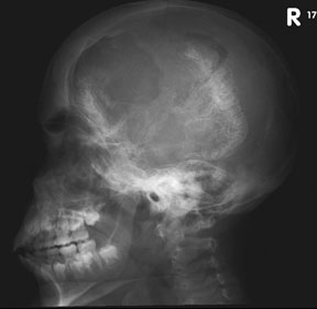
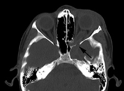
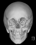
    • Congenital absence of the greater wing of the Sphenoid bone

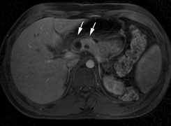
  2. Hypertension.22 There is increased incidence of aortic coarctation and pheochromocytomas. Retroperitoneal neurofibromas may compress the renal artery. These complications may manifest as hypertension in affected children

Ophthalmic Features

Virtually any part of the visual system may be affected by NF1: the bony orbit, extraocular muscles, orbital nerves, eyelids, conjunctiva, cornea, uvea, retina, optic nerves, chiasm, and optic radiations.

OCULAR FEATURES.

Conjunctiva.

Neurofibromas of the conjunctiva are infrequent, affecting about 2% of patients with NF1, often on the perilimbal conjunctiva.35

Cornea.

Enlarged corneal nerves have been reported in association with NF1 but are more commonly seen with multiple endocrine neoplasia syndrome (MEN-IIb).36,37

Uveal Tract.

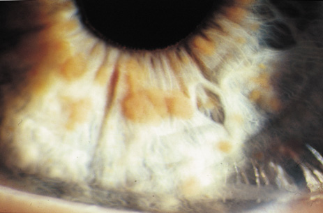
Lisch nodules are hamartomas (a tumor involving only those tissue elements normally found at the involved site) of the iris pigment epithelium. They are dome-shaped discrete lesions, are typically light brown in color, and may also be found in the angle.38 Lisch nodules appear earlier (33% at 2.5 years, 50% at 5 years of age)39 than neurofibromas. They are benign and can help to confirm diagnosis in children who may have café au lait spots as the only other clinical finding. Lisch nodules are present in nearly all adults with NF139,40 but are rare in NF2 (Fig. 3).41,42 Diffuse nodular iris nevi (also known as iris mamillations) should not be confused with Lisch nodules. Its clinical significance is not well established.43,44

Fig. 3. Neurofibromatosis type 1: Lisch nodules. These are hamartomas of the iris pigment epithelium. Lisch nodules are present in nearly all adults with NF1,39,40 but are rare in NF2.41,42

Like Lisch nodules of the iris, pigment epithelial hamartomas may affect the choroid. In one series, flat pigmented lesions of the choroid were found in 35% of patients with NF1. Fluorescein angiography suggests these are choroidal nevi.45The entire uveal tract may be thickened by a diffuse neurofibroma, thought to be hamartomatous hyperplasia of Schwann cells. This may result in the development of glaucoma.46,47
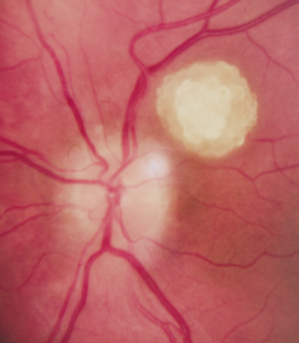
Retina.

Retinal involvement in NF1 is unusual. Astrocytic hamartomas (similar to those in tuberous sclerosis), retinal capillary hemangiomas, and combined hamartoma of retina and retinal pigment epithelium (RPE) occur.48,49

EYELID AND ORBIT.

Orbital neurofibromas may arise from any nerve in the orbit, especially the trigeminal nerve.50 Classically the triad of visual loss, optic atrophy and optociliary shunt vessels have been associated with optic nerve sheath meningiomas,51 but these findings may be nonspecific for optic nerve compression,52,53 particularly in children. If a child with a presumed diagnosis of NF1 is found to have an optic nerve meningioma, NF2 should be considered.

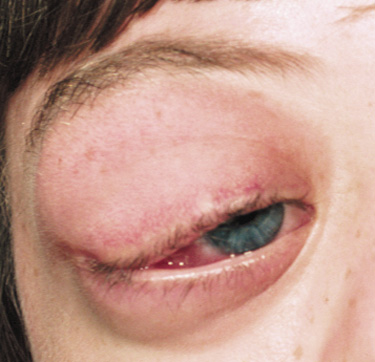
The eyelids are frequently involved in NF1. The lateral portion of the upper lid is prone to develop a plexiform neurofibroma, producing a sinusoidal or S-shaped deformity of the lid margin.54–56 Amblyopia develops from ptosis and astigmatism (Fig. 4).

Fig. 4. Neurofibromatosis type 1: plexiform neuroma of right upper eyelid. Note the S-shaped deformity.

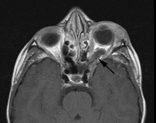
Growth of a plexiform neuroma may be directed posteriorly, infiltrating orbital tissues and involving the bony walls. Congenital plexiform neuromas of the orbit are often associated with absence of the sphenoid wing (Fig. 5). Intracranial pulsation can be transmitted to the orbit causing pulsatile proptosis or enophthalmos.57–59 Plexiform neuromas of the lid and orbit are difficult to treat; complete excision is difficult and potentially disfiguring. The tumors are vascular and may bleed copiously at surgery. They are not radiosensitive and there is also risk of inducing malignant transformation. Trials with antiangiogenic chemotherapeutic agents (thalidomide, interferon alpha) and mitotic-signaling pathway blockers (inhibition of Ras by Farnesyl protein tranferase inhibitor) are in progress.60,61
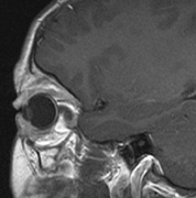
Fig. 5. Surveillance images from a 15-year-old boy with a history of visual loss, proptosis and a diagnosis of NF1. Postcontrast axial (a) and sagittal (b) T1-weighted images demonstrate a plexiform neurofibroma of the left upper and lower eyelids, which extends into the orbit and to the extraconal soft tissues through a widened superior orbital foramen, best seen in (c). An optic nerve glioma widens the optic canal. (d) A sphenoid wing dysplasia is visible and seen as asymmetry of the orbits on the 3D CT bone reconstruction images.

OPTIC NERVE GLIOMAS

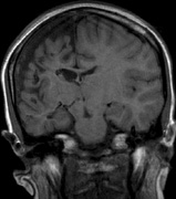
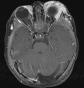
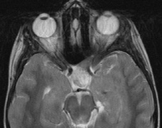
Optic pathway gliomas arise from the astrocytes of the optic nerve. Most are pilocytic astrocytomas and typically remain intradural, extending in the subdural space. Malignant transformation is rare. There appear to be two growth patterns62:

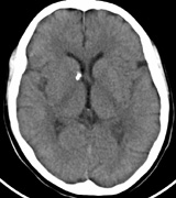
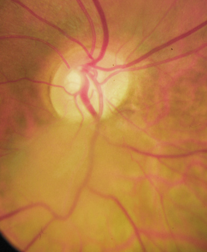
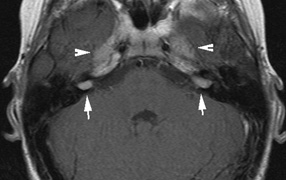
  1. Perineural growth pattern, correlating with a diagnosis of NF1. The tumor expands in the subarachnoid space and the optic nerve is compressed as a central ribbon. On T2-weighted MRI, this may be seen as a low-intensity core with surrounding high-intensity rim. Increased tortuosity of the optic nerve is also associated with this growth pattern (Fig. 5).63
  2. Intraneural growth pattern, correlating with the absence of NF1.

The incidence of optic pathway gliomas in NF1 is difficult to estimate, as the majority are asymptomatic and do not affect vision. In NF1, radiographic evidence of an optic nerve glioma occurs in 15% of patients.64,65 Most tumors are neither suspected historically nor detected by ophthalmic examination.65 Even when optic nerve gliomas become clinically detectable, visual function often remains stable in the absence of any intervention.66,67 Thus routine computed tomography (CT) imaging, even in patients with a known optic nerve glioma, is controversial. The potential risks of repeated radiation exposure in a child with a tumor-suppressor gene defect is a consideration. MRI studies are more revealing than CT but may require sedation or anesthetic and are costly. An MRI is often recommended at the time of diagnosis, but in most cases routine “follow-up” MRIs are unnecessary. Annual ophthalmic clinical examination for relative afferent pupillary defect (RAPD), visual acuity, visual fields, color vision, and funduscopy is usually all that is required, as no therapeutic action is likely to be taken unless there is significant and progressive visual involvement.

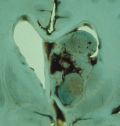
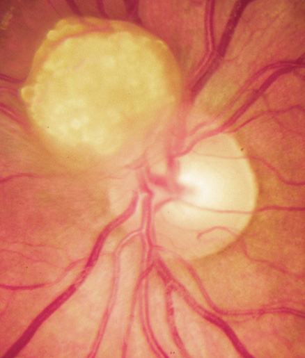
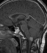
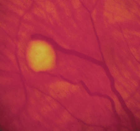
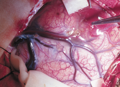
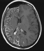
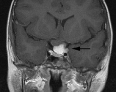
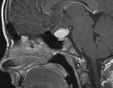
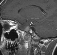
Optic nerve gliomas may involve the optic chiasm and be associated with endocrine disorders or nystagmus.68–70 Surgical excision of chiasmal gliomas (Fig. 6) carries a high risk of visual loss. Invasion of the hypothalamus or the third ventricle carries a poor prognosis, with greater than 50% 15-year mortality rate.71 One review of radiation treatment for chiasmal gliomas collated data from small case series and found no significant long-term improvement in visual function, progression, or mortality with radiation treatment.71 Other reports suggest that radiation doses over 4500cGy improve symptoms and slow progression of chiasmal gliomas over several years.72 Adequate tumor coverage by radiotherapy results in irradiation of normal brain and nearly all children need hormone replacement.73 Chemotherapy is an alternative.74,75

Fig. 6. Surveillance images of a 12-year-old girl with NF1 and an optic nerve glioma which has extended to involve the chiasm. Pre- (a) and postcontrast (b) T1-weighted coronal images reveal a large suprasellar mass with an enhancing component (arrow) seen separately from the normally enhancing pituitary gland. (c) Axial scans through the suprasellar cistern show the tumor is high signal on T2-weighted scans. (d) Enlargement of the chiasm, optic nerve and hypothalamus is visible on the post-contrast T1-weighted sagittal image. Although optic nerve glioma usually has a good prognosis (with visual function often remaining stable in the absence of any intervention66,67), chiasmal involvment is a poor prognostic indicator. A sign of chiasmal involvment may be new onset of endocrine disorders or nystagmus. Surgical excision of chiasmal gliomas carries a high risk of visual loss.68–70

Diagnostic Criteria

The NIH has defined 7 criteria,22 at least two of which must be present:

  1. A first-degree relative with NF1
  2. At least two Lisch nodules (Fig. 3)
  3. At least five café au lait spots ≥ 5 mm before puberty or at least 6 café au lait spots ≥ 15 mm after puberty (Fig. 1)
  4. Axillary or inguinal freckling (Fig. 2)
  5. Optic glioma (Fig. 5)
  6. A “distinctive” bone lesion: sphenoid bone dysplasia or thinning of long bone cortex (with or without pseudoarthrosis)
  7. At least two neurofibromas of any type or one plexiform neurofibroma

In young children, nearly half of sporadic cases fail to meet NIH criteria by 1 year of age, but meet them by age 8 to 12 years. The clinical features appear in the following order: café au lait spots, axillary freckling, Lisch nodules, neurofibromas.26 The inclusion of UBOs on MRI also aids the diagnosis of very young children25 but may require a general anesthetic.

Genetics and Screening

All family members of patients with NF1 should be screened. Lisch nodules are present in 90% or more of patients with the disease, so ophthalmic examination is essential. Annual ophthalmic examination with visual acuity, visual field, color vision, and fundoscopy is recommended for affected individuals.

The NF1 gene (17q11.2) was cloned in 1990.76,77 Its protein, neurofibromin, is expressed throughout the body but particularly in the central and peripheral nervous system.12 It is a tumor-suppressor gene with autosomal dominant inheritance, high penetrance, and variable expressivity. NF1 has been called a neurocristopathy, but the disease is not limited to neural crest derived tissues.12,78

Neurofibromin has structural similarity to GTPase, activating proteins that inhibit the function of G proteins such as Ras. Inhibition of Ras by neurofibromin has been shown in vitro and in vivo. NF1-deficient tumors show increased levels of Ras-GTP. Increased Ras function has been shown in a number of human tumors.12,79–81

NEUROFIBROMATOSIS TYPE 2

Neurofibromatosis type 2 (NF2) was described in 1822 by the Scottish surgeon Wishart.82 Initially, NF1 and NF2 were lumped together despite their differences.83 The identification of different genes re-established them as separate entities.84

Epidemiology

The NIH consensus conference22 estimated an incidence of 1:50,000 but there are varying estimates internationally.84 NF2 is autosomal dominant with full penetrance by age 60.85 In all, 10% of affected patients are symptomatic by age 10, 50% by 20 years, and 80% by 30 years.86 Some sporadic cases reflect parental mosaicism.87,88

Systemic Features

Several studies have established the clinical picture of NF2.85,89–91 Patients may become severely disabled because of the combination of deafness and imbalance from vestibular schwannomas, weakness from multiple spinal tumors, and poor sight from cataracts, optic nerve meningiomas, and retinal hamartomas.

CUTANEOUS FINDINGS.

Skin findings in NF2 may be subtle but not uncommon. Skin tumors were found in 60% and were the first presenting sign in more than 25% of patients.92 The skin tumors are predominantly schwannomas. A minority are neurofibromas and mixed types. Clinically differentiating the histological types may be difficult.92 They may present as dermal tumors or subcutaneous nodules of peripheral nerves.

NF2 plaques are well circumscribed, slightly raised, often pigmented, and hairy. They are a sensitive marker for NF222 and are more common on the limbs and trunk than the neck and face.92

Café au lait spots are not uncommon in NF2, but it is unusual to find more than five and most tend to be restricted to the trunk. Plexiform neurofibromas and axillary/inguinal freckling are uncommon.92,93

NEUROLOGIC FINDINGS.

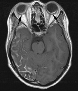
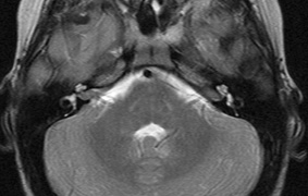
  1. Schwannomas most commonly affect the fifth cranial nerve (CN), but any cranial nerve may be affected. In contrast to vestibular schwannomas (CN VIII), these typically do not grow large.84,90 Vestibular schwannomas (acoustic neuromas) are the classic finding 
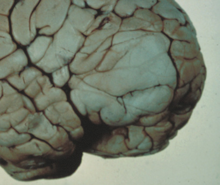
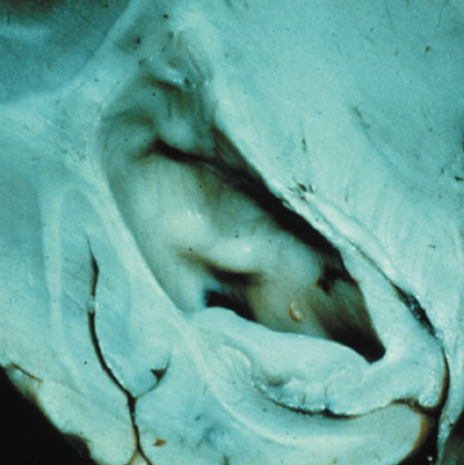
in NF2 (Fig. 7). The risk for malignant transformation is low, but may be higher with radiation exposure.84,94
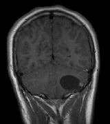
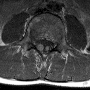
  2. Meningiomas are most likely to affect the spinal cord and supratentorial part of the cranium.84
  3. Other tumors: low-grade ependymomas and gliomas may affect the cervical spine and Brainstem (Fig. 7).84
  4. Neuropathy: A generalized polyneuropathy affects 3% to 5% of affected adults and is associated with an “onion-bulb” appearance on nerve biopsy. In children, mononeuropathy, particularly the facial nerve (CN VII), may be an early sign of NF2. Some children may present with a poliomyelitis-like illness with lower-limb muscle wasting.86,95,96

Fig. 7. Neurofibromatosis type 2: Images of a 12-year-old boy with deafness and weakness in his arms and legs, whose father has bilateral deafness. Axial T2-weighted (a) and postcontrast axial (b) T2-weighted images reveal bilateral vestibular schwannomas, which are also known as acoustic neuromas (arrows). This is the classic finding of NF2. (b, c) Bilateral schwannomas are seen in Meckel's cave (arrowheads) and a (d) lower left cranial nerve schwannoma extends into the pars nervosa of the jugular foramen (arrow). (e) A part cystic and part solid enhancing ependymoma in seen within the cervical cord and medulla and within the distal cord and conus. (e, f) Thoracic schwannomas are present at numerous levels (arrowheads). Marked enhancement and thickening of the roots within the cauda equina also represent multiple schwannomas.

VISCERAL FINDINGS.

NF2 does not appear to have significant visceral complications

Ophthalmic Features

The most common ocular findings in NF2 include premature cataracts and retinal hamartomas. Ocular motor nerve deficits may also be associated with NF2.

Cataracts in NF2 were recognized in 1986.97 They are common, reported to be found in 69%,98 81%,91 85%,99 and 87%100of cases. They are typically mild98 and a lens opacity may be the initial manifestation of disease, as found in 10% (5 of 49 patients) in one study.98

Retinal hamartomas affect about 10% to 20% of patients (8% in one study91and 22% in another98) and may be associated with a more severe phenotype of NF2.91,98 Combined pigment epithelial and retinal hamartomas (CPERH) and epiretinal membranes have been reported.101–103 Lisch nodules are rare in NF2. The absence of Lisch nodules, in association with the presence of posterior subcapsular or cortical cataracts (Fig. 8) may aid the differentiation of NF1 and NF2.97,104,105 Optic nerve sheath meningiomas may cause significant visual impairment in the first years of life (Table 2).84,106

Fig. 8. Neurofibromatosis type 2: cortical wedge cataract. Lens opacities (cortical wedge and posterior subcapsular cataract) may be the initial manifestation of disease in 10%.98

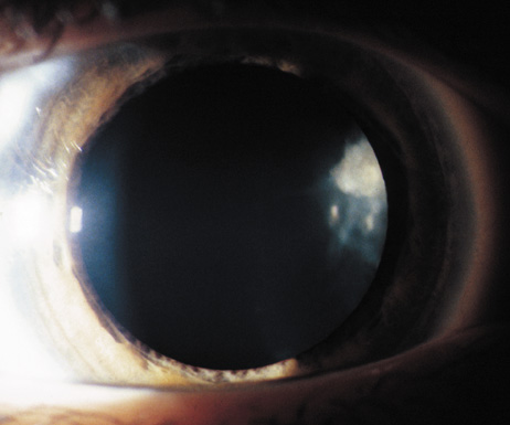
TABLE 2. Ocular Findings in Neurofibromatosis Type 298

Lens opacities (most common finding)
    Posterior subcapsular cataract
    Cortical wedge opacities
Lisch nodules (rarely)
Optic nerve sheath meningiomas
Retinal hamartomas
Combined pigment epithelial and retinal hamartomas
Epiretinal membranes
Optic disc gliomas
Motor nerve palsies


Diagnostic Criteria

The NIH scheme is most commonly used:95,107

  1. Bilateral eighth nerve schwannoma, or
  2. Positive family history of NF2, plus either unilateral eighth nerve schwannoma or two of the following:
    • Neurofibroma
    • Meningioma
    • Glioma
    • Schwannoma affecting other nerve(s)
    • Juvenile lens opacity

A significant proportion of cases (20% to 30%) may present with intracranial meningioma, spinal tumors, or cutaneous tumors. Vestibular schwannomas may account for as little as 15% to 30% of presenting signs in children. Thus, expanded criteria have been proposed to include individuals with multiple schwannomas/meningiomas who have not developed vestibular schwannomas as yet and do not have a clear family history.84

Genetics and Screening

Cataracts can affect vision in early life. For children at risk, annual ophthalmic examination is prudent. Central nervous system (CNS) tumors rarely become symptomatic before age 10 years. Vestibular schwannomas tend to grow faster in younger patients; therefore, asymptomatic at-risk individuals may benefit from MRI scanning every 2 years under age 20 and every 3 years over age 20. Epiretinal membranes and combined hamartomas should lead to investigation for NF2. An annual neurologic exam has also been recommended.84

The NF2 tumor-suppressor gene product is known as schwanomin108 or merlin109. The gene is located at 22q11.1–22q13.1 (Online Mendelian Inheritance in Man [OMIM] #607379).110 The NF2 gene shows homology to the highly conserved ezrin-radixin-moesin (ERM) family of membrane-cytoskeleton linking proteins. The NF2 protein is believed to interact with the actin cytoskeleton. Disruption of NF2 function has been linked to loss of cell-to-cell contact inhibition of growth and division.14

Back to Top
TUBEROUS SCLEROSIS COMPLEX
In 1880, Bourneville, a French neurologist, described a girl with the triad of mental retardation, seizures, and vesiculopapular rash on the face. At postmortem, tumors were found in the brain (cortical sclerotic lesions and tuberous extensions into the ventricles) and kidneys. Earlier, Rayer (1835) and Addison and Gull (1850) had described distinctive facial papules.111 Balzer and Menetrier (1885) described the microscopic appearance of the facial papules as “adenoma sebaceum,” thought to be benign tumors of sebaceous glands, although the skin lesions are actually angiofibromas.3,111,112 Sherlock (1911) coined the acronym epiloia—for epilepsy, low intelligence, and adenoma sebaceum—which is no longer used.113 Van der Hoeve (1920) described the retinal tumors of tuberous sclerosis.114

There have been exciting developments in regard to the molecular genetics of tuberous sclerosis complex (TSC). Two tumor suppressor genes have been identified: TSC1 and TSC2. Further insight into the role of these genes promises potential treatments to halt the progression of disease,115 thus emphasizing the need for early diagnosis.

EPIDEMIOLOGY

Tuberous sclerosis is autosomal dominant with 65% to 75% of new cases representing spontaneous mutations.116 Many “sporadic” cases may actually reflect somatic or germ-line mosaicism of a parent.117,118 Its prevalence is approximately 1:10,000 to 1:15,000.119,120 TSC has high penetrance but variable expressivity, even within the same family.121

SYSTEMIC FEATURES

Cutaneous Findings

  1. Facial angiofibromas typically manifest as a papular rash over the nose and cheek (Fig. 9). Angiofibromas may also be found on the chin but they spare the upper lip. Pain and bleeding in association with minor trauma is common.24,122 They eventually affect 75% to 85% of patients, but are infrequent before the age of 2 years and may not be present until late childhood or adolescence. Facial angiofibromas may be treated cosmetically with laser.122–125 A forehead fibrous plaque is raised, red, and waxy. It is found in 20% to 35% of affected individuals. This feature is important diagnostically as it may be present at birth and is a sensitive marker for TSC.122,125,126
  2. Subungal (Fig. 10) and periungal fibromas are histologically similar to facial angiofibromas. These are infrequent in childhood and become more prevalent with age (>50% of adults). Ungual fibromas, may be the only sign of TSC.122 Ungual fibromas are more common on the toenails than the fingernails. Gingival (gum) fibromas may also be found.24,122,125
  3. Skin tags occur on the neck, axilla, and upper back in 10% to 20% of affected individuals. Though common in normal elderly patients, extensive skin tags are uncommon in normal children and adolescents.122,125
  4. Hypomelanotic macules (or ash leaf spots) are depigmented patches (Fig. 11) that occur in 90% to 95% of affected children, but may become less apparent with age.122,125 They commonly take the form of “ash leaf spots,” appearing lanceolate, pointed at one end and round at the other like the leaves of the mountain ash tree. They are the feature of TSC most likely to occur in the first year of life. A Wood's ultraviolet light (which is selectively absorbed by skin melanin) helps in their detection: ash leaf spots stand out as white patches.119,122 Hypomelanotic macules also occur in normal infants.127,128 Histologically, they have a normal numbers of melanocytes, but the melanosomes are smaller and contain less melanin than the surrounding normal skin.3,122,125,128
  5. Shagreen patches are reported in about 50% of older children.122,125 This is a connective tissue nevus129,130 with a yellowish, plaque-like appearance like rough leather or shark's skin. The shagreen patch is typically found in the lumbosacral area (Fig. 12) but may also be found on the eyelids.3,25

Fig. 9. Tuberous Sclerosis Complex: facial angiofibroma. Previously known as “adenoma sebaceum.” Pain and bleeding in association with minor trauma is common. Facial angiofibromas are infrequent before the age of 2 years, but eventually affect 75% to 85%.25,122

Fig. 10. Tuberous Sclerosis Complex: subungual fibroma. Similar in histology to facial angiofibromas.25,122

Fig. 11. Tuberous Sclerosis Complex: hypomelanotic macules, also known as “Ash-leaf spots,” pointed at one end and round at the other end, like the leaves of the mountain ash tree. A Wood's ultraviolet light helps in their detection. They are the feature of TSC most likely to occur in the first year of life.122,125

Fig. 12. Tuberous Sclerosis Complex: Shagreen patch. This is a connective tissue nevus which affects about 50% of older children.122,125,129,130 It is named for its appearance like untanned rough leather or shark's skin. The shagreen patch is typically found in the lumbosacral area but may also be found on the eyelids.3,25

Neurologic Findings

INTRACRANIAL LESIONS.

Mental retardation and seizures in TSC are often associated with benign CNS astrocytic hamartomas.131 On imaging, the cerebral lesions show three patterns:132

  1. Superficial cortical sclerosis (parenchymal hamartoma) which distort the gyri. Microscopy shows large atypical fibrillary type astrocytes with few associated neurons and areas of calcification.133 The numerous abnormal glial processes and fibers make the tissue abnormally firm or “sclerotic” on palpation (Fig. 13).132
  2. Subependymal nodules (SEN) are typically found along the lateral borders of the ventricles and parehncymal brain lesion (“cortical tubers”) (Figs. 14 and 15). Calcification in the first year of life is rare.132
  3. White matter abnormalities are a characteristic “ventriculofugal” pattern corresponding to the embryological migratory paths of neurons and glia. These lesions may represent aberrant neuronal migration, a possible reflection of haploinsufficiency for the TSC gene.116,132,134
    On MRI imaging, the subependymal nodules and parenchymal brain lesions of infants (age ≤3 months) and adults show different signal characteristics. Infant CNS tubers are hyperintense on T1-weighted images and hypointense on T2-weighted images, which is the opposite of the pattern seen in adults.134 Malignant transformation of SEN occurs in about 10% to 15% of patients and the resultant subependymal giant cell astrocytoma accounts for 25% of premature deaths in TSC.135,136

Fig. 13. Tuberous Sclerosis Complex: “cortical sclerosis.” Numerous abnormal glial processes make the tissue abnormally firm or “sclerotic” on palpation.132

Fig. 14. Tuberous Sclerosis Complex: Sub-Ependymal nodules. These are typically found along the lateral borders of the ventricles.132

Fig. 15. Tuberous Sclerosis Complex. (a) Patient 1: Axial CT scans demonstrating typical calcification of subependymal nodules in a 13-year-old girl with a history of seizures. (b and c) Patient 2. (b) Axial T2-weighted images demonstrate calcified subependymal nodules (arrowheads) and cortical tubers typical of tuberous sclerosis. (c) Widespread cortical tubers are seen on a coronal FLAIR sequence as thickening of the cortex and high signal of the subcortical white matter.

SEIZURES.

Typically observed first in infancy, seizures are a common presenting sign of TSC and a frequent source of morbidity. The seizures often begin as “salaam spasms”—repetitive myoclonic spasms that produce head nodding with associated extension/flexion of the trunk and limbs. They often progress to grand mal seizures25,116 and may be difficult to control. Vigabatrin has been advocated for control of infantile spasms but it carries a risk for irreversible visual field loss. Other treatments include corticosteroids, adrenocorticotrophic hormone, and a ketogenic diet.25,137–139 Selective surgical excision of an “epileptogenic tuber” may be an option for intractable epilepsy.140

MENTAL RETARDATION.

Mental deficiency, part of Vogt's diagnostic triad (adenoma sebaceum, epilepsy, mental deficiency),3 affects about 50% of patients overall.25,141 TSC2 mutations may carry a higher risk than TSC1.142

AUTISM.

This may affect 50% to 60% of TSC patients, possibly correlating with numbers of cerebellar and temporal tubers.25,143,144

Visceral Findings

Patients with TSC may have renal, cardiac, pulmonary and other visceral involvement.

RENAL.

The TSC2 gene was localized by linkage with the polycystic kidney disease gene in patients with TSC and polycystic kidneys. This represents a contiguous gene syndrome in which adjacent genes (PKD1 and TSC2) are deleted together.145–147

Multiple angiomyolipomas of the kidney are benign hamartomas of blood vessels, smooth muscle, and fat. By 10 years of age, the frequency of angiolmyolipomas is 75%, similar to adults.148 The fat signal of angiomyolipomas helps to distinguish them from renal cell carcinoma.25

Renal insufficiency/failure may be the complication of both cystic disease and angiomyolipomas and is one of the leading causes of morbidity and mortality in TSC.138,149

CARDIAC.

Rhabdomyoma are usually multiple, typically asymptomatic, and often regress with age.150,151

PULMONARY.

Lymphangioleiomyomatosis is a progressive lung disease characterized by bronchiolar smooth muscle infiltration and cystic changes of the lung parenchyma. About 1% of adult females with TSC are affected.116,152,153

OPHTHALMIC FEATURES

The principal ophthalmic manifestations of TSC are in the posterior segment.

Astrocytic Retinal Hamartomas

Although in Van der Hoeve's original description the term phakoma or phakomata was a generic term for the tumors occurring in various organ systems,1 it has become associated with the retinal astrocytic tumors of TSC,3 which occur in about 50% of affected patients and are bilateral in 25% to 30% (Table 3, Fig. 16).154,156

Fig. 16. Tuberous Sclerosis Complex: astrocytic retinal hamartoma, also referred to as retinal phakoma. There are three types: translucent type (a), calcified, nodular or “mulberry” type (b), and intermediate type (c).156 These usually do not affect vision and require no treatment. About half of patients are affected and there are bilateral lesions in 25% to 30%.154–156

TABLE 3. Astrocytic Retinal Hamartomas in Tuberous Sclerosis

Type 1. In the most common type, relatively flat, smooth-surfaced, and translucent lesions often difficult to see (an abnormal light reflex may be the only clue). They are in the posterior pole, superficial to the retinal vessels.156
Type 2. Raised, calcified, multinodular (“mulberry-like”) tumors in the posterior pole (80% within 2 disk diameters of the optic nerve).156
Type 3. The least common, intermediate type shows features of types 1 and 2, often in the posterior pole.156


Historically, the presence of mixed (type 3) hamartomas led to the suggestion of evolution from the type 1 to the type 2.157 A long-term photographic study found that although the types remained unchanged, a minority of types 1 and 3 increased in calcification and a new lesion was noted that had not been evident on an earlier photograph.157 All three types of retinal hamartomas may be found in young children. There is no correlation between age and the predominant type of lesion.156

Histologically, phakomata are composed of glial astrocytes—elongated, fibrous astrocytes with small oval nuclei, arising from the retina or the optic disc. The type 2 lesions contain basophilic, calcified drusen-like deposits within a dense vascular network. On fluorescein angiography, the type 2 lesions show autofluorescence, leakage with transit of dye, and marked late hyperflorescence.3,113

Astrocytic retinal hamartomas usually do not affect vision and require no treatment. Vitreous seeding and vitreous hemorrhage have been reported in association with phakomata, but these are rare.158,159 Laser photocoagulation may be considered in rare cases were exudation induces serous retinal detachments.160 The differential diagnosis of “white retinal lesions” includes retinoblastoma, toxoplasmosis and toxocara.3,113

Pigmentary Abnormalities

Depigmented “punched-out” chorioretinal lesions in the midperiphery have been reported in up to 40% of patients. Similar lesions may be found in normal patients.156

Nonretinal Features

The adnexae may be involved by angiofibromas: eyelid skin and “salmon-colored” subconjunctival nodules. Sector iris depigmentation and iris coloboma have been reported in a minority of patients.113,156,161

DIAGNOSTIC CRITERIA

When TSC is classical (“Salaam seizures,” facial angiofibroma, developmental delay), the diagnosis is straightforward. However, children can be mildly affected, and many of the classical signs may become apparent later.

Based on the revised clinical diagnostic criteria set forth by the Tuberous Sclerosis Consensus Conference162, a diagnosis of TSC may be made if the patient has two major features, or one major feature plus two minor features. Genetic testing for TSC still has a high false-negative rate (Table 4).25,162,163

TABLE 4. Features of Tuberous Sclerosis Complex


Major features Minor features
1. Facial angiofibroma or forehead plaque (Fig. 9)1. Dental pits
2. Nontraumatic ungual fibroma (Fig. 10)2. Rectal polyps (hamartoma)
3. More than three hypomelanotic (ash-leaf) patches (Fig. 11)3. Bone cysts
4. Shagreen patch (Fig. 12)4. Cerebral while matter “migration lines”
5. Multiple retinal hamartomas (Fig. 16)5. Gingival fibroma
6. Cortical tuber/sclerosis (Fig. 13)6. Nonrenal hamartoma
7. Subependymal nodule (Fig. 14)7. Retinal achromic patch
8. Subependymal giant cell astrocytoma8. “Confetti” skin lesions
9. Cardiac rhabdomyoma9. Multiple renal cysts
10. Lymphangioleiomyomatosis 
11. Renal angiomylipoma 


GENETICS AND SCREENING

The Tuberous Sclerosis Consensus Conference162 recommendations for screening once a diagnosis of TSC is established are:

  1. Cranial neuroimaging every 1 to 3 years
  2. Renal ultrasound every 1 to 3 years
  3. Chest CT for females at least once upon reaching adulthood to screen for lymphangio-leiomyomatosis.

Two genes have been identified, TSC1 (9q34, OMIM #605284) and TSC2 (16p13.3, OMIM #191092), which appear to be tumor-suppressor genes. The gene product of TSC1 is hamartin and the gene product of TSC2 is tuberin. Of sporadic cases of TSC, 70% have been linked to TSC2 mutations. Among the familial cases, half are linked to the 9q34 locus and half to the 16p13.3 locus. Hamartin and tuberin appear to interact together in vivo, which may be why mutations of two different genes produce a similar phenotype. Together they form a cytoplasmic protein complex that has an inhibitory effect on cell growth and division. The tuberin-hamartin complex inhibits the activity of TOR (target of rapamycin). This has raised the possibility of reestablishing the regulatory effect of the tuberin-hamartin complex on TOR with drugs such as rapamycin.115,164

Back to Top
VON HIPPEL-LINDAU SYNDROME
Von Hippel described the fundoscopic appearance and histology of “angiomatosis retinae” in 1911165 and Lindau linked the retinal, cerebral, and visceral components of the syndrome in 1926. The term von Hippel-Lindau syndrome (VHL) was first used in 1936.166 The first major clinical criteria for the syndrome were proposed in 1964.167

The main manifestations of von Hippel-Linau syndrome are: histologically similar hemangiomas of the retina and CNS, renal cysts, renal cell carcinomas, pancreatic cysts, pheochromocytomas, and endolymphatic sac tumors.13

EPIDEMIOLOGY

The prevalence of VHL syndrome is about 1 in 35,000 to 40,000.168 It is autosomal dominant with greater than 95% penetrance by age 60 years.169 Its expression, even within the same family, may be quite variable, especially for renal disease and pheochromocytoma.170,171

SYSTEMIC FEATURES

Cutaneous Findings

Cutaneous findings are not part of the diagnostic criteria for VHL syndrome.

Neurologic Findings

CNS hemangiomas are the most common tumor of VHL, affecting 60% to 80%, with a predilection for the cerebellum and spinal chord. An enlarging cystic component is a frequent finding in symptomatic tumors. Patients typically present in their early 30s; headaches or neck pain in affected individuals should not be ignored.172,173 On microscopy, CNS hemangiomas resemble retinal capillary hemangiomas. Their malignant potential is low.174 The treatment is surgical (Fig. 17, A and B).13

Fig. 17. Images from a 13-year-old boy with Von Hippel-Lindau syndrome. (a) Coronal postcontrast T1-weighted imaging reveals a cystic lesion with an enhancing nodule at the pial surface typical of a hemangioblastoma. (b) A second solid enhancing hemangioblastoma is seen at the craniocervial junction on a sagittal postcontrast T1-weighted image. (c) Associated cystic lesions (arrows) are seen within the pancreas.

Endolymphatic sac cystadenomas are a sensitive marker of VHL. Patients may present with hearing loss, tinnitus, disequilibrium or facial nerve palsy.175,176

Visceral Findings

KIDNEY.

Renal cysts are often asymptomatic and do not require treatment. However, complex cysts may carry a risk for malignant transformation.

Renal carcinoma affects about a third of patients. Patients typically present in their late 30s. These tumors may be completely asymptomatic, underscoring the necessity for ultrasound screening.13,177

ADRENALS.

Pheochromocytomas are benign neural crest tumors of the adrenal medulla (chromaffin cell derived) and probably affect 10% to 20% of patients. Mean age at diagnosis is at about 30 years of age. Pheochromocytomas may be multiple and bilateral. They are catecholamine-secreting tumors that classically produce severe hypertension and anxiety attacks, but they may also be asymptomaic.178 The National Cancer Institute classification of VHL syndrome is in part based on the absence (type I) or presence (type II) of pheochromocytoma (Table 5).171

TABLE 5. Classification of von Hippel-Lindau Syndrome

Type CNS hemangioma Retinal hemangiomaRenal Cell cancerPheochromocytoma
IYesYesYesNo
IIAYesYesNoYes
IIBYesYesYesYes
IICNoNoNoYes


PANCREAS.

Pancreatic lesions may be nonsecretory (most commonly cysts or cystadenomas) or secretory (islet cell tumor).179 In one study of 52 patients, 56% (29 patients) were found to have pancreatic lesions. The majority (19 of 29) had cystic changes only (Fig. 17C). Pancreatic lesions were the only abdominal manifestation of disease in 6 of 52 patients.180 In a separate study, pancreatic cysts did not show significant progression on follow-up examinations over an average period of 5 years.181 Like pancreatic cysts, islet cell tumors appear to be frequently asymptomatic.182

EPIDIDYMIS AND BROAD LIGAMENT.

Papillary cystadenomas of the epididymis are typically benign and asymptomatic. In one study, about half of patients were affected.183 Papillary cystadenomas of the broad ligament are thought to be the female equivalent of papillary cystadenomas of the epididymis.184

OPHTHALMIC FEATURES

VHL syndrome does not appear to have significant anterior segment manifestations.

Posterior Segment

Retinal capillary hemangiomas are among the most frequent and earliest manifestations of VHL syndrome. Most patients present between 10 and 40 (mean age of 25) years of age and 5% present before age 10. Overall, retinal capillary hemangiomas affect about 60% of individuals with VHL syndrome. Development of new retinal capillary hemangiomas after age 60 years is unusual.169,170

Retinal capillary hemangiomas (Fig. 18) may be the only manifestation of the syndrome,185 and typically involve the midperipheral temporal retina.186 A pair of dilated and tortuous retinal vessels (artery and vein) classically supply the vascular tumor. Without fluorescein angiography, it may be difficult to distinguish artery from vein on fundus examination.170

Fig. 18. Von Hippel–Lindau syndrome: retinal capillary hemangioma. In contrast to the phakoma of tuberous sclerosis, they require early treatment to prevent exudation and associated retinal detachment.186

Retinal capillary hemangiomas may also occur in the absence of VHL syndrome, but the age at presentation is typically later (50 vs. 25 years).187 Small retinal capillary hemangiomas may remain stable for many years, but most tend to enlarge and may become symptomatic secondary to exudation and retinal detachment.

On microscopy, retinal capillary hemangiomas are similar in appearance to the CNS hemangiomas of VHL syndrome. Thin vascular channels are lined by endothelial cells and pericytes. The vascular channels are separated by foamy stromal cells. The foamy appearance is due to phagocytosed lipids that escape the fenestrated endothelium of the vessel walls. The lipid is plasma–derived cholesterol stearate.188–190 Hemangioma is more appropriate than hemangioblastoma.170,188,190

The cellular origin of capillary hemangiomas in VHL is controversial,170 but loss of heterozygosity of the VHL gene has been demonstrated in the stromal cells of capillary hemangiomas.191

There are various classification schemes based on morphology (endophytic, exophytic, and sessile) and the presence or absence of exudation. Endophytic tumors grow on the retinal surface, protruding into the vitreous cavity. Exophytic tumors tend to be nodular and orange-colored, growing the in outer layers of the retina. The sessile growth pattern implies a flat tumor, gray or orange in color, and grows within the middle layers of the retina.185 The “natural history” of the retinal capillary hemangioma has been divided into four192 or five193 stages170:

  • Stage 1: Preclassical, small capillary retinal angioma with or without feeder vessels
  • Stage 2: Classical, hemangioma enlarges and draining vein becomes more prominent
  • Stage 3: Exudation, often in association with enlargement of feeding artery and draining vein
  • Stage 4: Retinal detachment
  • Stage 5: End stage, rubeosis, glaucoma, phthisis

On occasion, an exophytic-type capillary hemangioma adjacent to the optic nerve can masquerade as chronic disc edema.194 The frequency of optic-disc angiomas was 15% in a group of 175 patients.186 Optic-disc hemangiomas may be the only manifestation of disease in VHL syndrome.186 In the absence of effective treatment, exudation from capillary hemangiomas may induce retinal detachment and macular star formation.170,186

Early detection (i.e., when the hemangioma is small) and treatment are associated with better visual outcomes.186 The exception may be small juxtapapillary hemangiomas, in which treatment may produce significant loss of acuity. Small juxtapapillary hemangiomas may remain stable.185

Both direct and feeder vessel photocoagulation with a yellow-dye laser have been shown to be effective for treating small (<1.5mm) capillary hemangiomas. Feeder vessel technique may require a greater number of treatment sessions.195 Direct treatment may be associated with a smaller scotoma than feeder vessel treatment, but direct photocoagulation carries a potential for hemorrhage and retinal detachment.186 Hemoglobin absorbs yellow laser more than green or blue wavelengths. Complete obliteration of the hemangioma may not be necessary to achieve resolution of symptoms.170

When the retinal capillary hemangioma is located in the far anterior periphery, or when it is larger than 3.0 mm in diameter, cryotherapy may be more effective than photocoagulation.170 To treat hemangiomas 4 to 5 mm in diameter, plaque radiotherapy has been recommended.196

The VHL gene product appears to inhibit the production of VEGF (vascular endothelial growth factor). Treatment with SU 5416, a VEGF-receptor inhibitor, has been reported to restore and maintain visual acuity and visual field in a patient with an optic-disc hemangioma.197

Retinal “twin vessels”—a paired retinal arteriole and venule of normal caliber, separated by less than the diameter of the venule—have been described as part of VHL198 but the finding has not been reproduced.186 Twin vessels are not associated with retinal capillary hemangiomas, and may be found in normal patients.199

Differential Diagnosis

In the presence of subretinal exudates, the peripheral hemangioma may be overlooked, giving a false impression of Coats disease (diffuse unilateral retinal telangiectasias with exudation in young boys). Dilated feeder vessels may be confused for the arteriovenous malformation of Wyburn Mason disease (racemose hemangioma). Exudation is not a feature of Wyburn Mason disease and there is no angioma. Retinal cavernous hemangiomas lack prominent feeder vessels and do not leak fluid into the subretinal space. Retinal macroaneurysms may leak fluid but do not have dilated feeder vessels. Uveal melanomas usually have only a dilated vein.170

DIAGNOSTIC CRITERIA:

If there is a positive family history of VHL syndrome, transmission is likely based on finding any one of the following13:

  • CNS hemangioma (including retinal capillary hemangioma)
  • Characteristc visceral lesion (e.g., renal carcinoma, pheochromocytoma)

If family history is negative, a diagnosis is likely based on:

  • At least two CNS hemangiomas (including retinal capillary hemangioma), or
  • A single CNS hemangioma (including retinal capillary hemangioma) and a characteristic visceral lesion (excluding epididymal and renal cysts, which are not uncommon in the general population)

The National Cancer Institute has classified VHL syndrome into two main categories on clinical grounds: type I without pheochromocytoma and type II with pheochromocytoma. Type II is further subdivided based on the presence or absence of renal cell carcinoma and CNS hemangioblastoma, and type IIC implies pheochromocytoma only (Table 5).170

GENETICS AND SCREENING

Subclinical lesions are common in VHL syndrome and there is more than one “accepted” protocol.13,169,200 In general, the protocols are similar, differing in the age to start screening for the different manifestations of the syndrome. Annual fundoscopy and urinary catecholamine testing should begin early in childhood (age 1 to 5 years). Spine and brain MRI should be carried out every 1 to 2 years and abdominal ultrasonography annually beginning age 10 years.

Autosomal dominant inheritance of multisystem tumors with malignant potential is the “trademark” of tumor-suppressor gene defects and the phakomatoses. New mutations are thought to represent about 10% of affected individuals. The VHL gene product (OMIM #193300) located at 3p25–26 appears to downregulate the transcription factors that turn on the production of VEGF as part of the cellular response to hypoxia. This may account for the predilection to form hemangiomas.201 Molecular analysis suggests that missense mutations of the gene predispose to pheochromocytomas, which is type II (96% of mutations in VHL syndromeand pheochromocytoma were found to be missense mutations in one study).202 No particular type of mutation has been associated with frequency or severity of retinal capillary hemangiomas.186

DNA testing for VHL gene mutations can achieve 99% detection rates.203 This may be valuable for counseling of young parents of an affected child who do not have a clear family history and may still be too young to manifest features of the syndrome themselves.

Back to Top
STURGE-WEBER SYNDROME AND OTHER VASCULAR DISORDERS

STURGE-WEBER SYNDROME

Sturge (1879) reported the case of a child with facial “port-wine stain,” epilepsy, buphthalmos, and a dark choroid.204 Schirmer had noted the association of the facial vascular malformation and glaucoma in 1860.3 Weber (1922) reported brain X-ray findings of parallel streaks (“railtrack” sign), which he interpreted to represent calcific changes (Fig. 19).205

Fig. 19. Sturge-Weber syndrome. Lateral skull X-ray of an adult with Sturge-Weber syndrome with the typical tram track calcification of the pial angioma.

Port-wine stains (PWS) affect 0.3% of live births. The majority of isolated PWS occur in the head and neck.206–208 The possibility of Sturge- Weber syndrome (SWS) is raised when a newborn is noted to have a facial PWS. SWS does not have any clear inheritance pattern. The minimum diagnostic criteria for SWS is a subject of debate.209–211 The complete syndrome includes:

  1. Facial capillary vascular malformation (“port-wine stain”)
  2. Intracranial leptomeningeal (the two innermost layers of the meninges, which consists of the dura mater, arachnoid, and pia mater) capillary vascular malformation
  3. Seizures
  4. Contralateral neuromuscular weakness
  5. Ipsilateral increased choroidal vascularity, often with increased retinal vessel tortuosity
  6. Glaucoma

Facial capillary vascular malformation (Fig. 20) is the most obvious feature of SWS. It consists of many dilated capillaries lined by a single layer of endothelial cells.3 The PWS is present at birth and partially blanches with pressure. Historically it has also been referred to as a “naevus flammeus.” The PWS is typically unilateral, and may follow the distribution of the trigeminal (CN V) nerve divisions. The conjunctiva and episclera ipsilateral to the PWS are often involved.

Fig. 20. Sturge-Weber syndrome: facial capillary vascular malformation or “port-wine stain.” Note this child has involvement of both V-I (upper eyelid) and V-II (lower eyelid) dermatomes, which place him at risk for intracranial involvement (correlating with VI involvement)208,218 and glaucoma (risk is highest when both VI and VII are involved).224

The PWS may not entirely respect the midline, possibly a reflection of transmedian innervation.212 The lesion grows proportionally with the child, often becoming darker in color. 213 The overlying skin may develop nodularity or hypertrophy in two-thirds of patients (by the fifth decade).214 Treatment with a pulsed-dye laser may significantly improve the appearance, but recurrence (i.e., redarkening) is possible.215,216 A deficit in the number of perivascular nerves in port-wine stains suggests that abnormal neural modulation of vascular flow may be involved in their pathogenesis.217

Intracranial leptomeningeal capillary vascular malformation (Figs. 21 and 22) is associated with seizures and contralateral neuromuscular weakness. Central nervous system involvement correlates highly with ipsilateral involvement of the V1 dermatome by a PWS.208,218

Fig. 21. Sturge-Weber syndrome: leptomeningeal vascular malformation.

Fig. 22. Axial T2-weighted (a) and coronal T1-weighted (b) images of a 16-year-old boy with a port-wine lesion over the right side of his face. The right hemisphere is markedly atrophic and abnormal draining veins are seen within the right lateral ventricle (arrowheads). (c, d) The entire right hemisphere is covered by an enhancing pial angioma and the choroid plexi are enlarged. Enhancing retinal angiomas (arrows), typical of Sturge-Weber syndrome, are seen in (d).

It has been hypothesized that the calcific changes reflect cellular anoxic injury secondary to venous stasis/poor drainage.219,220 Abnormal autonomic innervation has also been reported in association with the malformed cortical vessels—innervation only by noradrenergic fibers.221 Studies suggest that the normal increase in brain-blood flow to accommodate increased metabolic demand during seizure activity is blunted in affected brain regions of SWS.222 Thus, seizures in the setting of impaired perfusion may be exacerbating brain injury. Periods of contralateral weakness (days to weeks) can follow a prolonged seizure episode.206,223

When seizures begin early in life, the risk of developmental delay is much higher. In a review of 171 patients with a facial PWS and associated intracranial vascular malformation, 80% were found to have a seizure disorder. Of those, 75% with seizures had an onset during the first year of life, rising to 95% before age 5 years. Nearly all patients had a PWS that included (or was limited to) the V1 distribution. Developmental delay was strongly associated with seizures (71% of children with seizures as compared to 6% without seizures).224

Glaucoma affects 50% to 70% of SWS patients224,225 and it is a therapeutic challenge. From the time of diagnosis, the intraocular pressure, the optic discs, and the corneal diameters need to be measured intermittently, even if an occasional examination under anesthetic is required.

The development of glaucoma appears to follow a bimodal age distribution: early-onset/congenital-type and later-onset. The glaucomatous eye is nearly always on the same side as the PWS.224 The highest risk for glaucoma appears to be when the PWS involves both the upper and lower lid: 72% vs. 21% for VI-only (upper eyelid) involvement in one study.224

The bimodal age distribution of glaucoma suggests that more than one mechanism is involved. Angle abnormalities (“trabeculodysgenesis” with or without flat anterior iris insertion) have been reported in association with the early-onset/congential type.225 Other proposed mechanisms include uveal capillary dilation226 and elevated episcleral venous pressure. Reports of blood in Schlemm's canal on gonioscopy support the idea of increased episcleral venous pressure pathomechanics.227,228

Glaucoma in SWS often responds poorly to medical therapy alone. The “trabeculodysgenesis” of early onset Sturge-Webber glaucoma is reported to be similar in gonioscopic appearance to primary congenital glaucoma.229 On this basis, goniotomy was suggested as a treatment of choice in early-onset SWS glaucoma.

One group looked at the success of goniotomy, trabeculotomy, trabeculectomy, laser trabeculoplasty and medication in the management of SWS-associated glaucoma.230 In the early onset subgroup receiving goniotomy, median stable interval of intraocular pressure reduction after one procedure was 8 months; with multiple goniotomies and the addition of medical treatment, the median stable interval was extended to 9 years. Other investigators have found that goniotomy alone may be insufficient, requiring filtering surgery at some point. However, trabeculectomy carries a significant risk of choroidal expansion in association with hypotony. Theoretically, the risk may be higher when there is increased choroidal vascularity in association with the facial PWS—a “tomato ketchup” fundus.231

Often, there is associated increased tortuosity of the retinal vessels as well. Placement of a prophylactic posterior sclerotomy prior to filtering surgery to prevent choroidal expansion is controversial.232 Cryo- or thermocoagulation of the ciliary body in combination with medical therapy may be an alternative to surgery.233

KLIPPEL TRENAUNAY—WEBER SYNDROME

Klippel-Trenaunay syndrome consists of combined capillary, lymphatic, and venous malformations, in association with varicosities and limb enlargement (Fig. 23). In the majority of cases, the lower limb is affected.234,235

Fig. 23. Klippel Trenaunay-Weber syndrome: combined capillary, lymphatic, and venous malformations in association with varicosities and limb enlargement.234

The occurrence of “merged” Klippel-Trenaunay and Sturge-Weber syndromes has recently been questioned. The contention is that the presumed combined cases actually represent Sturge-Weber syndrome with capillary malformations below the head and neck, without the associated lymphatic malformations of Klippel-Trenaunay syndrome.235 Extracranial port-wine stains are relatively common in SWS (45% in a study of 171 patients).224

PHACES SYNDROME

PHACES236,237 is an acronym for the association of:

  • Posterior fossa brain malformation, most commonly Dandy-Walker type (which exists when there is marked dilation of the fourth ventricle—“posterior fossa cyst”—in association with cerebellar vermis hypoplasia; 70% to 90% may develop hydrocephalus)
  • Hemangioma, most commonly facial and “plaque-like”
  • Arterial malformation
  • Cardiac anomalies and coarctation of the aorta
  • Eye abnormalities
  • Sternal clefting

The majority of patients are female, and affected children may have only one extracutaneous manifestation. The facial vascular lesion is typically large, segmental, and plaque-like.238,239 Unlike the vascular malformation of Sturge-Weber Syndrome, the PHACES facial vascular lesion is likely a true hemangioma.240 Visceral and/or CNS hamangiomas may affect up to 50% of patients.241 The most common associated ocular abnormalities appear to be microphthalmos and increased vascularity on examination of the fundus. Glaucoma does not appear to be a common association of the syndrome (Fig. 24).242

Fig. 24. PHACES. Images from a 10-month-old girl with widespread hemangioma over the left forehead, upper eye lid, chest and right earlobe. Examination of the carotid arteries revealed bilateral bruits. (a) The enhancing hemangiomata (larger on the left) are predominantly preseptal with extraconal intraorbital extension on the postcontrast T1-weighted image. (b) Cerebellar hypoplasia is seen on the left.

Back to Top
REFERENCES

1. van der Hoeve J: The Doyne memorial lecture: Eye symptoms in phakomatoses. Trans Ophthalm Soc UK 52:380, 1932

2. Brouwer B, van der Hoeve J, Mahoney W: A fourth type of phakomatosis: Sturge-Weber Syndrome. Verh K Akad Wet Amst 36:1937

3. Albert DM, Jacobiec FA, Azar DT, Gragoudas ES: Principles and Practice of Ophthalmology. 2nd ed. Philadelphia: WB Saunders, 2000

4. Hogan MJ, Zimmerman LE: Ophthalmic Pathology: An Atlas and Textbook. 2nd ed. Philadelphia: WB Saunders, 1962:58

5. Albert DM, Jacobiec FA, Azar DT, Gragoudas ES. Principles and Practice of Ophthalmology, 2nd ed. Philadelphia: WB Saunders, 2000

6. Duke-Elder S: System of Ophthalmology. Normal and Abnormal Development, Vol. 3, Part 2, Congenital Deformities. St. Louis: Mosby, 1964:1153

7. Aita JA: Neurocutaneous Diseases. Springfield, IL: Charles C. Thomas, 1966

8. Barlow CF: Mental Retardation and Related Disorders. Philadelphia: FA Davis, 1978:75

9. Walsh FB: Clinical Neuro-Ophthalmology, Vol. 3. 3rd ed. Baltimore: Williams & Wilkins, 1969:1939

10. Dahan D, Fenichel GM, El Said R: Neurocutaneous syndromes. Adolesc Med 13:495, 2002

11. Gold DH: The Eye in Systemic Disease. Philadelphia: JB Lippincott, 1990:439

12. Cichowski K, Jacks T: NF1 tumor suppressor gene function: narrowing the GAP. Cell 104:593, 2001

13. Lonser RR, Glenn GM, Walther M, Chew EY, Libutti SK, Linehan WM, et al: von Hippel-Lindau disease. Lancet 361:2059, 2003

14. Xiao GH, Chernoff J, Testa JR: NF2: the wizardry of merlin. Genes Chromosomes. Cancer 38:389, 2003.

15. Yeung RS: Multiple roles of the tuberous sclerosis complex genes. Genes Chromosomes. Cancer 38:368, 2003

16. Riccardi VM: Neurofibromatosis: clinical heterogeneity. Curr Probl Cancer 7:1, 1982

17. Gorlin R, Cohen MM Jr, Levine M: The neurofibromatoses (NfI Reckllinghausen type, NfII acoustic type, other types). In Syndromes of the Head and Neck. Gorlin R, Cohen MM Jr, Levine M, Eds. Oxford: Oxford University Press, 1992: 392

18. National Institutes of Health: Consensus Development Conference Statement: Neurofibromatosis. Arch Neurology 45:575, 1988

19. Knudson AG Jr: Mutation and cancer: statistical study of retinoblastoma. Proc Natl Acad Sci U S A 68:820, 1971

20. Gutmann DH, Aylsworth A, Carey JC: The diagnostic evaluation and multidisciplinary management of neurofibromatosis 1 and 2. JAMA 278:51, 1997

21. National Institutes of Health Consensus Development Conference: Neurofibromatosis. Conference statement. Arch Neurol 45:575, 1988

22. Ruggieri M: The different forms of neurofibromatosis. Childs Nerv Syst 15:295, 1999

23. Lakkis MM, Tennekoon GI: Neurofibromatosis type 1. I. General overview. J Neurosci Res 62:755, 2000

24. Korf BR: Diagnostic outcome in children with multiple cafe au lait spots. Pediatrics 90:924, 1992

25. Kandt RS: Tuberous sclerosis complex and neurofibromatosis type 1: the two most common neurocutaneous diseases. Neurol Clin 21:983, 2003

26. DeBella K, Szudek J, Friedman JM: Use of the national institutes of health criteria for diagnosis of neurofibromatosis 1 in children. Pediatrics 105:608, 2000

27. Skuse GR, Kosciolek BA, Rowley PT: The neurofibroma in von Recklinghausen neurofibromatosis has a unicellular origin. Am J Hum Genet 49:600, 1991

28. Kerrison JB, Newman NJ: The phacomatoses. Neurosurg Clin N Am 10:775, 1999

29. Huson SM, Harper PS, Compston DA: Von Recklinghausen neurofibromatosis. A clinical and population study in south-east Wales. Brain 111:1355, 1988

30. Neville HL, Seymour-Dempsey K, Slopis J, Gill BS, Moore BD, Lally KP, et al: The role of surgery in children with neurofibromatosis. J Pediatr Surg 36:25, 2001

31. Pollack IF, Shultz B, Mulvihill JJ: The management of brainstem gliomas in patients with neurofibromatosis 1. Neurology 46:1652, 1996

32. Hofman KJ, Harris EL, Bryan RN, Denckla MB: Neurofibromatosis type 1: the cognitive phenotype. J Pediatr 124:S1, 1994

33. North K: Neurofibromatosis type 1. Am J Med Genet 97:119, 2000

34. North K, Hyman S, Barton B: Cognitive deficits in neurofibromatosis 1. J Child Neurol 17:605, 2002

35. Insler MS, Helm C, Napoli S: Conjunctival hamartoma in neurofibromatosis. Am J Ophthalmol 99:731, 1985

36. Knox DE: Thickened corneal nerves and eyelid signs of neurofibromatosis and medullary thyroid carcinoma. Progress in Neuro-ophthalmology. Excerpta Medica (Amsterdam) 262, 1969

37. Knox DE, Prout TE: Enlarged corneal nerves in the syndrome of medullary carcinoma of the thyroid, multiple neuromas and in some bilateral pheochromocytoma. Birth Defects 7:161, 1971

38. Lyons C. In Pediatric Ophthalmology. 2nd ed. Taylor D, Ed. London: Blackwell Science, 2004:309

39. Ragge NK, Falk RE, Cohen WE, Murphree AL: Images of Lisch nodules across the spectrum. Eye 7:95, 1993

40. Lubs ML, Bauer MS, Formas ME, Djokic B: Lisch nodules in neurofibromatosis type 1. N Engl J Med 324:1264, 1991

41. Charles SJ, Moore AT, Yates JR, Ferguson-Smith MA: Lisch nodules in neurofibromatosis type 2. Case report. Arch Ophthalmol 107:1571, 1989

42. Kaye LD, Rothner AD, Beauchamp GR, Meyers SM, Estes ML: Ocular findings associated with neurofibromatosis type II. Ophthalmology 99:1424, 1992

43. Ragge NK, Acheson J, Murphree AL: Iris mammillations: significance and associations. Eye 10:86, 1996

44. Ticho BH, Rosner M, Mets MB, Tso MO: Bilateral diffuse iris nodular nevi. Clinical and histopathologic characterization. Ophthalmology 102:419, 1995

45. Huson S, Jones D, Beck L: Ophthalmic manifestations of neurofibromatosis. Br J Ophthalmol 71:235, 1987

46. Brownstein S, Little JM: Ocular neurofibromatosis. Ophthalmology 90:1595, 1983

47. Kurosawa A, Kurosawa H. Ovoid bodies in choroidal neurofibromatosis. Arch Ophthalmol 100:1939, 1982

48. Destro M, D'Amico DJ, Gragoudas ES, Brockhurst RJ, Pinnolis MK, Albert DM, et al: Retinal manifestations of neurofibromatosis. Diagnosis and management. Arch Ophthalmol 109:662, 1991

49. Vianna RN, Pacheco DF, Vasconcelos MM, de Laey JJ: Combined hamartoma of the retina and retinal pigment epithelium associated with neurofibromatosis type-1. Int. Ophthalmol 24:63, 2001

50. Rose GE, Wright JE: Isolated peripheral nerve sheath tumours of the orbit. Eye 5:668, 1991

51. Frisen L HWTB: Optocilliary veins, disc pallor, and visual loss: a triad of signs indicating spheno-orbital meninigioma. Acta Ophthalmol 57:241, 1973

52. Miller NR, Solomon S: Retinochoroidal (optociliary) shunt veins, blindness and optic atrophy: a non-specific sign of chronic optic nerve compression. Aust N Z J Ophthalmol 19:105, 1991

53. Dutton JJ: Optic nerve sheath meningiomas. Surv Ophthalmol 37:167, 1992

54. Lee V, Ragge NK, Collin JR: The surgical management of childhood orbito-temporal neurofibromatosis. Br J Plast Surg 56:380, 2003

55. Lee V, Ragge NK, Collin JR: Orbitotemporal neurofibromatosis. Clinical features and surgical management. Ophthalmology 111:382, 2004

56. Smith B, English FP: Classical eyelid border sign of neurofibromatosis. Br J Ophthalmol 54:134, 1970

57. Freeman AG: Proptosis and neurofibromatosis. Lancet 1:1032, 1987

58. Reed D, Robertson WD, Rootman J, Douglas G: Plexiform neurofibromatosis of the orbit: CT evaluation. Am J Neuroradiol 7:259, 1986

59. Savino PJ, Glaser JS, Luxenberg MN: Pulsating enophthalmos and choroidal hamartomas: two rare stigmata of neurofibromatosis. Br J Ophthalmol 61:483, 1977

60. Gupta A, Cohen BH, Ruggieri P, Packer RJ, Phillips PC: Phase I study of thalidomide for the treatment of plexiform neurofibroma in neurofibromatosis 1. Neurology 60:130, 2003

61. Packer RJ, Gutmann DH, Rubenstein A, Viskochil D, Zimmerman RA, Vezina G, et al: Plexiform neurofibromas in NF1: toward biologic-based therapy. Neurology 58:1461, 2002

62. Stern J, Jakobiec FA, Housepian EM: The architecture of optic nerve gliomas with and without neurofibromatosis. Arch Ophthalmol 98:505, 1980

63. Imes RK, Hoyt WF: Magnetic resonance imaging signs of optic nerve gliomas in neurofibromatosis 1. Am J Ophthalmol 111:729, 1991

64. Listernick R, Charrow J, Greenwald MJ, Esterly NB: Optic gliomas in children with neurofibromatosis type 1. J Pediatr 114:788, 1989

65. Lewis RA, Gerson LP, Axelson KA, Riccardi VM, Whitford RP: von Recklinghausen neurofibromatosis. II. Incidence of optic gliomata. Ophthalmology 91:929, 1984

66. Glaser JS, Hoyt WF, Corbett J: Visual morbidity with chiasmal glioma. Long-term studies of visual fields in untreated and irradiated cases. Arch Ophthalmol 85:3, 1971

67. Imes RK, Hoyt WF: Childhood chiasmal gliomas: update on the fate of patients in the 1969 San Francisco Study. Br J Ophthalmol 70:179, 1986

68. Cnossen MH, Stam EN, Cooiman LC, Simonsz HJ, Stroink H, Oranje AP, et al: Endocrinologic disorders and optic pathway gliomas in children with neurofibromatosis type 1. Pediatrics 100:667, 1997

69. Listernick R, Louis DN, Packer RJ, Gutmann DH: Optic pathway gliomas in children with neurofibromatosis 1: consensus statement from the NF1 Optic Pathway Glioma Task Force. Ann Neurol 41:143, 1997

70. Listernick R, Darling C, Greenwald M, Strauss L, Charrow J: Optic pathway tumors in children: the effect of neurofibromatosis type 1 on clinical manifestations and natural history. J Pediatr 127:718, 1995

71. Dutton JJ: Gliomas of the anterior visual pathway. Surv Ophthalmol 38:427, 1994

72. Tao ML, Barnes PD, Billett AL, Leong T, Shrieve DC, Scott RM, et al: Childhood optic chiasm gliomas: radiographic response following radiotherapy and long-term clinical outcome. Int J Radiat Oncol Biol Phys 39:579, 1997

73. Brauner R, Malandry F, Rappaport R, Zucker JM, Kalifa C, Pierre-Kahn A, et al: Growth and endocrine disorders in optic glioma. Eur J Pediatr 149:825, 1990

74. Gururangan S, Cavazos CM, Ashley D, Herndon JE, Bruggers CS, Moghrabi A, et al: Phase II study of carboplatin in children with progressive low-grade gliomas. J Clin Oncol 20:2951, 2002

75. Packer RJ, Ater J, Allen J, Phillips P, Geyer R, Nicholson HS, et al: Carboplatin and vincristine chemotherapy for children with newly diagnosed progressive low-grade gliomas. J Neurosurg 86:747, 1997

76. Cawthon RM, Weiss R, Xu GF, Viskochil D, Culver M, Stevens J, et al: A major segment of the neurofibromatosis type 1 gene: cDNA sequence, genomic structure, and point mutations. Cell 62:193, 1990

77. Wallace MR, Marchuk DA, Andersen LB, Letcher R, Odeh HM, Saulino AM, et al: Type 1 neurofibromatosis gene: identification of a large transcript disrupted in three NF1 patients. Science 249:181, 1990

78. Cohen BH, Rothner AD: Incidence, types, and management of cancer in patients with neurofibromatosis. Oncology (Huntingt) 3:23, 1989

79. Basu TN, Gutmann DH, Fletcher JA, Glover TW, Collins FS, Downward J: Aberrant regulation of ras proteins in malignant tumour cells from type 1 neurofibromatosis patients. Nature 356:713, 1992

80. Martin GA, Viskochil D, Bollag G, McCabe PC, Crosier WJ, Haubruck H, et al: The GAP-related domain of the neurofibromatosis type 1 gene product interacts with ras p21. Cell 63:843, 1990

81. Xu GF, O'Connell P, Viskochil D, Cawthon R, Robertson M, Culver M, et al: The neurofibromatosis type 1 gene encodes a protein related to GAP. Cell 62:599, 1990

82. Wishart JH: Case of tumours in the skull, dura mater, and brain. Edinborough Med Surg J 18:393, 1882

83. Gardner WJ FC: Bilateral acoustic neurofibromas: a clinical study and field survey of a family of five generations with bilateral deafness in 38 members. Arch Neurol Psychiatry 23:266, 1930

84. Evans DG, Sainio M, Baser ME: Neurofibromatosis type 2. J Med Genet 37:897, 2000

85. Evans DG, Huson SM, Donnai D, Neary W, Blair V, Newton V, et al: A clinical study of type 2 neurofibromatosis. Q J Med 84:603, 1992

86. Evans DG, Huson SM, Donnai D, Neary W, Blair V, Teare D, et al: A genetic study of type 2 neurofibromatosis in the United Kingdom. I. Prevalence, mutation rate, fitness, and confirmation of maternal transmission effect on severity. J Med Genet 29:841, 1992

87. Evans DG, Wallace AJ, Wu CL, Trueman L, Ramsden RT, Strachan T: Somatic mosaicism: a common cause of classic disease in tumor-prone syndromes? Lessons from type 2 neurofibromatosis. Am J Hum Genet 63:727, 1998

88. Moyhuddin A, Baser ME, Watson C, Purcell S, Ramsden RT, Heiberg A, et al: Somatic mosaicism in neurofibromatosis 2: prevalence and risk of disease transmission to offspring. J Med Genet 40:459, 2003

89. Kanter WR, Eldridge R, Fabricant R, Allen JC, Koerber T: Central neurofibromatosis with bilateral acoustic neuroma: genetic, clinical and biochemical distinctions from peripheral neurofibromatosis. Neurology 30:851, 1980

90. Mautner VF, Lindenau M, Baser ME, Hazim W, Tatagiba M, Haase W, et al: The neuroimaging and clinical spectrum of neurofibromatosis 2. Neurosurgery 38:880, 1996

91. Parry DM, Eldridge R, Kaiser-Kupfer MI, Bouzas EA, Pikus A, Patronas N: Neurofibromatosis 2 (NF2): clinical characteristics of 63 affected individuals and clinical evidence for heterogeneity. Am J Med Genet 52:450, 1994

92. Mautner VF, Lindenau M, Baser ME, Kluwe L, Gottschalk J: Skin abnormalities in neurofibromatosis 2. Arch Dermatol 133:1539, 1997

93. Barbagallo JS, Kolodzieh MS, Silverberg NB, Weinberg JM: Neurocutaneous disorders. Dermatol Clin 20:547, 2002

94. Baser ME, Evans DG, Jackler RK, Sujansky E, Rubenstein A: Neurofibromatosis 2, radiosurgery and malignant nervous system tumours. Br J Cancer 82:998, 2000

95. Evans DG, Birch JM, Ramsden RT: Paediatric presentation of type 2 neurofibromatosis. Arch Dis Child 81:496, 1999

96. Thomas PK, King RH, Chiang TR, Scaravilli F, Sharma AK, Downie AW: Neurofibromatous neuropathy. Muscle Nerve 13:93, 1990

97. Pearson-Webb MA, Kaiser-Kupfer MI, Eldridge R: Eye findings in bilateral acoustic (central) neurofibromatosis: association with presenile lens opacities and cataracts but absence of Lisch nodules. N Engl J Med 315:1553, 1986

98. Ragge NK, Baser ME, Klein J, Nechiporuk A, Sainz J, Pulst SM, et al: Ocular abnormalities in neurofibromatosis 2. Am J Ophthalmol 120:634, 1995

99. Kaiser-Kupfer MI, Freidlin V, Datiles MB, Edwards PA, Sherman JL, Parry D, et al: The association of posterior capsular lens opacities with bilateral acoustic neuromas in patients with neurofibromatosis type 2. Arch Ophthalmol 107:541, 1989

100. Bouzas EA, Freidlin V, Parry DM, Eldridge R, Kaiser-Kupfer MI: Lens opacities in neurofibromatosis 2: further significant correlations. Br J Ophthalmol 77:354, 1993

101. Bouzas EA, Parry DM, Eldridge R, Kaiser-Kupfer MI: Familial occurrence of combined pigment epithelial and retinal hamartomas associated with neurofibromatosis 2. Retina 12:103, 1992

102. Good WV, Erodsky MC, Edwards MS, Hoyt WF: Bilateral retinal hamartomas in neurofibromatosis type 2. Br J Ophthalmol 75:190, 1991

103. Landau K, Dossetor FM, Hoyt WF, Muci-Mendoza R: Retinal hamartoma in neurofibromatosis 2. Arch Ophthalmol 108:328, 1990

104. Ragge NK: Clinical and genetic patterns of neurofibromatosis 1 and 2. Br J Ophthalmol 77:662, 1993

105. Ragge NK, Baser ME, Riccardi VM, Falk RE: The ocular presentation of neurofibromatosis 2. Eye 11:12, 1997

106. Bouzas EA, Parry DM, Eldridge R, Kaiser-Kupfer MI: Visual impairment in patients with neurofibromatosis 2. Neurology 43:622, 1993

107. National Institutes of Health Consensus Development Conference Statement: Neurofibromatosis. Bethesda, MD, July 13–15, 1987. Neurofibromatosis 1:172, 1988

108. Rouleau GA, Merel P, Lutchman M, Sanson M, Zucman J, Marineau C, et al: Alteration in a new gene encoding a putative membrane-organizing protein causes neuro-fibromatosis type 2. Nature 363:515, 1993

109. Trofatter JA, MacCollin MM, Rutter JL, Murrell JR, Duyao MP, Parry DM, et al: A novel moesin-, ezrin-, radixin-like gene is a candidate for the neurofibromatosis 2 tumor suppressor. Cell 72:791, 1993

110. Rouleau GA, Wertelecki W, Haines JL, Hobbs WJ, Trofatter JA, Seizinger BR, et al: Genetic linkage of bilateral acoustic neurofibromatosis to a DNA marker on chromosome 22. Nature 329:246, 1987

111. Morgan JE., Wolfort F: The early history of tuberous sclerosis. Arch Dermatol 115:1317, 1979

112. Pringle JJ: A case of congential adenoma sebaceum. Br J Dermatol 2:1, 1890

113. Williams R, Taylor D: Tuberous sclerosis. Surv Ophthalmol 30:143, 1985

114. van der Hoeve J: Eye symptoms in tuberous sclerosis of the brain. Trans Ophthalm Soc UK 40:329, 1920

115. Narayanan V: Tuberous sclerosis complex: genetics to pathogenesis. Pediatr Neurol 29:404, 2003

116. Sparagana SP, Roach ES: Tuberous sclerosis complex. Curr Opin Neurol 13:115, 2000

117. Rose VM, Au KS, Pollom G, Roach ES, Prashner HR, Northrup H: Germ-line mosaicism in tuberous sclerosis: how common? Am J Hum Genet 64:986, 1999

118. Verhoef S, Bakker L, Tempelaars AM, Hesseling-Janssen AL, Mazurczak T, Jozwiak S, et al: High rate of mosaicism in tuberous sclerosis complex. Am J Hum.Genet 64:1632, 1999

119. Hunt A, Lindenbaum RH: Tuberous sclerosis: a new estimate of prevalence within the Oxford region. J Med Genet 21:272, 1984

120. Wiederholt WC, Gomez MR, Kurland LT: Incidence and prevalence of tuberous sclerosis in Rochester, Minnesota, 1950 through 1982. Neurology 35:600, 1985

121. Van dorp DB KML: Tuberous sclerosis: diagnostic problems in a family. Ophthalmic Paediatr Genet 11:95, 1990

122. Webb DW, Clarke A, Fryer A, Osborne JP: The cutaneous features of tuberous sclerosis: a population study. Br J Dermatol 135:1, 1996

123. Bioxeda PS: CO2, argon and pulsed dye laser treatment of angiofibromas. J Dermatol Surg Ocol 20:808, 1994

124. Hunt A: Tuberous sclerosis: a survey of 97 cases. II: Physical findings. Dev Med Child Neurol 25:350, 1983

125. Jozwiak S, Schwartz RA, Janniger CK, Michalowicz R, Chmielik J: Skin lesions in children with tuberous sclerosis complex: their prevalence, natural course, and diagnostic significance. Int J Dermatol 37:911, 1998

126. Fryer AE, Osborne JP, Schutt W: Forehead plaque: a presenting skin sign in tuberous sclerosis. Arch Dis Child 62:292, 1987

127. Vanderhooft SL, Francis JS, Pagon RA, Smith LT, Sybert VP: Prevalence of hypopigmented macules in a healthy population. J Pediatr 129:355, 1996

128. Alper JC, Holmes LB: The incidence and significance of birthmarks in a cohort of 4,641 newborns. Pediatr Dermatol 1:58, 1983

129. Trau H, Dayan D, Hirschberg A, Hiss Y, Bubis JJ, Wolman M: Connective tissue nevi collagens. Study with picrosirius red and polarizing microscopy. Am J Dermatopathol 13:374, 1991

130. Konomi H, Arima M, Tanaka H, Nagai Y: Immunohistochemical and biochemical studies on collagen types in angiofibromas and shagreen patches from patients with tuberous sclerosis. Brain Dev 4:367, 1982

131. Shepherd CW, Houser OW, Gomez MR: MR findings in tuberous sclerosis complex and correlation with seizure development and mental impairment. AJNR Am J Neuroradiol 16:149, 1995

132. Inoue Y, Nemoto Y, Murata R, Tashiro T, Shakudo M, Kohno K, et al: CT and MR imaging of cerebral tuberous sclerosis. Brain Dev 20:209, 1998

133. Trombley IK MS: Ultrastructure of tuberous sclerosis: Cortical tuber and subepidermaltuber. Ann Neurol 9:174, 1981

134. Baron Y, Barkovich AJ: MR imaging of tuberous sclerosis in neonates and young infants. AJNR Am J Neuroradiol 20:907, 1999

135. Monaghan HP, Dea KBM: Tuberous Sclerosis complex in children. Am J Dis Child 135:912, 1981

136. Shepherd CW, Jea GML: Causes of death in patients with tuberose sclerosis. Mao Clin Proc 66:792, 1991

137. Best JL, Acheson JF: The natural history of Vigabatrin associated visual field defects in patients electing to continue their medication. Eye 19:41, 2005

138. Hancock E, Osborne J, Milner P: Treatment of infantile spasms. Cochrane Database Syst Rev CD001770, 2003

139. Mitchell WG, Shah NS: Vigabatrin for infantile spasms. Pediatr Neurol 27:161, 2002

140. Koh S, Jayakar P, Dunoyer C, Whiting SE, Resnick TJ, Alvarez LA, et al: Epilepsy surgery in children with tuberous sclerosis complex: presurgical evaluation and outcome. Epilepsia 41:1206, 2000

141. Lagos JC, Gomez MR: Tuberous sclerosis: reappraisal of a clinical entity. Mayo Clin Proc 42:26, 1967

142. Jones AC, Shyamsundar MM, Thomas MW, Maynard J, Idziaszczyk S, Tomkins S, et al: Comprehensive mutation analysis of TSC1 and TSC2-and phenotypic correlations in 150 families with tuberous sclerosis. Am J Hum Genet 64:1305, 1999

143. Weber AM: Autism and the cerebellum: evidence from tuberous sclerosis. J Autism Dev Disord 30:511, 2000

144. Bolton PF, Griffiths PD: Association of tuberous sclerosis of temporal lobes with autism and atypical autism. Lancet 349:392, 1997

145. Brook-Carter PT, Peral B, Ward CJ, Thompson P, Hughes J, Maheshwar MM, et al: Deletion of the TSC2 and PKD1 genes associated with severe infantile polycystic kidney disease—a contiguous gene syndrome. Nat Genet 8:328, 1994

146. Longa L, Scolari F, Brusco A, Carbonara C, Polidoro S, Valzorio B, et al: A large TSC2 and PKD1 gene deletion is associated with renal and extrarenal signs of autosomal dominant polycystic kidney disease. Nephrol Dial Transplant 12:1900, 1997

147. Sampson JR, Maheshwar MM, Aspinwall R, Thompson P, Cheadle JP, Ravine D, et al: Renal cystic disease in tuberous sclerosis: role of the polycystic kidney disease 1 gene. Am J Hum Genet 61:843, 1997

148. Ewalt DH, Sheffield E, Sparagana SP, Delgado MR, Roach ES: Renal lesion growth in children with tuberous sclerosis complex. J Urol 160:141, 1998

149. Neumann HP, Schwarzkopf G, Henske EP: Renal angiomyolipomas, cysts, and cancer in tuberous sclerosis complex. Semin Pediatr Neurol 5:269, 1998

150. Bader RS, Chitayat D, Kelly E, Ryan G, Smallhorn JF, Toi A, et al: Fetal rhabdomyoma: prenatal diagnosis, clinical outcome, and incidence of associated tuberous sclerosis complex. J Pediatr 143:620, 2003

151. Pipitone S, Mongiovi M, Grillo R, Gagliano S, Sperandeo V: Cardiac rhabdomyoma in intrauterine life: clinical features and natural history. A case series and review of published reports. Ital Heart J 3:48, 2002

152. Hancock E, Osborne J: Lymphangioleiomyomatosis: a review of the literature. Respir Med 96:1, 2002

153. Hancock E, Tomkins S, Sampson J, Osborne J: Lymphangioleiomyomatosis and tuberous sclerosis. Respir Med 96:7, 2002

154. Nyboer JH: Retinal lesions in Tuberous Sclerosis. Arch Ophthalmol 94:1277, 1976

155. Robertson DM: Ophthalmic manifestations of tuberous sclerosis. Ann N Y Acad Sci 615:17, 1991

156. Rowley SA, O'Callaghan FJ, Osborne JP: Ophthalmic manifestations of tuberous sclerosis: a population based study. Br J Ophthalmol 85:420, 2001

157. Zimmer-Galler IE, Robertson DM: Long-term observation of retinal lesions in tuberous sclerosis. Am J Ophthalmol 119:318, 1995

158. de Juan E Jr, Green WR, Gupta PK, Baranano EC: Vitreous seeding by retinal astrocytic hamartoma in a patient with tuberous sclerosis. Retina 4:100, 1984

159. Kroll AJ, Ricker DP, Robb RM, Albert DM: Vitreous hemorrhage complicating retinal astrocytic hamartoma. Surv Ophthalmol 26:31, 1981

160. Bloom SM, Mahl CF: Photocoagulation for serous detachment of the macula secondary to retinal astrocytoma. Retina 11:416, 1991

161. Lucchese NJ, Goldberg MF: Iris and fundus pigmentary changes in tuberous sclerosis. J Pediatr Ophthalmol Strabismus 18:45, 1981

162. Roach ES: Tuberous sclerosis complex consensus conference: Revised clinical diagnostic criteria. J Child Neurol 13:624, 1998

163. Roach ES, DiMario FJ, Kandt RS, Northrup H: Tuberous Sclerosis Consensus Conference: Recommendations for diagnostic evaluation. National Tuberous Sclerosis Association. J Child Neurol 14:401, 1999

164. Li Y, Corradetti MN, Inoki K, Guan KL: TSC2: Filling the GAP in the mTOR signaling pathway. Trends Biochem Sci 29:32, 2004

165. Richard S, Graff J, Lindau J, Resche F: Von Hippel-Lindau disease. Lancet 363:1231, 2004

166. Davison C, Brock S, Dyke CG: Retinal and central nervous system hemangioblastomas with visceral changes (von Hippel-Lindau's disease). Bull Neurol Instit 5:72, 1936

167. Melmon KL, Rosen SW: Lindau's Disease. Review of the Literature and Study of a Large Kindred. Am J Med 36:595, 1964

168. Neumann HP, Wiestler OD: Clustering of features of von Hippel-Lindau syndrome: Evidence for a complex genetic locus. Lancet 337:1052, 1991

169. Maher ER, Yates JR, Harries R, Benjamin C, Harris R, Moore AT, et al: Clinical features and natural history of von Hippel-Lindau disease. Q J Med 77:1151, 1990

170. Singh AD, Shields CL, Shields JA: von Hippel-Lindau disease. Surv Ophthalmol 46:117, 2001

171. Kaelin WG Jr, Maher ER: The VHL tumour-suppressor gene paradigm. Trends Genet 14:423, 1998

172. Wanebo JE, Lonser RR, Glenn GM, Oldfield EH: The natural history of hemangioblastomas of the central nervous system in patients with von Hippel-Lindau disease. J Neurosurg 98:82, 2003

173. Filling-Katz MR, Choyke PL, Oldfield E, Charnas L, Patronas NJ, Glenn GM, et al: Central nervous system involvement in Von Hippel-Lindau disease. Neurology 41:41, 1991

174. Neumann HP, Eggert HR, Weigel K, Friedburg H, Wiestler OD, Schollmeyer P: Hemangioblastomas of the central nervous system. A 10-year study with special reference to von Hippel-Lindau syndrome. J Neurosurg 70:24, 1989

175. Manski TJ, Heffner DK, Glenn GM, Patronas NJ, Pikus AT, Katz D, et al: Endolymphatic sac tumors. A source of morbid hearing loss in von Hippel-Lindau disease. JAMA 277:1461, 1997

176. Choo D, Shotland L, Mastroianni M, Glenn G, van Waes C, Linehan WM, et al: Endolymphatic sac tumors in von Hippel-Lindau disease. J Neurosurg 100:480, 2004

177. Choyke PL, Glenn GM, Walther MM, Zbar B, Weiss GH, Alexander RB, et al: The natural history of renal lesions in von Hippel-Lindau disease: A serial CT study in 28 patients. AJR Am J Roentgenol 159:1229, 1992

178. Walther MM, Reiter R, Keiser HR, Choyke PL, Venzon D, Hurley K, et al: Clinical and genetic characterization of pheochromocytoma in von Hippel-Lindau families: Comparison with sporadic pheochromocytoma gives insight into natural history of pheochromocytoma. J Urol 162:659, 1999

179. Hammel PR, Vilgrain V, Terris B, Penfornis A, Sauvanet A, Correas JM, et al: Pancreatic involvement in von Hippel-Lindau disease. The Groupe Francophone d'Etude de la Maladie de von Hippel-Lindau. Gastroenterology 119:1087, 2000

180. Hough DM, Stephens DH, Johnson CD, Binkovitz LA: Pancreatic lesions in von Hippel-Lindau disease: prevalence, clinical significance, and CT findings. AJR Am J Roentgenol 162:1091, 1994

181. Neumann HP, Dinkel E, Brambs H, Wimmer B, Friedburg H, Volk B, et al: Pancreatic lesions in the von Hippel-Lindau syndrome. Gastroenterology 101:465, 1991

182. Binkovitz LA, Johnson CD, Stephens DH. Islet cell tumors in von Hippel-Lindau disease: Increased prevalence and relationship to the multiple endocrine neoplasias. AJR Am J Roentgenol 155:501, 1990

183. Korn WT, Schatzki SC, DiSciullo AJ, Scully RE: Papillary cystadenoma of the broad ligament in von Hippel-Lindau disease. Am J Obstet Gynecol 163:596, 1990

184. Choyke PL, Glenn GM, Wagner JP, Lubensky IA, Thakore K, Zbar B, et al: Epididymal cystadenomas in von Hippel-Lindau disease. Urology 49:926, 1997

185. McCabe CM, Flynn HW, Jr., Shields CL, Shields JA, Regillo CD, McDonald HR, et al: Juxtapapillary capillary hemangiomas. Clinical features and visual acuity outcomes. Ophthalmology 107:2240, 2000

186. Webster AR, Maher ER, Moore AT: Clinical characteristics of ocular angiomatosis in von Hippel-Lindau disease and correlation with germline mutation. Arch Ophthalmol 117:371, 1999

187. Chang JH, Spraul CW, Lynn ML, Drack A, Grossniklaus HE: The two-stage mutation model in retinal hemangioblastoma. Ophthalmic Genet 19:123, 1998

188. Goldberg MF, Duke JR: Von Hippel-Lindau disease. Histopathologic findings in a treated and an untreated eye. Am J Ophthalmol 66:693, 1968

189. Jakobiec FA, Font RL, Johnson FB: Angiomatosis retinae. An ultrastructural study and lipid analysis. Cancer 38:2042, 1976

190. Nicholson DH, Green WR, Kenyon KR: Light and electron microscopic study of early lesions in angiomatosis retinae. Am J Ophthalmol 82:193, 1976

191. Lee JY, Dong SM, Park WS, Yoo NJ, Kim CS, Jang JJ, et al: Loss of heterozygosity and somatic mutations of the VHL tumor suppressor gene in sporadic cerebellar hemangioblastomas. Cancer Res 58:504, 1998

192. Vail D: Angiomatosis retinae, 11 years after diathermy coagulation. Am J Ophthalmol 46:525, 1958

193. Hardwig P, Robertson DM: von Hippel-Lindau disease: A familial, often lethal, multi-system phakomatosis. Ophthalmology 91:263, 1984

194. Gass JD, Braunstein R: Sessile and exophytic capillary angiomas of the juxtapapillary retina and optic nerve head. Arch Ophthalmol 98:1790, 1980

195. Blodi CF, Russell SR, Pulido JS, Folk JC: Direct and feeder vessel photocoagulation of retinal angiomas with dye yellow laser. Ophthalmology 97:791, 1990

196. Kreusel KM, Bornfeld N, Lommatzsch A, Wessing A, Foerster MH: Ruthenium-106 brachytherapy for peripheral retinal capillary hemangioma. Ophthalmology 105:1386, 1998

197. Aiello LP, George DJ, Cahill MT, Wong JS, Cavallerano J, Hannah AL, et al: Rapid and durable recovery of visual function in a patient with von Hippel-Lindau syndrome after systemic therapy with vascular endothelial growth factor receptor inhibitor su5416. Ophthalmology 109:1745, 2002

198. de Jong PT, Verkaart RJ, van de Vooren MJ, Majoor-Krakauer DF, Wiegel AR: Twin vessels in von Hippel-Lindau disease. Am J Ophthalmol 105:165, 1988

199. de Jong PT, Verkaart RJ, van de Vooren MJ, Majoor-Krakauer DF, Wiegel AR: Twin vessels in von Hippel-Lindau disease. Am J Ophthalmol 105:165, 1988

200. Choyke PL, Glenn GM, Walther MM, Patronas NJ, Linehan WM, Zbar B: von Hippel-Lindau disease: Genetic, clinical, and imaging features. Radiology 194:629, 1995

201. Kaelin WG Jr: Cancer. Many vessels, faulty gene. Nature 399:203, 1999

202. Zbar B, Kishida T, Chen F, Schmidt L, Maher ER, Richards FM, et al: Germline mutations in the Von Hippel-Lindau disease (VHL) gene in families from North America, Europe, and Japan. Hum Mutat 8:348, 1996

203. Stolle C, Glenn G, Zbar B, Humphrey JS, Choyke P, Walther M, et al: Improved detection of germline mutations in the von Hippel-Lindau disease tumor suppressor gene. Hum Mutat 12:417, 1998

204. Sturge WA: A case of partial epilepsy, apparetly due to a lesion of one of the vasomotor centres of the brain. Trans Clin Soc London 12:162, 1879

205. Weber FP: Right sided hemi-hypertrophy resulting from right sided congenital spastic hemiplegia, with a morbid condition of the left side of the brain, revealed by radiograms. J Neurol Psychopathol 3:134, 1922

206. Comi AM: Pathophysiology of Sturge-Weber syndrome. J Child Neurol 18:509, 2003

207. Jacobs AH, Walton RG: The incidence of birthmarks in the neonate. Pediatrics 58:218, 1976.

208. Tallman B, Tan OT, Morelli JG, Piepenbrink J, Stafford TJ, Trainor S, et al: Location of port-wine stains and the likelihood of ophthalmic and/or central nervous system complications. Pediatrics 87:323, 1991

209. Bebin EM, Gomez MR: Prognosis in Sturge-Weber disease: Comparison of unihemispheric and bihemispheric involvement. J Child Neurol 3:181, 1988

210. Roach ES: Diagnosis and management of neurocutaneous syndromes. Semin Neurol 8:83, 1988

211. Smirniotopoulos JG, Murphy FM: The phakomatoses. AJNR Am J Neuroradiol 13:725, 1992

212. Fitzgerald MJ: Transmedian cutaneous innervation. J Anat 97:313, 1963

213. Batta K: Management of large birthmarks. Semin Neonatol 5:325, 2000

214. Geronemus RG, Ashinoff R: The medical necessity of evaluation and treatment of port-wine stains. J Dermatol Surg Oncol 17:76, 1991

215. Greve B, Raulin C: Prospective study of port wine stain treatment with dye laser: comparison of two wavelengths (585 nm vs. 595 nm) and two pulse durations (0.5 milliseconds vs. 20 milliseconds). Lasers Surg Med 34:168, 2004

216. Michel S, Landthaler M, Hohenleutner U: Recurrence of port-wine stains after treatment with the flashlamp-pumped pulsed dye laser. Br J Dermatol 143:1230, 2000

217. Smoller BR, Rosen S: Port-wine stains. A disease of altered neural modulation of blood vessels? Arch Dermatol 122:177, 1986

218. Enjolras O, Riche MC, Merland JJ: Facial port-wine stains and Sturge-Weber syndrome. Pediatrics 76:48, 1985

219. Norman MG, Schoene WC: The ultrastructure of Sturge-Weber disease. Acta Neuropathol (Berl) 37:199, 1977

220. Lee JS, Asano E, Muzik O, Chugani DC, Juhasz C, Pfund Z, et al: Sturge-Weber syndrome: Correlation between clinical course and FDG PET findings. Neurology 57:189, 2001

221. Cunha e Sa, Barroso CP, Caldas MC, Edvinsson L, Gulbenkian S: Innervation pattern of malformative cortical vessels in Sturge-Weber disease: An histochemical, immunohistochemical, and ultrastructural study. Neurosurgery 41:872, 1997

222. Aylett SE, Neville BG, Cross JH, Boyd S, Chong WK, Kirkham FJ: Sturge-Weber syndrome: Cerebral haemodynamics during seizure activity. Dev Med Child Neurol 41:480, 1999

223. Kramer U, Kahana E, Shorer Z, Ben Zeev B: Outcome of infants with unilateral Sturge-Weber syndrome and early onset seizures. Dev Med Child Neurol 42:756, 2000

224. Sujansky E, Conradi S: Sturge-Weber syndrome: Age of onset of seizures and glaucoma and the prognosis for affected children. J Child Neurol 10:49, 1995

225. Sullivan TJ, Clarke MP, Morin JD: The ocular manifestations of the Sturge-Weber syndrome. J Pediatr Ophthalmol Strabismus 29:349, 1992

226. Dunphy EB: Glaucoma accompanying nevus flammeus. Trans Am Ophthalmol Soc 32:143, 1934

227. Weiss DI: Dual origin of glaucoma in encephalotrigeminal haemangiomatosis. Trans Ophthalmol Soc UK 93:477, 1973

228. Phelps CD: The pathogenesis of glaucoma in Sturge-Weber syndrome. Ophthalmology 85:276, 1978

229. Hoskins HD, Shaffer RN, Hetherington J: Anatomical classification of the developmental glaucomas. Arch Ophthalmol 102:1331, 1984

230. Iwach AG, Hoskins HD Jr, Hetherington J Jr, Shaffer RN: Analysis of surgical and medical management of glaucoma in Sturge-Weber syndrome. Ophthalmology 97:904, 1990

231. Susac JO, Smith JL, Scelfo RJ. The “tomato-catsup” fundus in Sturge-Weber syndrome. Arch Ophthalmol 92:69, 1974

232. Eibschitz-Tsimhoni M, Lichter PR, Del Monte MA, Archer SM, Musch DC, Schertzer RM, et al: Assessing the need for posterior sclerotomy at the time of filtering surgery in patients with Sturge-Weber syndrome. Ophthalmology 110:1361, 2003

233. van Emelen C, Goethals M, Dralands L, Casteels I: Treatment of glaucoma in children with Sturge-Weber syndrome. J Pediatr Ophthalmol Strabismus 37:29, 2000

234. Vikkula M, Boon LM, Mulliken JB, Olsen BR: Molecular basis of vascular anomalies. Trends Cardiovasc Med 8:281, 1998

235. Cohen MM Jr: Klippel-Trenaunay syndrome. Am J Med Genet 93:171, 2000

236. Frieden IJ, Reese V, Cohen D: PHACE syndrome. The association of posterior fossa brain malformations, hemangiomas, arterial anomalies, coarctation of the aorta and cardiac defects, and eye abnormalities. Arch Dermatol 132:307, 1996

237. Hersh JH, Waterfill D, Rutledge J, Harrod MJ, O'sheal SF, Verdi G, et al: Sternal malformation/vascular dysplasia association. Am J Med Genet 21:177, 1985

238. Metry DW, Hebert AA: Benign cutaneous vascular tumors of infancy: When to worry, what to do. Arch Dermatol 136:905, 2000

239. Metry DW, Dowd CF, Barkovich AJ, Frieden IJ: The many faces of PHACE syndrome. J Pediatr 139:117, 2001

240. James PA, McGaughran J: Complete overlap of PHACE syndrome and sternal malformation-vascular dysplasia association. Am J Med Genet 110:78, 2002

241. Metry DW, Hawrot A, Altman C, Frieden IJ: Association of solitary, segmental hemangiomas of the skin with visceral hemangiomatosis. Arch Dermatol 140:591, 2004

242. Coats DK, Paysse EA, Levy ML: PHACE: A neurocutaneous syndrome with important ophthalmologic implications: case report and literature review. Ophthalmology 106:1739, 1999

Back to Top