
|
|
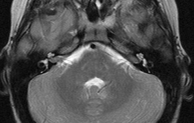
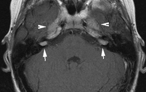
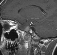
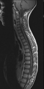
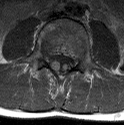
| Fig. 7. Neurofibromatosis type 2: Images of a 12-year-old boy with deafness and weakness in his arms and legs, whose father has bilateral deafness. Axial T2-weighted (a) and postcontrast axial (b) T2-weighted images reveal bilateral vestibular schwannomas, which are also known as acoustic neuromas (arrows). This is the classic finding of NF2. (b, c) Bilateral schwannomas are seen in Meckel's cave (arrowheads) and a (d) lower left cranial nerve schwannoma extends into the pars nervosa of the jugular foramen (arrow). (e) A part cystic and part solid enhancing ependymoma in seen within the cervical cord and medulla and within the distal cord and conus. (e, f) Thoracic schwannomas are present at numerous levels (arrowheads). Marked enhancement and thickening of the roots within the cauda equina also represent multiple schwannomas. |