|
|
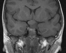
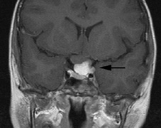
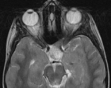
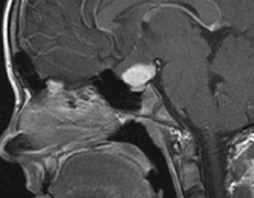
| Fig. 6. Surveillance images of a 12-year-old girl with NF1 and an optic nerve glioma which has extended to involve the chiasm. Pre- (a) and postcontrast (b) T1-weighted coronal images reveal a large suprasellar mass with an enhancing component (arrow) seen separately from the normally enhancing pituitary gland. (c) Axial scans through the suprasellar cistern show the tumor is high signal on T2-weighted scans. (d) Enlargement of the chiasm, optic nerve and hypothalamus is visible on the post-contrast T1-weighted sagittal image. Although optic nerve glioma usually has a good prognosis (with visual function often remaining stable in the absence of any intervention66,67), chiasmal involvment is a poor prognostic indicator. A sign of chiasmal involvment may be new onset of endocrine disorders or nystagmus. Surgical excision of chiasmal gliomas carries a high risk of visual loss.68–70 |