|
|
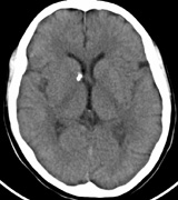
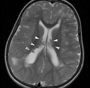
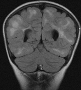
| Fig. 15. Tuberous Sclerosis Complex. (a) Patient 1: Axial CT scans demonstrating typical calcification of subependymal nodules in a 13-year-old girl with a history of seizures. (b and c) Patient 2. (b) Axial T2-weighted images demonstrate calcified subependymal nodules (arrowheads) and cortical tubers typical of tuberous sclerosis. (c) Widespread cortical tubers are seen on a coronal FLAIR sequence as thickening of the cortex and high signal of the subcortical white matter. |