|
|
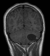
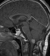
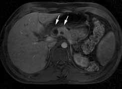
| Fig. 17. Images from a 13-year-old boy with Von Hippel-Lindau syndrome. (a) Coronal postcontrast T1-weighted imaging reveals a cystic lesion with an enhancing nodule at the pial surface typical of a hemangioblastoma. (b) A second solid enhancing hemangioblastoma is seen at the craniocervial junction on a sagittal postcontrast T1-weighted image. (c) Associated cystic lesions (arrows) are seen within the pancreas. |