|
|
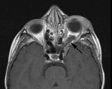
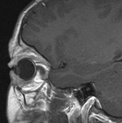
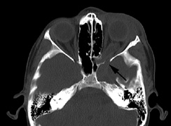
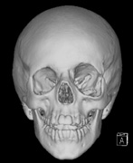
| Fig. 5. Surveillance images from a 15-year-old boy with a history of visual loss, proptosis and a diagnosis of NF1. Postcontrast axial (a) and sagittal (b) T1-weighted images demonstrate a plexiform neurofibroma of the left upper and lower eyelids, which extends into the orbit and to the extraconal soft tissues through a widened superior orbital foramen, best seen in (c). An optic nerve glioma widens the optic canal. (d) A sphenoid wing dysplasia is visible and seen as asymmetry of the orbits on the 3D CT bone reconstruction images. |