|
|
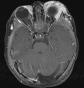
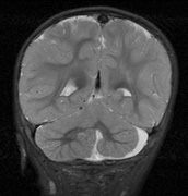
| Fig. 24. PHACES. Images from a 10-month-old girl with widespread hemangioma over the left forehead, upper eye lid, chest and right earlobe. Examination of the carotid arteries revealed bilateral bruits. (a) The enhancing hemangiomata (larger on the left) are predominantly preseptal with extraconal intraorbital extension on the postcontrast T1-weighted image. (b) Cerebellar hypoplasia is seen on the left. |