Chapter 66
Degeneration and Atrophy of the Choroid
Richard G. Weleber and Peter J. Francis
Main Menu   Table Of Contents

Search

DEFINITION
RELATIONSHIP TO AGING MACULAR DEGENERATION
PRIMARY ATROPHIES
PRIMARY CHOROIDAL ATROPHIES
ADVANCED-STAGE RETINAL DEGENERATIONS
END-STAGE MACULAR DYSTROPHIES
PERIPHERIN/RDS
DISORDERS WITH ANGIOID STREAKS
SECONDARY ATROPHIES
GLAUCOMA
DRUG TOXICITY
TREATMENT
CONCLUSION
REFERENCES

The goal of this chapter is to present an overview of degeneration and atrophy of the choroid. As such, we present a differential diagnosis of the different types of degeneration and atrophy of the choroid and briefly discuss some disorders that will be covered more extensively in other chapters, such as gyrate atrophy, choroideremia, the inherited macular dystrophies, and retinitis pigmentosa. In this chapter, we will focus on the choroidal manifestations of these allied disorders and will leave the reader to refer to other chapters for more complete discussions.
Back to Top
DEFINITION
The term choroidal degeneration refers to a group of disorders that present with acquired, usually progressive cellular or tissue dysfunction resulting eventually in cell death and consequent structural changes involving the choroid. Atrophy of the choroid is a more specific term that applies to the anatomic changes that arise from loss of cells and tissue subsequent to cell death. Atrophy of the choroid can be either congenital or acquired and can occur from any process that involves cell loss. Dystrophy is the term that applies to acquired cell or tissue degeneration with subsequent atrophy that is the result of a genetic defect, such as a deficiency of an enzyme (i.e., ornithine aminotransferase deficiency in gyrate atrophy of the choroid and retina) or a mutated gene product (i.e., atrophy that can be seen with patients who carry mutations of the rhodopsin gene or the peripherin/RDS gene).

One cannot assume that the basic underlying defect in choroidal atrophies and dystrophies resides in the choroid alone. Both gyrate atrophy and choroideremia are the result of defects of gene products that are widespread throughout the body but, for different reasons, produce pathology limited mostly to the eye. The choriocapillaris has an intimate association with the pigmented retinal epithelium, and any disorder, genetic or otherwise, that produces a loss of one will be expected to also eventually result in a loss of the other. The recent discoveries of peripherin/RDS mutations in patients with either diffuse or regional choroidal atrophy indicate that even defects of proteins limited in expression to the photoreceptors can result in pigment epithelial and choroidal atrophic changes.

Back to Top
RELATIONSHIP TO AGING MACULAR DEGENERATION
Degeneration and atrophy of the choroid can occur as part of age-related macular degeneration. Often the end stage of specific disorders, such as Best's macular dystrophy, Stargardt's macular dystrophy, pattern dystrophy of the retinal pigment epithelium, Sorsby's fundus dystrophy, North Carolina macular dystrophy, and juvenile X-linked retinoschisis, will present with an atrophic fundus appearance that is mistaken for that resulting from advanced typical aging macular degeneration. Several investigators have suggested that many common forms of aging macular degeneration are the result of genetic disease.1,2 Stone and Sheffield have proposed that some forms of aging macular degeneration may be mutations allelic to known macular dystrophies but differently expressed with regard to age of onset because the mutations involved affect a different portion of the gene product.3

One of the problems that has impeded searches for genes associated with age-related macular degenerations has been the lack of multigeneration families to study because of the late age of onset. Classical studies of linkage by descent are most productive in large families with three or more generations of individuals available for study. Newer genetic techniques, such as affected pedigree member analysis4 and sib pair analysis and association studies5 represent powerful tools to discover and map new genes that cause macular degeneration.

In general, specificity of any degenerative or dystrophic process is reduced or lost as the disorder progresses. In the end stages, the fundus appearance may resemble that of any of a number of diseases. Seeing the range of various expression and stages of progression in multiple affected family members is very helpful in recognizing and differentiating one form of choroidal atrophy or degeneration from another. Special tests, such as the fluorescein angiogram, optical coherence tomography (OCT), the electrooculogram (EOG), and the electroretinogram (ERG), may give additional information that will help to define a precise diagnosis. For example, an older individual with Best's vitelliform macular dystrophy can present with an atrophic lesion of the macula involving marked loss of choriocapillaris or choroid. The EOG will disclose the subnormality of the slow oscillation of the resting potential of the eye that is characteristic of this disease.6–8 Males with X-linked juvenile retinoschisis may present with only marked macular atrophy as a result of long-standing foveomacular schisis. The ERG for these individuals will show the marked, selective loss of the scotopic b-wave of the ERG (the so-called electronegative ERG configuration) that is typical of this disorder.9,10 However, for most forms of choroidal atrophy or degeneration, accurate diagnosis depends not on electrophysiology alone but on the assimilation of information from history, fundus examination, visual fields, fluorescein angiograms, and other special tests, including plasma amino acids or tests on DNA for specific genetic mutations.

Examination of other family members who may be similarly affected is often crucial for recognition of the disorder in its earlier or more specific stage and for appreciation of the range of expression. For those individuals who do not have a family history of others similarly affected, valuable information can be obtained from previous examination records, especially if photographs were taken or other specific tests were performed. Often, examining other family members or obtaining old records is the only way to arrive at the correct diagnosis.

Back to Top
PRIMARY ATROPHIES
In this chapter, we will discuss briefly all primary atrophies or dystrophies that include significant choroidal atrophy in at least one stage of the disease. Primary atrophies that can lead to choroidal atrophy include primary choroidal atrophies, retinal degenerations (retinitis pigmentosa group), end-stage macular dystrophies, mutations of the peripherin/RDS gene, and disorders that cause angioid streaks, such as pseudoxanthoma elasticum.
Back to Top
PRIMARY CHOROIDAL ATROPHIES
Primary choroidal atrophies are those genetically determined degenerations that involve prominent choroidal atrophy at an early stage in the disease. Primary choroidal atrophies can be divided into regional choroidal atrophies and diffuse choroidal atrophies, which, in turn, can be further subdivided into choriocapillaris and total vascular choroidal atrophies, depending on whether the atrophy involves predominantly the choriocapillaris or the entire choroid.11

REGIONAL CHOROIDAL ATROPHIES

Regional Choriocapillaris Atrophies

CENTRAL AREOLAR CHOROIDAL DYSTROPHY (CENTRAL AREOLAR CHOROIDAL SCLEROSIS).

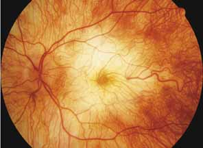
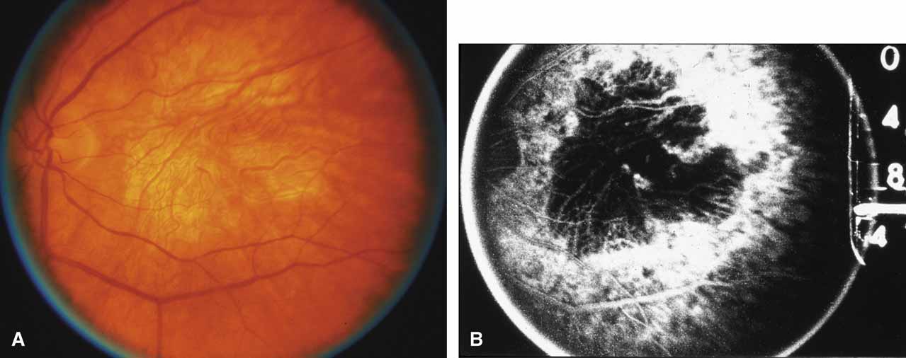
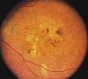
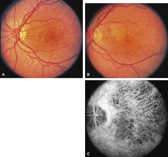
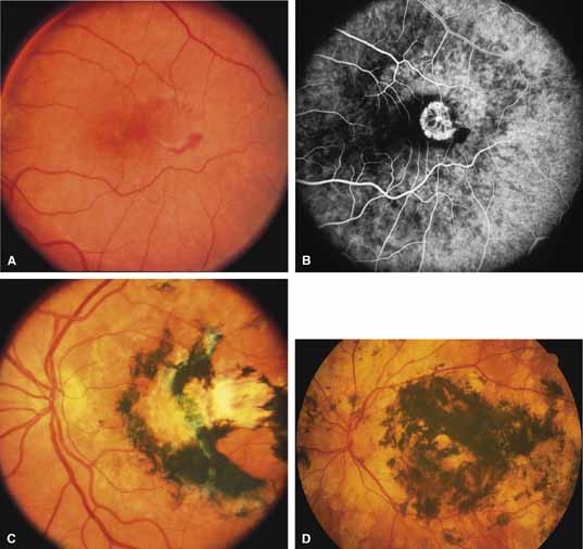
First described by Nettleship12 in 1884, central areolar choroidal dystrophy (CACD) (MIM No. 215500, Phenotype catalog number (MIM) from McKusick VA: Mendelian Inheritance in Man. Catalogs of Human Genes and Genetic Disorders. 12th ed. Baltimore: Johns Hopkins University Press, 1998 [http://www.ncbi.nlm.nih.gov/Omim/]), as it is most appropriately called,13 has been described under many names, including central areolar choroidal sclerosis, central areolar choroidal atrophy, central angiosclerosis, and central senile choroiditis.14,15 The disorder can be autosomal dominant or autosomal recessive, and phenocopies can occur from many other diseases, including mutations of peripherin/RDS16–19 and advanced stages of macular dystrophies (see later text). A locus for CACD has been identified on chromosome 17p.20 The earliest symptoms result from pericentral scotomas and include difficulty reading, poor dark adaptation, reduced visual acuity, and glare sensitivity. The earliest fundus findings are subtle and include pigment epithelial and choriocapillaris lesions in the macula (Fig. 1A) that enlarge and eventually form the punched-out central atrophic lesions typical of this disease (Fig. 1B). Histopathology shows fibrotic scarring with absence of choriocapillaris, retinal pigment epithelium, and overlying photoreceptors in the affected areas.21 The Ganzfeld electroretinogram is usually normal early in the course but may become mildly to moderately abnormal for cone and rod responses late in the course of disease when extensive atrophy of the choroid and secondarily the pigment epithelium and neurosensory retina occurs. Recent studies using the multifocal ERG have indicated that the abnormality of retinal function extends beyond the borders of the visible atrophy and is consistent with presynaptic photoreceptor dysfunction.22 The EOG can be normal or mildly abnormal depending on the extent of associated retinal pigment epithelial dysfunction.

Fig. 1 A. Right eye of a 22-year-old woman with central areolar choroidal dystrophy in early stage showing discrete, oval macular lesion. Visual acuity was 20/100. B. Left eye of a 57-year-old woman (mother of patient shown in A) with typical large depigmented atrophic macular lesion. Visual acuity was 20/300. (From Carr RE: Central areolar choroidal dystrophy. Arch Ophthalmol 73:32, 1965)

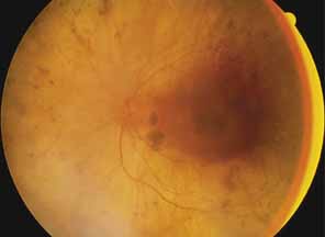
Other forms of central choroidal dystrophy exist that do not show the discrete oval lesions of typical central areolar choroidal sclerosis. These forms of central choroidal choriocapillaris atrophy often present with progressive pigment epithelial mottling and patchy choriocapillaris atrophy initially limited to the macula (Figs. 2A and 2B). With time, the atrophy enlarges and eventually encompasses the entire posterior pole (Fig. 2C). For this form of central choroidal atrophy, a gradual transition usually occurs from atrophic central pigment epithelium and choriocapillaris to essentially normal retina and choroid in the peripheral fundus.

Fig. 2 Central choroidal dystrophy of a less discrete form than central areolar choroidal dystrophy, showing in early stages the focal pigment epithelial and choriocapillaris defects in a male at 30 years of age (A) and the subsequent progression evident at 45 years of age (B). C. Late stage of central choroidal dystrophy showing atrophic macular choroid in a 54-year-old woman (mother of patient shown in A and B). The father of the patient shown in C was similarly affected, suggesting autosomal dominant inheritance.

BIETTI'S CRYSTALLINE DYSTROPHY.

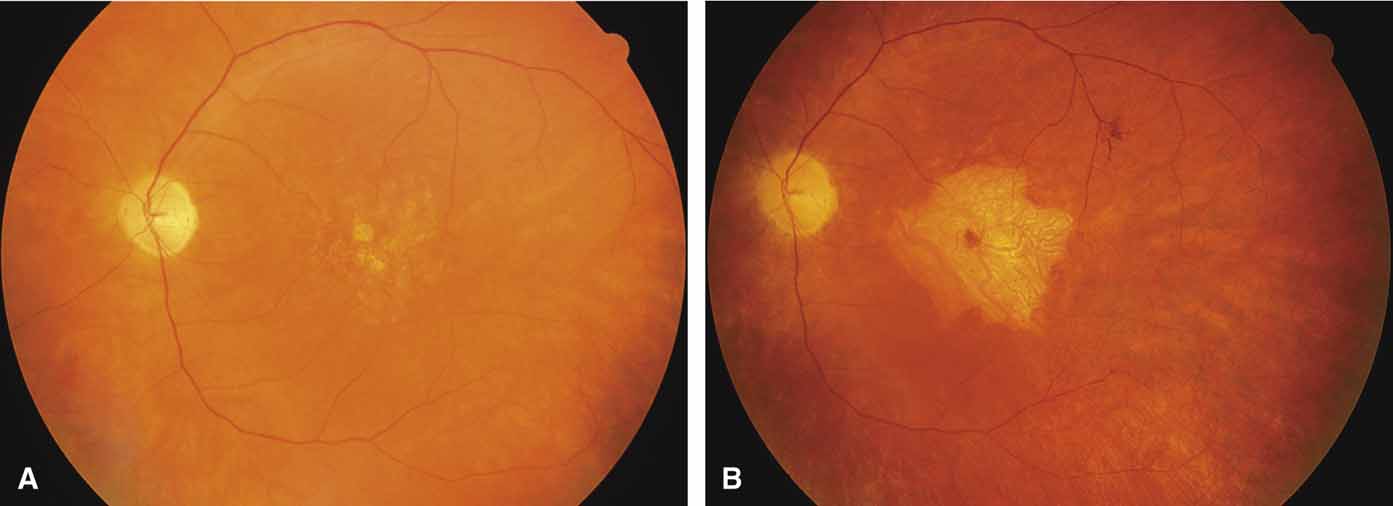
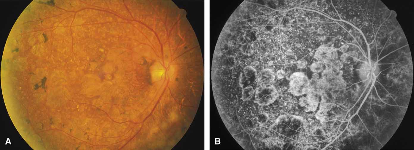
Bietti's crystalline dystrophy (MIM No. 210370) of the cornea and retina is an autosomal recessive disorder that is characterized by the presence of crystals of unknown composition in the stroma of the peripheral cornea and at several layers of the retina (Figs. 3A and 3B).23 The disease can be subdivided into regional and diffuse forms, and the lack of any reports of the two patterns in the same family suggests genetic heterogeneity and not just variable expressivity.24,25 The regional form begins in midlife as pericentral scotomas that cause difficulty reading and reduced central visual acuity. Peripheral retinal function is retained and the electroretinogram and electrooculogram are normal or near normal even in moderately advanced disease. The fundus appearance and fluorescein angiogram reveal regional loss of pigment epithelium and choriocapillaris limited to the posterior pole (Figs. 3C and 3D). The finding of abnormal crystals in leukocytes indicates that this is a systemic metabolic disorder.24

Fig. 3 Right fundus (A) and fluorescein angiogram (B) of 52-year-old man with regional form of Bietti's crystalline corneal retinal dystrophy. Note retinal crystals adjacent to areas of dystrophy. Left fundus (C) and fluorescein angiogram (D) of 61-year-old brother of patient shown in A, showing marked regional atrophy of choroid and retina limited to the posterior pole and peripapillary regions. (A and B from Wilson DJ, Weleber RG, Klein ML, et al: Bietti's crystalline dystrophy: A clinicopathologic correlative study. Arch Ophthalmol 107:213–221, 1989; Copyright 1989, American Medical Association)

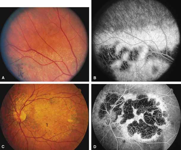
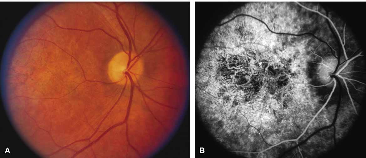
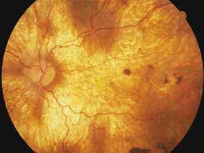
The diffuse form presents with loss of peripheral visual field, symptoms of poor night vision as well as reduced visual acuity, and diffuse loss of pigment epithelium and choriocapillaris on funduscopy (Fig. 4) and on fluorescein angiogram. The ERG is profoundly abnormal early in the course of the disease, and visual impairment eventually becomes much more severe in the diffuse than in the regional form of the disease.

Fig. 4 Posterior pole (A) and inferior retina (B) of right eye of a 45-year-old woman with diffuse form of Bietti's crystalline corneal retinal dystrophy, showing widespread atrophy (same patient as in reference 24).

REGIONAL TOTAL VASCULAR CHOROIDAL ATROPHIES.

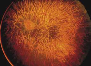
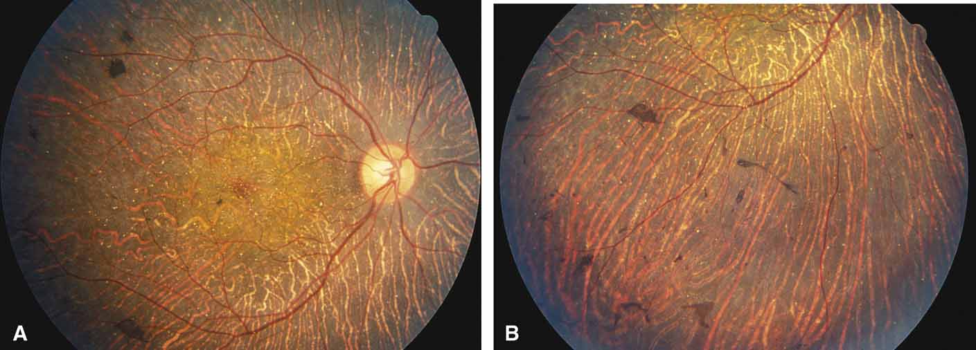
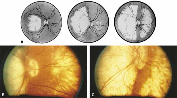
Helicoidal peripapillary chorioretinal degeneration (also called atrophia areata and peripapillary chorioretinal degeneration, Iceland type). First described in Iceland by Sveinsson26 in 1939 as choroiditis areata and renamed as helicoidal peripapillary chorioretinal degeneration (MIM No. 108985),27,28 this autosomal dominant disorder is characterized by peripapillary tongue-shaped patches of total vascular choroidal atrophy that extend radially away from the disc. Signs of edema or inflammation have not been described, distinguishing this disorder from serpiginous choroidopathy. Brazitikos and Safran29 believe that the peculiar fundus lesions are the result of tearing and retraction of the retinal pigment epithelium, and possibly Bruch's membrane, followed by atrophy of the choroid and retina. The areas of atrophy bear no relation to the pattern of the retinal vessels. The ERG suggests focal rather than diffuse retinal dysfunction initially at the level of the RPE and later affecting the sensory retina.30 Magnússon,31 who used the term atrophia areata, studied a large pedigree of 26 affected individuals in whom the disorder was inherited as an autosomal dominant trait (Fig. 5). Myopia and astigmatism were usually present, and the visual acuity was extremely poor in later years. Occasionally, the disorder has been called central gyrate atrophy, but this name should be discouraged because of the confusion with gyrate atrophy of the choroid and retina with hyperornithinemia. Serum amino acid levels are normal in helicoidal peripapillary chorioretinal degeneration.31

Fig. 5 Fundi of a 24-year-old woman with helicoidal peripapillary chorioretinal degeneration. Ten years later, central vision was lost in the right eye. (From Magnússon L: Atrophia areata: A variant of peripapillary chorioretinal degeneration. Acta Ophthalmol 59:659, 1981)

This clearly dominantly inherited disorder should not be confused with the nonfamilial condition called serpiginous choroidopathy, which has unfortunately been reported in the past (and in some reviews is still classified) under the name geographic helicoid peripapillary choroidopathy.32

PROGRESSIVE BIFOCAL CHORIORETINAL ATROPHY.

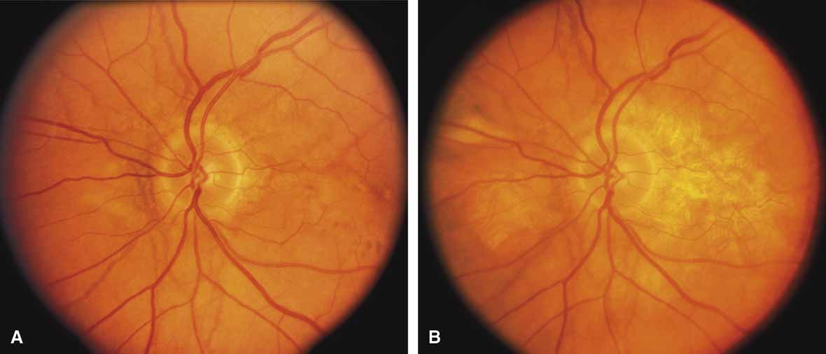
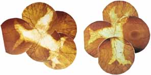
Progressive bifocal chorioretinal atrophy is a rare autosomal dominant disorder mapped to chromosome 6q33,34 that was first described by Douglas, Waheed, and Wyse35 in a large Scottish family in 1968. Thirty-three of 91 family members were affected. The authors classified the disease into three stages (Fig. 6). Stage 1, which lasts from birth to age 14 years, involves a temporal focus of atrophy of retinal and choroidal tissue in the macula that continues to slowly enlarge with time. The upper, nasal, and lower edges of the temporal lesion are well defined, whereas the temporal edge is serrated and indistinct. Stage 2, which lasts from age 15 years to age 45 years, is associated with a second focus of atrophy nasal to the disc, hence the term bifocal in the name. At the end of this stage, the nasal focus is about three disc diameters in size. Stage 3, which occurs after 45 years of age, is associated with further expansion of the temporal and, especially, the nasal focus, leaving only a vertically oriented one- to two-disc-diameter swath or band of intact retina and choroid extending from the disc to each equator superiorly and inferiorly. Visual acuity was markedly subnormal compared with early years, but no patients lost all vision. All patients had coarse nystagmoid eye movements that precluded measurement of visual fields. Most patients were myopic. The ERG b-wave amplitude was subnormal but not unrecordable by a non-Ganzfeld single-flash technique.

Fig. 6 A. Artist's drawing of the three stages of progressive bifocal chorioretinal atrophy: Stage 1 (left), Stage 2 (center), and Stage 3 (right). B. Right eye of 17-year-old patient with late Stage 2, early Stage 3 disease. Visual acuity was 20/200. C. Right eye of older patient with Stage 3 disease. (From Douglas AA, Waheed I, Wyse CT: Progressive bifocal chorio-retinal atrophy: A rare familial disease of the eyes. Br J Ophthalmol 52:742, 1968)

SORSBY'S FUNDUS DYSTROPHY.

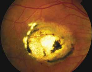
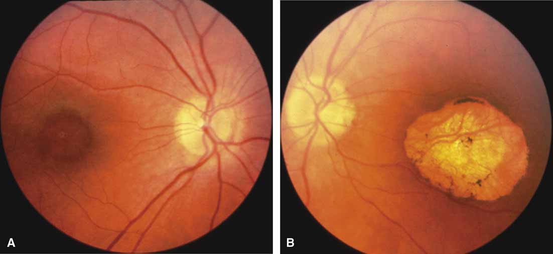
Sorsby's fundus dystrophy (MIM No. 136900), previously called Sorsby's pseudoinflammatory macular dystrophy, is a highly penetrant, autosomal dominantly inherited disorder characterized by a tritan color vision defect, drusen-like subretinal deposits, and pigment epithelial atrophy in younger individuals followed by choroidal neovascularization, hemorrhage, subretinal fibrosis, and choroidal atrophy in later years (Fig. 7).14,36–38 Loss of vision usually commences after the age of 50 years. The disease in late stages progresses to involve the peripheral retina. More recent studies of members of Sorsby's original family demonstrated angioid streaks and yellow plaque-like subretinal deposits, features distinguishing this disorder from dominant drusen.39,40 Visual prognosis is poor because of the tendency of the disease process to involve the macula. The ERG is generally normal except in the most advanced stages when extensive areas of the retina are involved.41 Linkage studies assigned the gene for Sorsby's dystrophy to 22q13-qter.42 Weber et al.43 found mutations in the gene for the tissue inhibitor of metalloproteinase-3 (TIMP3), which is located on the long arm of chromosome 22, in patients with Sorsby's fundus dystrophy. This finding suggests that a defect in maintenance and renewal of Bruch's membrane by the altered gene product is an important underlying pathologic event in Sorsby's fundus dystrophy.

Fig. 7 Left fundus of a woman from a multigeneration family with Sorsby's fundus dystrophy at 36 years of age (A, B) and at 40 years of age (C) demonstrating scarring from recurrent neovascularization and hemorrhage after laser photocoagulation. D. Untreated left eye of the 67-year-old mother of the patient in A through C. Visual acuity was counting fingers at 2 feet. (Illustrations courtesy of Michael L. Klein, M.D., Portland, OR)

SERPIGINOUS CHOROIDOPATHY.

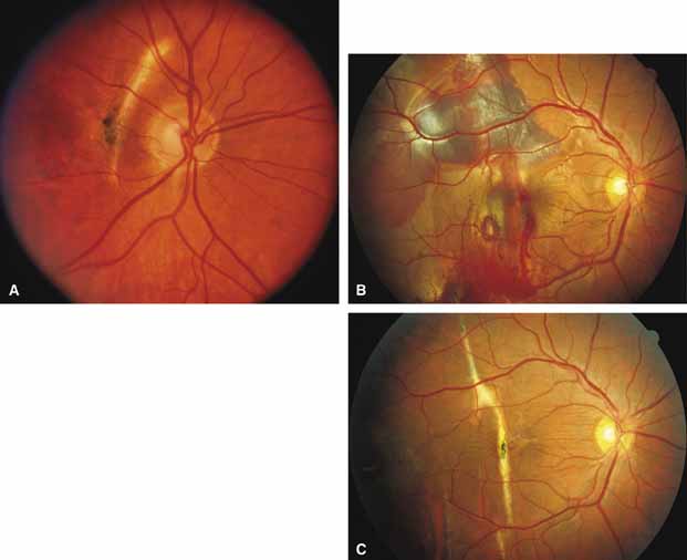
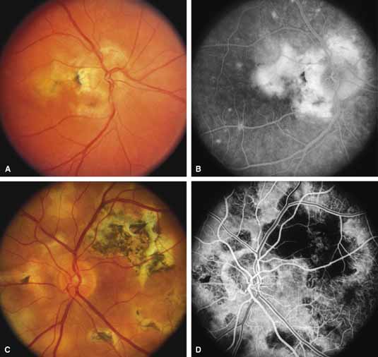
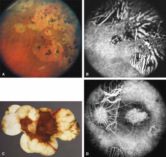
Serpiginous choroidopathy, also called serpiginous choroiditis, is a recurrent, progressive, destructive chronic degeneration of the choroid and retinal pigment epithelium that begins around the optic nerve and extends through the posterior pole.44 Unfortunately, some cases of this disorder have been reported under the name geographic helicoid peripapillary choroidopathy, and this has led to confusion with the clearly dominantly inherited genetic disorder first described in Iceland by Sveinsson26 in 1939 as choroiditis areata and subsequently renamed helicoidal peripapillary chorioretinal degeneration.27,28 The etiology of serpiginous choroidopathy is unknown, but the disease is not thought to be genetic. The disease starts as a gray, cream, or greenish discoloration and edema of the retinal pigment epithelium, followed by extension of the lesion, usually away from the disc, in a stepwise fashion (Figs. 8A and 8B). Vitritis is present in one-third of cases. Periods of quiescence or activity can be separated by months to years. Subretinal fibrous scarring, atrophy of the choroid, and hyperpigmentation in adjacent tissues can be prominent features (Figs. 8C and 8D). The prognosis for retention of central vision is poor because the disease process will often involve the macula. Systemic immunosuppression may be effective in prolonging remission and improving the visual outcome.45

Fig. 8 Right fundus (A) and late-phase fluorescein angiogram (B) of a 35-year-old woman with serpiginous choroidopathy. The visual acuity was 20/200. Left fundus (C) and fluorescein angiogram (D) of a 47-year-old man with advanced serpiginous choroidopathy, demonstrating several stages of lesions and subretinal gliosis. Visual acuity was 14/400. (A and B courtesy of Michael L. Klein, M.D., Portland, OR)

Although originally described as separate disorders, serpiginous choroidopathy and geographic choroidopathy are believed by most authors to be variations of a single disease process.32,46–48 Thus, there appear to be two distinct conditions only—serpiginous choroidopathy, which is an acquired nongenetic disorder, and helicoidal peripapillary chorioretinal degeneration (MIM No. 108985), which is a dominantly inherited disorder.

CENTRAL GYRATE ATROPHY.

Some clinicians have used the term central gyrate atrophy as a label for the fundus appearance of marked total vascular choroidal atrophy in the posterior pole. Central gyrate atrophy appears to not be a distinct entity but probably represents the atrophic changes that can occur in the posterior pole in advanced malignant myopic degeneration, the end stage of serpiginous choroidopathy, or the end stage of helicoidal peripapillary chorioretinal degeneration. Use of the term central gyrate atrophy for such fundus appearances should be avoided because it leads to confusion with the metabolic disorder gyrate atrophy with hyperornithinemia.

DIFFUSE CHOROIDAL ATROPHIES

Diffuse choriocapillaris atrophies

DIFFUSE CHORIOCAPILLARIS ATROPHY.

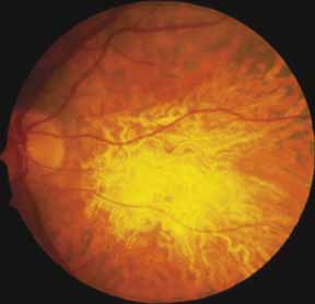
Diffuse choriocapillaris atrophy, also called diffuse choroidal sclerosis and diffuse choroidal angiosclerosis, can be inherited either as an autosomal dominant or, less commonly, as a recessive trait. Certain mutations of the peripherin/RDS gene have also been associated with diffuse choriocapillaris atrophy.18 The disorder is characterized by progressive thinning and loss of choriocapillaris beginning in midlife and resulting in severe loss of vision by later years (Fig. 9). Although the disease in most families is quite consistent among affected individuals, both diffuse and regional central choriocapillaris atrophy have been reported in the same family, suggesting that these two disorders are interrelated.11 Symptoms may include night blindness, loss of central vision, and loss of peripheral vision—the latter feature distinguishing this disorder from the regional form of central choriocapillaris atrophy. Fluorescein angiograms show thinned or absent choriocapillaris with prominent medium and large choroidal vessels throughout the retina. Eventually, mild pigment dispersion and clumping occur in the peripheral retina as the retinal pigment epithelium and overlying retina become affected, but the picture is still dominated by the atrophy of the choroid. The electroretinogram, except in the earliest stages, is usually severely abnormal and eventually becomes unrecordable. The EOG is abnormal early in the disease.

Fig. 9 Right fundus (A) and fluorescein angiogram (B) of a 65-year-old woman with diffuse choriocapillaris atrophy, showing widespread thinning of retinal pigment epithelium and loss of choriocapillaris.

DIFFUSE TOTAL VASCULAR CHOROIDAL ATROPHIES.

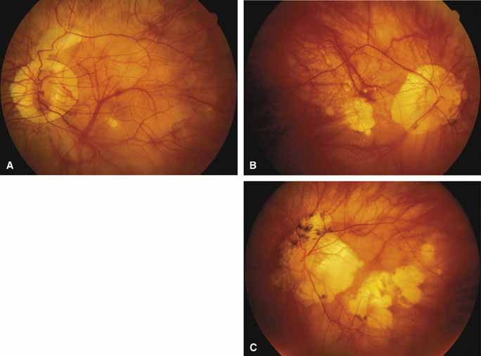
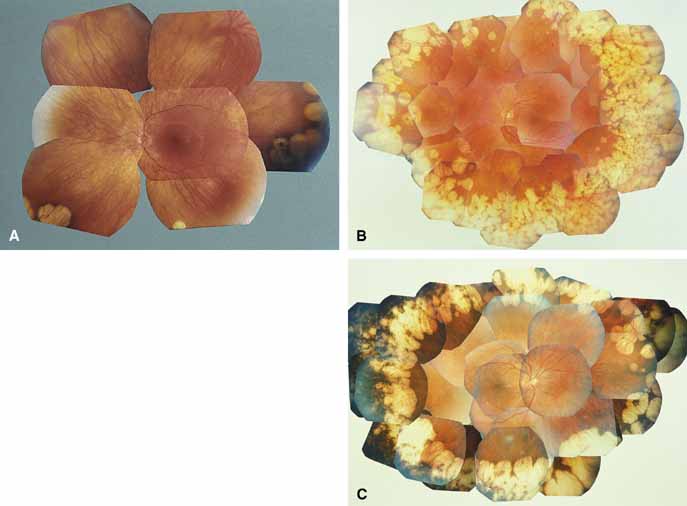
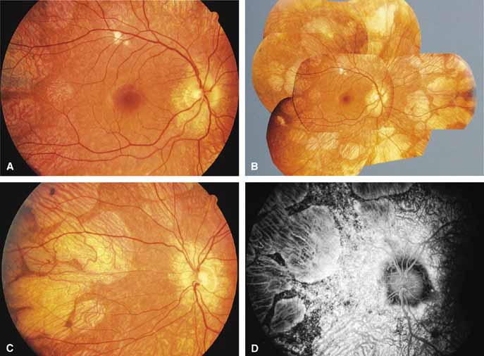
Gyrate atrophy of the choroid and retina with hyperornithinemia from ornithine aminotransferase deficiency. Gyrate atrophy of the choroid and retina (MIM No. 258870) is an autosomal recessive disease that is associated with a 10- to 20-fold elevation of plasma and tissue levels of ornithine.49–52 Vitamin B6 responsive and nonresponsive forms exist. Although the disease is found worldwide, the greatest number of patients, nearly 50% of those reported (all B6 nonresponsive), are of Finnish descent.52 In the first or second decade of life, patients experience night blindness and begin to develop irregular round areas of total vascular choroidal atrophy (Fig. 10A).50 These lesions enlarge and coalesce with time, forming extensive atrophy in the periphery that is associated with constriction of the peripheral visual field (Figs. 10B and 10C). The classic appearance of the fundus is that of a sharp transition from more normal retina to nearly complete atrophy of the choroid and retina (Figs. 11A and 11B).50,53 Central vision may be lost from cystoid macular edema, epiretinal proliferation, or macular involvement in the atrophic process (Figs. 11C and 11D).50 Many patients will develop clinically significant posterior subcapsular cataracts. The ERG and EOG are markedly abnormal from early in the course of the disease.54

Fig. 10 A. Left eye of a 12-year-old girl with B6-nonresponsive gyrate atrophy of the choroid and retina. One allele of the ornithine aminotransferase gene was inactivated by a (TAC)Tyr299-TAG stop mutation in exon 8, whereas the other allele was inactivated by an Arg180Thr (AGG→ACG) mutation in exon 6 (patient 4 in reference 61). B. Left eye of a 30-year-old woman with B6-responsive gyrate atrophy. One allele of the ornithine aminotransferase gene was the Glu318Lys mutation (GAG → AAG mutation at codon 318 in exon 9), whereas the other allele was inactivated by a splice site mutation of intron 4.62 C. Right eye of a 40-year-old man with B6-nonresponsive gyrate atrophy, showing accumulation of dense, black pigment in the temporal periphery in region of choroidal atrophy. One allele was inactivated by a deletion within exon 6, whereas the other was inactivated by formation of a stop codon in exon 6 (TAT)Tyr209TAA.61 (A, B, and C from Weleber RG, Kennaway NG: Gyrate atrophy of the choroid and retina. In Heckenlively JR [ed]: Retinitis Pigmentosa. Philadelphia: JB Lippincott, 1988:198–220)

Fig. 11 A. Transition zone between atrophic and more intact midperipheral retina in 37-year-old patient with B6-responsive gyrate atrophy. One allele is the Glu318Lys mutation of the ornithine aminotransferase gene, but the mutation affecting the other allele has yet to be defined (Inana G, Weleber RG, personal communication). B. Fluorescein angiogram of same region, showing pigment epithelial window defects and loss of choriocapillaris. C. Left fundus (C) and fluorescein angiogram (D) of advanced gyrate atrophy associated with macular atrophy in a 64-year-old woman homozygous for the most common Finnish mutation, the Leu402Pro mutation of exon 11 of the ornithine aminotransferase gene (Inana G, Weleber RG, unpublished finding). (B from Weleber RG, Kennaway NG: Gyrate atrophy of the choroid and retina. In Heckenlively JR [ed]: Retinitis Pigmentosa. Philadelphia: JB Lippincott, 1988:198–220. Images C and D courtesy of Robert Kalina, M.D., Seattle, WA)

The underlying defect of gyrate atrophy involves ornithine aminotransferase (OAT), which is a mitochondrial matrix enzyme that is pyridoxal phosphate dependent. The OAT gene is located at chromosome 10q26. Although the majority of patients have the form of gyrate atrophy that does not respond to vitamin B6 or pyridoxine, some individuals (less than 5%) show partial vitamin B6 responsiveness. This is shown in vitro by an increase in enzyme activity when assayed with additional pyridoxine and in vivo with a 50% reduction of plasma ornithine levels with oral vitamin B6 supplementation.55,56 Histologic abnormalities occur also in muscle (subsarcolemmal deposits in type II muscle fibers) and other tissues (abnormal-appearing mitochondria in liver and iris).57–59 However, the predominant pathology and clinical symptoms occur in the choroid and retina (see elsewhere in these volumes).50,52 The exact mechanism of pathology for the ocular lesions in gyrate atrophy is unknown, but the three major theories are that, secondary to the OAT deficiency: (1) intracellular proline in the retina may be deficient; (2) creatine phosphate stores in the retina and choroid may be deficient, leading to cell dysfunction and cell death; and/or (3) elevated levels of ornithine may be toxic to the retinal pigment epithelium.60 The abnormal muscle inclusions appear to be the result of deficiency of creatine phosphate, but the role of creatine phosphate as an energy store in the eye is currently undefined.

The molecular basis of ornithine aminotransferase deficiency has been defined for many patients with gyrate atrophy.61–68 Two mutations of the OAT gene (Leu402-Pro and Arg180Thr) account for nearly all of the Finnish patients with nonpyridoxine-responsive gyrate atrophy.66 Loss of gene function occurs from deletions, insertions, splice site base-pair changes, and missense mutations. The rare pyridoxine-responsive patients with gyrate atrophy are homozygous for the Glu318SLys mutation or heterozygous for either the Val332Met mutation or the Glu318Lys mutation of the OAT gene, with the other allele inactivated by another mutation that may or may not be pyridoxine responsive.62,63,69

CHOROIDEREMIA (CHM)—TAPETOCHOROIDAL DYSTROPHY (TCD).

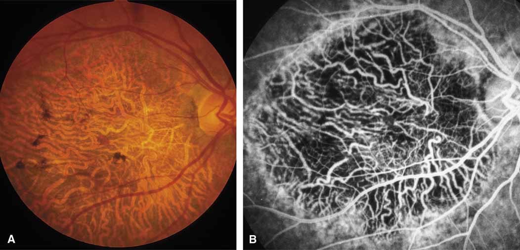
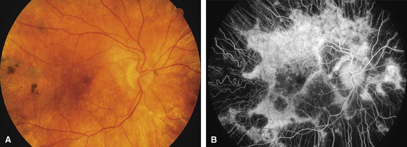
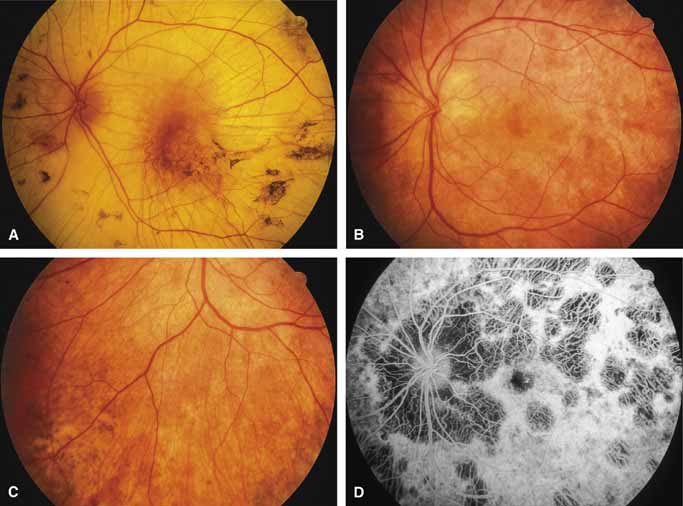
Choroideremia (gene symbol CHM; MIM No. 303100) is an X-linked disorder that is characterized by the onset of night blindness in the first or second decade of life followed by slowly progressive atrophy of the choroid and retina that usually results in legal blindness by midlife and near virtual blindness in later years. The disorder begins with a diffuse atrophic process involving retinal pigment epithelium and choriocapillaris (Figs. 12A, 12B, 12C, and 12D) but eventually results in near total vascular choroidal atrophy (Fig. 13A). The fundus does not show the sharp border or transition area that is characteristically seen with gyrate atrophy. The peripheral visual field is depressed and eventually becomes severely constricted. The ERG and EOG are abnormal early in the course of the disease.

Fig. 12 Posterior pole of right eye (A, B), retina nasal to left disc (C), and corresponding fluorescein angiogram of left fundus (D) of a 15-year-old boy with early choroideremia.

Fig. 13 Left eye of a 53-year-old man with advanced choroideremia (A). Posterior fundus (B), inferior nasal retina (C), and fluorescein angiogram of posterior pole (D) of the left eye of a 30-year-old woman with carrier state for choroideremia, showing the characteristic mottled pigment epithelium and extensive patchy choroidal atrophy. At age 36, her visual acuity had fallen to 20/30 in each eye, and she has become significantly visually disabled by loss of pericentral visual field. She is the daughter of the patient shown in Figure A and the granddaughter of the carrier woman whose retinal histology was reported in reference 70.

Women who are carriers for choroideremia will nearly always show evidence of this on examination with patchy mottling and atrophy of the fundus with hyperpigmentation (Figs. 13B, 13C, and 13D). Random inactivation of one of the two X chromosomes, also called lyonization, results in the expression of the defect in a certain population of a carrier woman's retinal and choroidal cells, causing the carrier manifestations. These changes can occur in a patchy distribution throughout the fundus. Usually, carrier women are asymptomatic or, at most, experience only mild symptoms of poor night vision or pericentral scotomas, if the changes affect the posterior pole. Uncommonly, the carrier manifestations may cause considerable visual impairment, if the macular regions are involved. Histology has showed patchy loss of choroid, pigment epithelium, and photoreceptor outer segments, with abrupt transition from normal to abnormal areas.70 More diffuse abnormalities of the retinal pigment epithelium have been found throughout the fundus, suggesting that the defect may have a primary effect on this tissue.

The gene for choroideremia had been localized by linkage studies to Xq21.2 and has been found to encode a gene product, Rab escort protein-1 (REP-1),71 previously referred to as component A of Rab geranylgeranyl transferase (also called the CHM protein).72–74 REP-1 is involved in geranylgeranylation, which is the post-translational process of insertion of hydrophobic lipid side chains onto proteins to help them attach to membranes and to help them interact with other proteins.71,75 The gene for another similar protein, REP-2 (also called CHML protein), was subsequently found to be on the long arm of chromosome 1.76,77 The gene products of both are widespread throughout the body, but REP-1 appears necessary for the prenylation of a specific subset of Rab proteins essential for the retina and is the protein that is defective in choroideremia. Exactly how deficiency of REP-1 results in degeneration of the choroid is unknown, but loss of this protein would be expected to disrupt intracellular cycling of proteins between cytosol and membranes.71,75

A single point mutation (insertion of a T within a splice donor site of the intron downstream of exon C, changing the normal sequence of AGgtaag to AGgttaag) accounts for one-fifth of the world's population of patients with choroideremia.78 Five other point mutations have been reported, each of which results in the occurrence of a stop codon that leads to a truncated gene product.79 Deletions of the CHM gene account for approximately 12% of cases.78

LONG-CHAIN 3-HYDROXYACYL-COA DEHYDROGENASE (LCHAD) DEFICIENCY.

LCHAD deficiency is a recently described autosomal recessive disorder of mitochondrial fatty acid β-oxidation in which neonates or infants present with a Reye-like syndrome, cardiomyopathy, life-threatening episodes of hypoketotic hypoglycemia and cardiorespiratory arrest.80 Up to 90% of individuals share the same G1528C allele in the LCHAD gene.81 Patients can have recurrent muscle cramps with rhabdomyolysis during acute illnesses and can develop peripheral neuropathy. At least half, if not the majority, of the patients who survive infancy develop a severe, debilitating atrophy of the choroid and retina.82 The fundus and ERG may be normal at birth (Stage 1). Soon, however, pigment dispersion occurs in the RPE (Stage 2), followed by circumscribed chorioretinal atrophy, occlusion of choroidal vessels with accompanying reductions in acuity, visual field, and ERG amplitudes (Stage 3). Eventually, the posterior pole undergoes near total vascular choroidal atrophy (Fig. 14); however, the peripheral fundus is usually relatively spared. Finally, posterior staphylomas may develop (Stage 4). Cataract and progressive axial myopia are also features.82,83 Histopathologically, diffuse choroidal atrophy with loss of the choriocapillaris is seen, suggesting a primary fault at the level of the RPE and choriocapillaris with a secondary macrophage response.84 Dietary management of the disease involves avoidance of fasting, a high-carbohydrate diet, limitation of dietary long chain fatty acid intake, and supplementation of the diet with medium-chain triglycerides.85 The cause of the chorioretinal atrophy is unknown but may be the result of chronic damage to the retinal pigment epithelium and subsequently the choroid by the accumulation of toxic long-chain 3-hydroxyl fatty acids and/or acylcarnitines.85

Fig. 14 Diffuse total vascular atrophy of the choroid and retina of the left fundus in a 7-year-old girl with LCHAD deficiency.

Back to Top
ADVANCED-STAGE RETINAL DEGENERATIONS

RETINITIS PIGMENTOSA

Advanced retinitis pigmentosa can lead to secondary atrophy of the choroid similar to that seen with primary choroidal dystrophies, such as choroideremia. Although many forms of retinitis pigmentosa can show such choroidal atrophy in older years, a specific form that can result in such a fundus appearance is advanced disease from the Pro→His mutation of the 23rd codon of the rhodopsin gene (Fig. 15).86–91

Fig. 15 Marked retinal and choroidal atrophy in left eye of a 75-year-old man with advanced retinitis pigmentosa secondary to the Pro23His mutation of rhodopsin. (From Weleber RG, Gregory-Evans: Retinitis pigmentosa and allied disorders. In Ryan SJ [ed]: Retina, vol 1. Basic Science and Inherited Retinal Disease, 3rd ed. St. Louis: Mosby-Year Book, 2001:362–460)

CONE AND CONE-ROD DYSTROPHIES

Cone and cone-rod retinal dystrophies usually present with a normal-appearing macula, macular pigment epithelial defects, or a bull's-eye maculopathy.92–94 Advanced cone or cone-rod dystrophies can produce an atrophic appearance to the macula and posterior pole.92 Bardet-Biedl syndrome is associated with a cone-rod retinal dystrophy that often leads to marked atrophic lesions within the macular region (Fig. 16).91,93,95–99

Fig. 16 A. Left eye of a male with cone-rod dystrophy from Bardet-Biedl syndrome, showing macular pigment epithelial mottling at 37 years of age. B. Marked central choriocapillaris atrophy at 43 years of age. (Patient presented in case 48 of reference 91)

Back to Top
END-STAGE MACULAR DYSTROPHIES

STARGARDT'S MACULAR DYSTROPHY

Stargardt's macular dystrophy (MIM No. 248200, 153900) is a heritable disease that usually presents within the first 2 decades of life as a reduction of visual acuity followed by the appearance of typical pisciform yellow-white deep-retinal lesions surrounding the macula. Although the inheritance is usually autosomal recessive, rare families have been reported with autosomal dominant inheritance,100–102 and the phenotype can even be seen with mutations of peripherin/RDS103,104 and mitochondrial DNA.105 Mutations in at least four genes are associated with the Stargardt's phenotype.103 The autosomal recessive form has been linked to markers on the short arm of chromosome 1.106 In 1997, the gene for autosomal recessive Stargardt's dystrophy was found to be ABCA4,107 the product of which is essential for transport of a reactive metabolite of retinal out of the rod photoreceptor outer segments, leading to accumulation of toxic levels of A2-E within the retinal pigment epithelium.108,109 The phenotype of ABCA4 mutations can be Stargardt's macular dystrophy, fundus flavimaculatus, cone-rod dystrophy, or retinitis pigmentosa, depending upon the mutations involved.110,111 Also, mutations in ABCA4 may contribute to the risk of age-related macular degeneration.112 Autosomal dominant Stargardt's macular dystrophy was linked to chromosome 6q102,113 and results from mutation of the gene ELOVL4.114

The advanced stages of Stargardt's macular dystrophy can appear as a regional partial atrophy of choroid and retina in the posterior pole (Fig. 17).93 Although the ERG is usually normal in the early to middle course of typical autosomal recessive Stargardt's macular dystrophy, the ERG is often abnormal by the advanced stage of disease, as shown in Figure 17.

Fig. 17 Fundus (A) and fluorescein angiogram (B) of left eye, showing central choriocapillaris choroidal atrophy secondary to advanced Stargardt macular dystrophy. (From Weleber RG, Eisner A: Cone degeneration (“bull's eye dystrophies”) and color vision defects. In Newsome DA [ed]: Retinal Dystrophies and Degenerations. New York: Raven Press, 1988:233–256)

BEST'S VITELLIFORM MACULAR DYSTROPHY

Best's vitelliform macular dystrophy (VMD2; MIM No. 153700) is an autosomal macular dystrophy that usually presents early in life with dome-shaped yellowish macular lesions that eventually partially resorb, develop scarring, or are replaced by choroidal atrophy, which, if extensive, can simulate choroidal sclerosis (Fig. 18).6,115–117 The ERG in Best's macular dystrophy is normal, but the EOG shows a diagnostic pattern of subnormal, slow oscillations and normal, fast oscillations of the resting potential.8 The gene for Best's disease has been linked to chromosome 11q13.118 The gene, VMD2, has been discovered119 and its product, bestrophin, appears to be a member of a new family of chloride channels that is important for generation of the light rise of the electrooculogram.120,121

Fig. 18 Left eye of a 78-year-old woman with Best's vitelliform macular dystrophy, showing central choroidal atrophy with loss of retinal pigment epithelium and choriocapillaris, similar to what has been called central choroidal sclerosis. The visual acuity was 20/400. This woman is individual II-2 in the pedigree reported by Stone et al.118 that demonstrated linkage of Best's disease with markers on chromosome 11q13. (From Stone EM: Heritable disorders of RPE, Bruch's membrane, and the choriocapillaris. In Wright K, Ellis F, Mets M, et al [eds]: Pediatric Ophthalmology. Philadelphia: JB Lippincott, 1995)

PATTERN MACULAR DYSTROPHY

Pattern macular dystrophy (MIM No. 169150) is an autosomal dominant, extremely variable disorder that involves presumed accumulation of lipofuscin within retinal pigment epithelium with subsequent cell degeneration and secondary choriocapillaris loss. The lesions often begin in middle life and can be associated with mild to moderate visual acuity loss.116,122–124 Pattern dystrophy can, in late stages, produce an atrophic macular lesion that resembles central areolar choroidal atrophy (Fig. 19).122

Fig. 19 Fundus (A) and fluorescein angiogram (B) of left eye of an 80-year-old woman with advanced pattern dystrophy with partial choroidal atrophy similar in appearance to central areolar choroidal dystrophy. (From Marmor MF, Byers B: Pattern dystrophy of the pigment epithelium. Am J Ophthalmol 84:32–44, 1977; Published with permission from The American Journal of Ophthalmology. Copyright by the Ophthalmic Publishing Company.)

Juvenile X-Linked Retinoschisis

X-linked retinoschisis (MIM No. 312700) is believed to be a disorder of Müller's cells that results in a splitting of the retina with either flat or bullous elevation of the inner layer. Outer layer retinal holes or breaks can occur, predisposing to retinal detachment. Virtually all patients have macular or foveal retinoschisis, even in the absence of peripheral schisis. After several decades, the typical cartwheel appearance of the foveal schisis fades and is replaced by a more atrophic, nondistinct macular appearance. In late stages, atrophic lesions of the central macula can occur that mimic central choroidal atrophy.125 The electroretinogram is markedly abnormal, showing a normal a-wave and markedly subnormal b-wave, the so-called electronegative ERG configuration.9,10,126,127 Although the electroretinogram is a key test for the condition, a normal scotopic b-wave has been noted in molecularly confirmed X-linked retinoschisis, suggesting that an ERG cannot be relied upon solely to exclude the diagnosis.128 The gene for X-linked retinoschisis, RS1, resides at Xp22.13.129 Although its expression is limited to the photoreceptors, the gene product, retinoschisin, is secreted into the inner retina.130

North Carolina Macular Dystrophy

North Carolina macular dystrophy (MCDR1; MIM No. 136550) is an autosomal dominantly inherited dystrophy that has been recently mapped to the long arm of chromosome 6.131 The expression varies considerably and ranges from only central drusen or confluent drusen to macular atrophy and staphyloma. Choroidal neovascularization is a rare complication (Fig. 20). Although termed dystrophy, the lesions are present at birth and appear nonprogressive.132 This dystrophy has been reported under several names, including dominant macular dystrophy of Lefler-Wadsworth-Sidbury,133 dominant progressive foveal dystrophy,134 and central areolar pigment epithelial dystrophy.135 Gass136 renamed the disorder North Carolina macular dystrophy because of the strong founder effect linking many of the North American families. Recently, an identical ocular phenotype associated with sensorineural hearing loss has been mapped to chromosome 14.137

Fig. 20 Right eye of a patient with North Carolina macular dystrophy. Visual acuity was 20/60. (Illustration courtesy of Kent Small, M.D., University of Florida, Gainesville, FL)

Back to Top
PERIPHERIN/RDS
The mouse model of retinal degeneration slow (RDS) is caused by a defect involving the gene for an outer segment photoreceptor glycoprotein called peripherin/RDS (MIM No. 179605),138 which is involved as an adhesion molecule in the formation and stability of photoreceptor outer segment discs.139 The human homologue of this gene is located on the short arm of chromosome 6.140 Mutations of the peripherin/RDS gene cause autosomal dominant disease that can be expressed as retinitis pigmentosa,141,142 pattern macular dystrophy,16,143 or a fundus flavimaculatus-like picture.104 In the late stage of disease, extensive atrophy of the pigment epithelium and choriocapillaris has been observed in association with several mutations of the peripherin/RDS gene. Central choriocapillaris atrophy similar to central areolar choroidal dystrophy has been reported with the Arg172Trp mutation (Fig. 21)16,17 and the Lys193-del mutation18 of peripherin/RDS (latter mutation designation revised to codon 193[2 bp del]→FS216ter).144 Diffuse choriocapillaris atrophy (Fig. 22) similar to diffuse choroidal sclerosis was reported with the Met67del mutation of peripherin/RDS18 (mutation later revised to codons 67–69[5 bp del/8 bp ins]→Met67Arg, Gly68His, and Arg ins at position 69).144 Extensive regional partial atrophy of the choroid of the posterior pole was seen in a woman with adult-onset retinitis pigmentosa from deletion of either codon 153 or 154 of the peripherin/RDS gene (Fig. 23).104

Fig. 21 Fundus (A) and fluorescein angiogram (B) of right eye, showing macular choriocapillaris and retinal atrophy in a 63-year-old male patient with the Arg172Trp mutation of the peripherin/RDS gene. (From Wells J, Wroblewski J, Keen J, et al: Mutations in the human retinal degeneration slow (RDS) gene can cause either retinitis pigmentosa or macular dystrophy. Nat Genet 3:213–218, 1993)

Fig. 22 Left eye of a 63-year-old woman with the complex codon 67–69 del/ins mutation of the peripherin/RDS gene, showing diffuse choriocapillaris and retinal atrophy. The visual acuity was 20/50. (Courtesy of Sam G. Jacobson, M.D., Ph.D, Bascom Palmer Eye Institute, Miami, FL; and Edwin M. Stone, M.D., Ph.D, Iowa City, IA)

Fig. 23 Left eye of a 76-year-old woman with deletion of codon 153/154 of the peripherin/RDS gene, showing extensive partial atrophy of retina and choroid. (From Weleber RG, Carr RE, Murphey WH, et al: Phenotypic variation including retinitis pigmentosa, pattern dystrophy, and fundus flavimaculatus in a single family with a deletion of codon 153 or 154 of the peripherin/RDS gene. Arch Ophthalmol 111:1531, 1993; Copyright 1993, American Medical Association.)

Back to Top
DISORDERS WITH ANGIOID STREAKS

PSEUDOXANTHOMA ELASTICUM

Pseudoxanthoma elasticum (PXE; MIM No. 177850, 264800) is a genetic disorder of elastin characterized by skin, ocular, and systemic findings. Both autosomal recessive and autosomal dominant forms exist, both of which can be caused by mutations of the ABC transporter gene ABCC6.145–147 The term peau d'orange has been used to describe both the skin lesions, which appear like the skin of a plucked chicken and the early ocular fundus appearance, which demonstrates irregular patches (usually temporal to the macula) of mottled retinal pigment epithelium with a myriad of tiny orange dots deep within the retina. Breaks in Bruch's membrane occur and expand, forming the characteristic angioid streak.148,149 Systemic hypertension, intestinal bleeding, and significant cardiovascular disease can develop. Patients may experience metamorphopsia in the fourth to fifth decades from development of angioid streaks that run through the macular regions. Loss of vision can occur from the development of subretinal neovascularization with pigment epithelial detachment or hemorrhage or secondary atrophy. In later years, the angioid streaks become less distinct and the posterior pole becomes involved in diffuse atrophy of pigment epithelium and choriocapillaris (Fig. 24).

Fig. 24 Left eye of a male with pseudoxanthoma elasticum with angioid streaks at age 53 (A) and at age 60 (B). Note development of marked, diffuse atrophy of pigment epithelium and choriocapillaris in posterior pole and nasal to the optic nerve head.

OTHER DISEASES WITH ANGIOID STREAKS

Although PXE is by far the most common system disorder associated with angioid streaks, these streaks can be seen with Paget's disease (osteitis deformans), hemochromatosis, thalassemia, sickle hemoglobinopathies, and a host of less common entities.148–150

Myopic Choroidal Atrophy

High myopic degeneration is associated with stretching and thinning of the retinal pigment epithelium.151 Lacquer cracks may occur and appear similar to angioid streaks (Fig. 25A). Round atrophic “coin” lesions develop, enlarge, and coalesce. Degenerative high myopia can produce atrophic macular lesions in later years with secondary atrophy of the choroid and retina in the posterior pole (Figs. 25B and 25C).

Fig. 25 Fundus (A) of left eye of a 13-year-old boy with degenerative high myopia (-24.5 +2.50 axis 110 degrees for 20/80-visual acuity) showing marked peripapillary absence of pigment epithelium and a lacquer crack with pigment epithelial and choroidal atrophy extending superior from the disc. Fundi (B, C) of a 50-year-old man with myopic peripapillary and macular choroidal degeneration secondary to high myopia and blue cone monochromatism. Visual acuity was 20/300 OD with -18 D sphere and 20/400 OS with -19 D sphere. Note in all three photographs the myopic “coin” lesions, which for C have enlarged and coalesced.

Back to Top
SECONDARY ATROPHIES

TRAUMATIC CHOROIDAL ATROPHY

Blunt trauma to the globe may produce rupture of the choroid resulting in distinctive crescent-shaped choroidal atrophy (Fig. 26). Unfortunately, these lesions often transect the macular region, destroying central acuity.152 Subretinal neovascularization and chorioretinal anastomosis may occur as sequelae.153,154 Photocoagulation for such neovascularization is controversial but has been successful in some cases.155

Fig. 26 A. Choroidal atrophy secondary to blunt trauma with choroidal rupture. B. Paintball injury to right eye with choroidal contusion and hemorrhage. C. Appearance 3 months later, showing extent of choroidal rupture and development of epiretinal membrane.

Another hallmark of severe ocular contusion is traumatic chorioretinal rupture, also called chorioretinitis sclopetaria, which is characterized by extensive rupture or tearing of choroid and retina.156–158 Sclopetaria occurs most frequently when a high-velocity missile strikes or passes adjacent to, but does not penetrate, the globe. Repair often involves the formation of proliferative scar tissue within the region of injury with surrounding hyperpigmentation.

INFECTION

Rubella

Rubella retinitis is occasionally associated with significant secondary chorioretinal atrophy (Fig. 27).

Fig. 27 Left eye of a 40-year-old man with rubella syndrome, showing macular chorioretinal atrophy. Visual acuity was 20/200 J-10. The right eye was microphthalmic with hand motion vision.

Usually, the other features of congenital rubella (deafness, cataracts, and “salt and pepper” retinopathy) are present and aid in the diagnosis.159 Juvenile-onset glaucoma can develop.160 Rubella retinopathy is believed to be an ongoing disease capable of developing subretinal neovascularization, greater pigment epithelial mottling, and progressive retinal changes, including choroidal atrophy.161–163

Other Infectious or Inflammatory Diseases

Herpes and cytomegalovirus infections of the retina, after resolution of the inflammatory stage, can result in marked loss of retina and choroid. Here the appearance early in the course of the disease is diagnostic.

Syphilitic retinal infections, both congenital and acquired, can include significant components of atrophy of the choroid and retina (Fig. 28).91,164–166

Fig. 28 Right fundus (A) and fluorescein angiogram (B) of a 73-year-old woman with secondary syphilitic retinopathy, demonstrating patchy areas of atrophy of retinal pigment epithelium and underlying choriocapillaris. Visual acuity was 20/40. (From Weleber RG: Retinitis pigmentosa and allied disorders. In Ryan SJ [ed]: Retina, vol 1. Basic Science and Inherited Retinal Disease, 2nd ed. St. Louis: CV Mosby, 1994:335–420)

Back to Top
GLAUCOMA
Peripapillary chorioretinal atrophy can occur in glaucoma but it remains controversial whether it is more frequent in those with normal tension glaucoma.167 Because of the history of chronic glaucoma and advanced disc cupping, differentiation from other causes of choroidal atrophy is usually not difficult.
Back to Top
DRUG TOXICITY

THIORIDAZINE (MELLARIL)

Thioridazine (Mellaril) is a phenothiazine that, because of the presence of a piperidyl group on the molecule, has a distinct retinal toxicity in high doses given over prolonged periods. Early, the retina is mottled and the toxic changes can be confused with retinitis pigmentosa and other pigmentary retinopathies. Late in the stage of the disorder, marked choroidal atrophy results in a fundus appearance that resembles a widespread, if not patchy, choroidal atrophy, at times similar to choroideremia (Fig. 29).91,168–171

Fig. 29 Left eye of a 32-year-old man with atrophy secondary to chronic thioridazine (mellaril) toxicity. (From Weleber RG: Retinitis pigmentosa and allied disorders. In Ryan SJ [ed]: Retina, vol 1. Basic Science and Inherited Retinal Disease, 2nd ed. St. Louis: CV Mosby, 1994:335–420)

Back to Top
TREATMENT

ANGIOID STREAKS

Pseudoxanthoma elasticum (PXE) may be associated with the growth of new choroidal vessels through cracks in Bruch's membrane, known as angioid streaks. Because of the tendency of Bruch's membrane in PXE to fragment, laser photocoagulation of choroidal neovascularization may be followed by recurrence. Nevertheless, successful laser can eradicate the subretinal neovascularization in some patients.172 Treatment close to the fovea is to be avoided because the growth of treatment scars will envelop the macula, resulting in further loss of vision.

DEGENERATIVE MYOPIA

Many treatments have been advocated for degenerative or high myopia, including various surgical procedures, the most notable of which are scleral or fascial slings. Although atropinization during childhood has advocates who believe that this form of therapy may slow down the progression of simple myopia,173 this treatment is far from universally accepted, and most would agree that it has no role to play in the management of degenerative or high degrees of myopia. Scleral reinforcement with strips of tendon, fascia lata, or sclera has been claimed to slow or halt progression and actually improve myopia and acuity in some.174–178 However, this form of therapy is extremely controversial and benefit has yet to be proved by an appropriately designed prospective, randomized, controlled study.

GYRATE ATROPHY OF CHOROID AND RETINA

B6-Responsive Gyrate Atrophy

The defective enzyme, ornithine aminotransferase, in gyrate atrophy of the choroid and retina uses pyridoxal phosphate as its cofactor. Moderately large doses of supplemental vitamin B6 have been associated with reductions of serum or plasma ornithine levels by half.55,56,179 Whether such incomplete improvement is beneficial in the long term is unknown, but short-term improvement has been noted in a few patients.180 There is no rational basis to give vitamin B6 to patients with gyrate atrophy who have not been shown to respond to pyridoxine in vivo or in vitro.

B6-Nonresponsive Gyrate Atrophy

Extreme reduction of dietary arginine, the precursor of ornithine, through protein restriction has resulted in normal or near normal ornithine levels in B6-nonresponsive patients with gyrate atrophy. Short-term improvement and long-term slowing of progression have been reported when the ornithine level is returned to normal or near normal.181,182 Berson et al.183 have documented continued progression when lesser degrees of reduction of ornithine levels are achieved. Achievement of such low levels requires such extreme reduction of dietary protein that negative nitrogen balance is a major concern, and these diets should be used only under close metabolic monitoring. Creatine supplementation decreases the tubular aggregates in type II muscle fibers but does not appear to slow or halt the chorioretinal disease.184,185

Back to Top
CONCLUSION
Many disease processes, both primary and secondary, can result in degeneration and atrophy of choroidal structures. The determination of as specific a diagnosis as possible is essential for optimal patient care. Many forms of acquired choroidal atrophy, such as those due to thioridazine toxicity and atrophies secondary to infectious processes, such as syphilis, can often be prevented or successfully managed with specific treatment. The correct specific diagnosis for the inherited types of choroidal atrophy allows for better prognostic and genetic counseling. Gene therapy offers hope for successful treatment of hereditary forms of choroidal atrophy. Such genetic therapies may involve replacement of defective genes (for recessive and X-linked null mutations), production of antisense messages that block translation of missense RNA (for dominant mutations and mutations where the gene product is damaging to cell function or health), modification of gene expression and regulation, and alteration of the metabolic defects through diet or supplementation.

Funded in part by research grants from The National Foundation Fighting Blindness and from Research to Prevent Blindness.

Back to Top
REFERENCES

1. Klien BA: Some aspects of classification and differential diagnosis of senile macular degeneration. Am J Ophthalmol 58:927, 1964

2. Deutman AF: Macular dystrophies. In Ryan SJ (ed): Retina, vol 2. Medical Retina. St. Louis: CV Mosby, 1989:243–298

3. Stone EM, Sheffield VC: The molecular genetic approach to macular degeneration. In Wright AF, Jay B (eds): Molecular Genetics of Inherited Eye Disorders. Chur, Switzerland: Harwood Academic Publishers, 1994:173–195

4. Ward PJ: Some developments on the affected-pedigree member method of linkage analysis. Am J Hum Genet 52:1200, 1993

5. Klein M, Francis PJ: Genetics of age-related macular degeneration. Ophthalmol Clin North Am 16:567 , 2003 (In press)

6. Krill AE, Morse PA, Potts AM, et al: Hereditary vitelliruptive macular degeneration. Am J Ophthalmol 61:1405, 1966

7. François J, De Rouck A, Fernandez-Sasso D: Electro-oculography in vitelliform macular dystrophy. Arch Ophthalmol 77:726, 1967

8. Weleber RG: Fast and slow oscillations of the electrooculogram in Best's macular dystrophy and retinitis pigmentosa. Arch Ophthalmol 107:530, 1989

9. Hirose T, Wolf E, Hara A: Electrophysiological and psychophysiological studies in congenital retinoschisis of X-linked recessive inheritance. Doc Ophthalmol Proc Series 13:173, 1977

10. Peachey NS, Fishman GA, Derlacki DJ, et al: Psychophysical and electroretinographic findings in X-linked juvenile retinoschisis. Arch Ophthalmol 105:513, 1987

11. Krill AE, Archer D: Classification of the choroidal atrophies. Am J Ophthalmol 72:562, 1971

12. Nettleship E: Central senile areolar choroidal atrophy. Trans Ophthalmol Soc UK 4:165, 1884

13. Carr RE: Central areolar choroidal dystrophy. Arch Ophthalmol 73:32, 1965

14. Sorsby A, Mason MEJ: A fundus dystrophy with unusual features. Br J Ophthalmol 33:67, 1949

15. Sandvig K: Familial central areolar choroidal atrophy of dominant inheritance. Acta Ophthalmol 33:71, 1955

16. Wells J, Wroblewski J, Keen J, et al: Mutations in the human retinal degeneration slow (RDS) gene can cause either retinitis pigmentosa or macular dystrophy. Nat Genet 3:213, 1993

17. Wroblewski JJ, Wells JA, Eckstein A, et al: Macular dystrophy associated with mutations at codon 172 in the human retinal degeneration slow (RDS) gene. Ophthalmology 101:12, 1994

18. Jacobson SG, Kemp CM, Cideciyan AV, et al: Spectrum of functional phenotypes in RDS gene mutations. Invest Ophthalmol Vis Sci 35(4):1479, 1994

19. Yanagihashi S, Nakazawa M, Kurotaki J, et al: Autosomal dominant central areolar choroidal dystrophy and a novel Arg195Leu mutation in the peripherin/RDS gene. Arch Ophthalmol 121:1458, 2003

20. Lichanska A, McGibbon D, Silvestri G, Hughes A: A physical and expression map of the D17S1810-D17S1353 region spanning the central areolar choroidal dystrophy locus. Cytogenet Cell Genet 93:43, 2001

21. Ashton N: Central areolar choroidal sclerosis: A histopathological study. Br J Ophthalmol 37:140, 1953

22. Nagasaka K, Horiguchi M, Shimada Y, Yuzawa M: Multifocal electroretinograms in cases of central areolar choroidal dystrophy. Invest Ophthalmol Vis Sci 44:1673, 2003

23. Bietti G: Su alcune forme atipiche o rare di degenerazione retinica (degenerazioni tappeto-retiniche e quadri morbosi similari). Boll Oculist 16:1159, 1937

24. Wilson DJ, Weleber RG, Klein ML, et al: Bietti's crystalline dystrophy: A clinicopathologic correlative study. Arch Ophthalmol 107:213, 1989

25. Weleber RG, Wilson DJ: Bietti's crystalline dystrophy of the cornea and retina. In Heckenlively JR, Arden GB (eds): Principles and Practice of Clinical Electrophysiology of Vision. St. Louis: Mosby Year Book, 1991:683–691

26. Sveinsson K: Choroiditis areata. Acta Ophthalmol 17:73, 1939

27. Franceschetti A: A curious affection of the fundus oculi. Helicoid peripapillary chorioretinal degeneration. Its relation to pigmentary paravenous chorioretinal degeneration. Doc Ophthalmol 16:81, 1962

28. Sveinsson K: Helicoidal peripapillary chorioretinal degeneration. Acta Ophthalmol 57:69, 1979

29. Brazitikos PD, Safran AB: Helicoid peripapillary chorioretinal degeneration. Am J Ophthalmol 109:290, 1990

30. Eysteinsson T, Jónasson F, Jónsson V, Bird A: Helicoidal peripapillary chorioretinal degeneration: Electrophysiology and psychophysics in 17 patients. Br J Ophthalmol 82:280, 1998

31. Magnússon L: Atrophia areata: A variant of peripapillary chorioretinal degeneration. Acta Ophthalmol 59:659, 1981

32. Schatz H, McDonald HR: Geographic helicoid peripapillary choroidopathy (serpiginous choroiditis). In Ryan SJ (ed): Retina, vol 2. Medical Retina. St. Louis: CV Mosby, 1989:705–713

33. Kelsell RE, Godley BF, Evans K, et al: Localization of the gene for progressive bifocal chorioretinal atrophy (PBCRA) to chromosome 6q. Hum Mol Genet 4:1653, 1995

34. Godley BF, Tiffin PA, Evans K, et al: Clinical features of progressive bifocal chorioretinal atrophy: A retinal dystrophy linked to chromosome 6q. Ophthalmology 103:893, 1996

35. Douglas AA, Waheed I, Wyse CT: Progressive bifocal chorio-retinal atrophy: A rare familial disease of the eyes. Br J Ophthalmol 52:742, 1968

36. Ashton N, Sorsby A: Fundus dystrophy with unusual features: A histological study. Br J Ophthalmol 35:751, 1951

37. Hamilton WK, Ewing CC, Ives EJ, et al: Sorsby's fundus dystrophy. Ophthalmology 96:1755, 1989

38. Polkinghorne PJ, Capon MRC, Berninger T, et al: Sorsby's fundus dystrophy: A clinical study. Ophthalmology 96:1763, 1989

39. Hoskin A, Sehmi K, Bird AC: Sorsby's pseudoinflammatory macular dystrophy. Br J Ophthalmol 65:859, 1981

40. Capon MRC, Marshall J, Krafft JI, et al: Sorsby's fundus dystrophy: A light and electron microscopic study. Ophthalmology 96:1769, 1989

41. Deutman AF: The Hereditary Dystrophies of the Posterior Pole of the Eye. Assen, The Netherlands: Van Gorcum, 1971:400–408

42. Weber BHF, Vogt G, Wolz W, et al: Sorsby's fundus dystrophy is genetically linked to chromosome 22q13-qter. Nat Genet 7:158, 1994

43. Weber BHF, Vogt G, Pruett RC, et al: Mutations in the tissue inhibitor of metalloproteinases-3 (TIMP3) in patients with Sorsby's fundus dystrophy. Nat Genet 8:352, 1994

44. Gass JDM: Stereoscopic Atlas of Macular Diseases: A Funduscopic and Angiographic Presentation. St. Louis: CV Mosby, 1970:66–67

45. Araujo A, Wells A, Dick A, Forrester J: Early treatment with cyclosporin in serpiginous choroidopathy maintains remission and good visual outcome. Br J Ophthalmol 84:979, 2000

46. Hamilton AM, Bird AC: Geographical choroidopathy. Br J Ophthalmol 58:784, 1974

47. Blumenkranz MS, Gass JDM, Clarkson JG: Atypical serpiginous choroiditis. Arch Ophthalmol 100:1773, 1982

48. Jiménez-Sierra JM, Ogden TE, Van Boemel GB: Inherited Retinal Disease: A Diagnostic Guide. St. Louis: CV Mosby, 1989:40, 41

49. Takki K: Gyrate atrophy of the choroid and retina associated with hyperornithinemia. Br J Ophthalmol 58:3, 1974

50. Weleber RG, Kennaway NG: Gyrate atrophy of the choroid and retina. In Heckenlively JR (ed): Retinitis Pigmentosa. Philadelphia: JB Lippincott, 1988:198–220

51. Valle D, Simell O: The hyperornithinemias. In Scriver CR, Beaudet AL, Sly WS, et al (eds): The Metabolic Basis of Inherited Disease, vol 1 New York: McGraw-Hill, 1989:599–627

52. Valle D, Simell O: The hyperornithinemias. In Scriver CR, Beaudet AL, Sly WS, et al (eds): The Metabolic and Molecular Bases of Inherited Disease, vol 2, 8th ed. New York: McGraw-Hill, 2001:1857–1895

53. Wilson DJ, Weleber RG, Green WR: Ocular clinicopathologic study of gyrate atrophy. Am J Ophthalmol 111:24, 1991

54. Weleber RG: Gyrate atrophy of the choroid and retina. In Heckenlively JR, Arden GB (eds): Principles and Practice of Clinical Electrophysiology of Vision. St. Louis: Mosby Year Book, 1991:649–658

55. Berson EL, Schmidt SY, Shih VE: Ocular and biochemical abnormalities in gyrate atrophy of the choroid and retina. Ophthalmology 85:1018, 1978

56. Kennaway NG, Stankova L, Wirtz MK, et al: Gyrate atrophy of the choroid and retina: Characterization of mutant ornithine aminotransferase and mechanism of response to vitamin B6. Am J Hum Genet 44:344, 1989

57. McCulloch C, Marliss EB: Gyrate atrophy of the choroid and retina with hyperornithinemia. Am J Ophthalmol 80:1047, 1975

58. Sipilä I, Simell O, Rapola J, et al: Gyrate atrophy of the choroid and retina with hyperornithinemia: Tubular aggregates and type II fiber atrophy in muscle. Neurology 29:996, 1979

59. Vannas-Sulonen K, Vannas A, O'Donnell JJ, et al: Pathology of iridectomy specimens in gyrate atrophy of the retina and choroid. Acta Ophthalmol 61:9, 1983

60. Nakauchi T, Ando A, Ueda-Yamada M, et al: Prevention of ornithine cytotoxicity by nonpolar side chain amino acids in retinal pigment epithelial cells. Invest Ophthalmol Vis Sci 44:5023, 2003

61. Mashima Y, Murakami A, Weleber RG, et al: Nonsense-codon mutations of the ornithine aminotransferase gene with decreased levels of mutant mRNA in gyrate atrophy. Am J Hum Genet 51:81, 1992

62. Mashima Y, Weleber RG, Kennaway NG, et al: A single-base change at a splice acceptor site in the ornithine aminotransferase gene causes abnormal RNA splicing in gyrate atrophy. Hum Genet 90:305, 1992

63. Ramesh V, McClatchey AI, Ramesh N, et al: Molecular basis of ornithine aminotransferase deficiency in B-6-responsive and -nonresponsive forms of gyrate atrophy. Biochemistry 85:3777, 1988

64. Inana G, Hotta Y, Zintz C, et al: Expression defect of ornithine aminotransferase gene in gyrate atrophy. Invest Ophthalmol Vis Sci 29:1001, 1988

65. Hotta Y, Kennaway NG, Weleber RG, et al: Inheritance of ornithine aminotransferase gene, mRNA, and enzyme defect in a family with gyrate atrophy of the choroid and retina. Am J Hum Genet 44:353, 1989

66. Mitchell GA, Brody LC, Sipilä I, et al: At least two mutant alleles of ornithine δ-aminotransferase cause gyrate atrophy of the choroid and retina in Finns. Proc Natl Acad Sci U S A 86:197, 1989

67. Akaki Y, Hotta Y, Mashima Y, et al: A deletion in the ornithine aminotransferase gene in gyrate atrophy. J Biol Chem 267:12950, 1992

68. Brody LC, Mitchell GA, Obie C, et al: Ornithine-δ-aminotransferase mutations causing gyrate atrophy: Allelic heterogeneity. J Biol Chem 267:3302, 1992

69. Mashima YG, Weleber RG, Kennaway NG, et al: Genotype-phenotype correlation of a pyridoxine-responsive form of gyrate atrophy. Ophthalmic Genet 20:219, 1999

70. Flannery JG, Bird AC, Farber DB, et al: A histopathological study of a choroideremia carrier. Invest Ophthalmol Vis Sci 31:229, 1990

71. Andres DA, Seabra MC, Brown MS, et al: cDNA cloning of component A of Rab geranylgeranyl transferase and demonstration of its role as a Rab escort protein. Cell 73:1091, 1993

72. Seabra MC, Brown MS, Goldstein JL: Retinal degeneration in choroideremia: Deficiency of Rab geranylgeranyl transferase. Science 259:377, 1993

73. Seabra MC, Brown MS, Slaughter CA, et al: Purification of component A of Rab geranylgeranyl transferase: Possible identity with the choroideremia gene product. Cell 70:1049, 1992

74. Merry DE, Jänne PA, Landers JE, et al: Isolation of a candidate gene for choroideremia. Proc Natl Acad Sci U S A 89:2135, 1992

75. Marshall CJ: Protein prenylation: A mediator of protein–protein interactions. Science 259:1865, 1993

76. Cremers FPM, Molloy CM, van de Pol DJR, et al: An autosomal homologue of the choroideremia gene colocalizes with the Usher syndrome type II locus on the distal part of chromosome Iq. Hum Mol Genet 1:71, 1992

77. Cremers FPM, Armstrong SA, Seabra MC, et al: REP-2, a Rab escort protein encoded by the choroideremia-like gene. J Biol Chem 269:2111, 1994

78. Sankila E-M, Tolvanen R, van den Hurk JAJM, et al: Aberrant splicing of the CHM gene is a significant cause of choroideremia. Nat Genet 1:109, 1992

79. van den Hurk JAJM, van de Pol TJR, Molloy CM, et al: Detection and characterization of point mutations in the choroideremia candidate gene by PCR-SSCP analysis and direct DNA sequencing. Am J Hum Genet 50:1195, 1992

80. den Boer ME, Wanders RJ, Morris AA, et al: Long-chain 3-hydroxyacyl-CoA dehydrogenase deficiency: Clinical presentation and follow-up of 50 patients. Pediatrics 109:99, 2002

81. IJlst L, Ruiter JPN, Hoovers JMN, et al: Common missense mutation G1528C in long-chain 3-hydroxyacyl-CoA dehydrogenase deficiency. Characterization and expression of the mutant protein, mutation analysis on genomic DNA and chromosomal localization of the mitochondrial trifunctional protein alpha subunit gene. J Clin Invest 98:1028, 1996

82. Tyni T, Kivela T, Lappi M, et al: Ophthalmologic findings in long-chain 3-hydroxyacyl-CoA dehydrogenase deficiency caused by the G1528C mutation: A new type of hereditary metabolic chorioretinopathy. Ophthalmology 105:810, 1998

83. Schrijver-Wieling I, van Rens GH, Wittebol-Post D, et al: Retinal dystrophy in long chain 3-hydroxy-acyl-coA dehydrogenase deficiency. Br J Ophthalmol 81:291, 1997

84. Tyni T, Pihko H, Kivela T: Ophthalmic pathology in long-chain 3-hydroxyacyl-CoA dehydrogenase deficiency caused by the G1528C mutation. Curr Eye Res 17:551, 1998

85. Gillingham MB, Connor WE, Matern D, et al: Optimal dietary therapy of long-chain 3-hydroxyacyl-CoA dehydrogenase deficiency. Mol Genet Metab 79:114, 2003

86. Kolb H, Gouras P: Electron microscopic observations of human retinitis pigmentosa, dominantly inherited. Invest Ophthalmol Vis Sci 13:489, 1974

87. Dryja TP, McGee TL, Reichel E, et al: A point mutation of the rhodopsin gene in one form of retinitis pigmentosa. Nature 343:364, 1990

88. Berson EL: Ocular findings in a form of retinitis pigmentosa with rhodopsin gene defect. Trans Am Ophthalmol Soc 88:355, 1990

89. Berson EL, Rosner B, Sandberg MA, et al: Ocular findings in patients with autosomal dominant retinitis pigmentosa and a rhodopsin gene defect (pro-23-his). Arch Ophthalmol 109:92, 1991

90. Heckenlively JR, Rodriguez JA, Daiger SP: Autosomal dominant sectoral retinitis pigmentosa: Two families with transversion mutation in codon 23 of rhodopsin. Arch Ophthalmol 109:84, 1991

91. Weleber RG: Retinitis pigmentosa and allied disorders. In Ryan SJ (ed): Retina, vol 1, 2nd ed. Basic Science and Inherited Retinal Disease. St. Louis: CV Mosby, 1994:335–466

92. Krill AE: Cone degenerations. In Krill AE (ed): Krill's Hereditary Retinal and Choroidal Diseases, vol 2. Clinical Characteristics. New York: Harper & Row, 1977:421–478

93. Weleber RG, Eisner A: Cone degeneration (“bull's eye dystrophies”) and color vision defects. In Newsome DA (ed): Retinal Dystrophies and Degenerations. New York: Raven Press, 1988:233–256

94. Szlyk JP, Fishman GA, Alexander KR, et al: Clinical subtypes of cone-rod dystrophy. Arch Ophthalmol 111:781, 1993

95. Bardet G: Sur un syndrome d'obésité infantile avec polydactylie et rétinite pigmentaire (contributions à l'étude des formes cliniques de l'obésité hypophysaiare). Thesis, University of Paris, 1920

96. Biedl A: Ein Geschwisterpaar mit adiposgenitaler Dystrophie. Dtsch Med Wochenschr 48:1630, 1922

97. Berson EL, Gouras P, Gunkel RD: Progressive cone-rod degeneration. Arch Ophthalmol 80:68, 1968

98. Campo RV, Aaberg TM: Ocular and systemic manifestations of the Bardet-Biedl syndrome. Am J Ophthalmol 94:750, 1982

99. Heckenlively JR: RP syndromes. In Heckenlively JR (ed): Retinitis Pigmentosa. Philadelphia: JB Lippincott, 1988:221–252

100. Cibis GW, Morey M, Harris DJ: Dominantly inherited macular dystrophy with flecks (Stargardt). Arch Ophthalmol 98:1785, 1980

101. Zhang K, Bither PP, Park R, et al: A dominant Stargardt's macular dystrophy locus maps to chromosome 13q34. Arch Ophthalmol 112:759, 1994

102. Stone EM, Nichols BE, Kimura AE, et al: Clinical features of a Stargardt-like dominant progressive macular dystrophy with genetic linkage to chromosome 6q. Arch Ophthalmol 112:765, 1994

103. Weleber RG: Stargardt's macular dystrophy. Arch Ophthalmol 112:752, 1994

104. Weleber RG, Carr RE, Murphey WH, et al: Phenotypic variation including retinitis pigmentosa, pattern dystrophy, and fundus flavimaculatus in a single family with a deletion of codon 153 or 154 of the peripherin/RDS gene. Arch Ophthalmol 111:1531, 1993

105. Heher KL, Johns DR: A maculopathy associated with the 15,257 mitochondrial DNA mutation. Arch Ophthalmol 111:1495, 1993

106. Kaplan J, Gerber S, Larget-Piet D, et al: A gene for Stargardt's disease (fundus flavimaculatus) maps to the short arm of chromosome 1. Nat Genet 5:308, 1993

107. Allikmets R, Singh N, Sun H, et al: A photoreceptor cell-specific ATP-binding transporter gene (ABCR) is mutated in recessive Stargardt macular dystrophy. Nat Genet 15:236, 1997

108. Weng J, Mata NL, Azarian SM, et al: Insights into the function of Rim protein in photoreceptors and etiology of Stargardt's disease from the phenotype in ABCR knockout mice. Cell 98:13, 1999

109. Sun H, Smallwood PM, Nathans J: Biochemical defects in ABCR protein variants associated with human retinopathies. Nat Genet 26:242, 2000

110. Cremers FPM, van de Pol DJR, van Driel M, et al: Autosomal recessive retinitis pigmentosa and cone-rod dystrophy caused by splice site mutations in the Stargardt's disease gene ABCR. Hum Mol Genet 7:355, 1998

111. Maugeri A, Klevering BJ, Rohrschneider K, et al: Mutations in the ABCA4 (ABCR) gene are the major cause of autosomal recessive cone-rod dystrophy. Am J Hum Genet 67:960, 2000

112. Allikmets R: Further evidence for an association of ABCR alleles with age-related macular degeneration. The International ABCR Screening Consortium. Am J Hum Genet 67:487, 2000

113. Edwards AO, Miedziak A, Vrabac T, et al: Autosomal dominant Stargardt-like macular dystrophy: I. Clinical characterization, longitudinal follow-up, and evidence for a common ancestry in families linked to chromosome 6q14. Am J Ophthalmol 127:426, 1999

114. Zhang K, Kniazeva M, Han M, et al: A 5-bp deletion in ELOVL4 is associated with two related forms of autosomal dominant macular dystrophy. Nat Genet 27:89, 2001

115. Deutman AF: The Hereditary Dystrophies of the Posterior Pole of the Eye. Vitelliform Dystrophy of the Fovea. Assen, The Netherlands: Van Gorcum, 1971:198–299

116. Deutman AF: Macular dystrophies. In Ryan SJ (ed): Retina, vol 2, 2nd ed. Medical Retina. St. Louis: CV Mosby, 1994:1186–1240

117. Stone EM: Heritable disorders of RPE, Bruch's membrane, and the choriocapillaris. In Wright K, Ellis F, Mets M, et al (eds): Pediatric Ophthalmology. Philadelphia: JB Lippincott, 1995

118. Stone EM, Nichols BE, Streb LM, et al: Genetic linkage of vitelliform macular degeneration (Best's disease) to chromosome 11q13. Nat Genet 1:246, 1992

119. Petrukhin K, Koisti MJ, Bakall B, et al: Identification of the gene responsible for Best's macular dystrophy. Nat Genet 19:241, 1998

120. Sun H, Tsunenari T, Yau KW, et al: The vitelliform macular dystrophy protein defines a new family of chloride channels. Proc Natl Acad Sci U S A 99:4008, 2002

121. Marmorstein LY, McLaughlin PJ, Stanton JB, et al: Bestrophin interacts physically and functionally with protein phosphatase 2A. J Biol Chem 277:30591, 2002

122. Marmor MF, Byers B: Pattern dystrophy of the pigment epithelium. Am J Ophthalmol 84:32, 1977

123. Watzke RC, Folk JC, Lang RM: Pattern dystrophy of the retinal pigment epithelium. Ophthalmology 89:1400, 1982

124. de PTVM, Delleman JW: Pigment epithelial pattern dystrophy: Four different manifestations in a family. Arch Ophthalmol 100:1416, 1982

125. George NDL, Yates JRW, Moore AT: Clinical features in affected males with X-linked retinoschisis. Arch Ophthalmol 114:274, 1996

126. Weleber RG, Eisner A: Retinal function and physiological studies. In Newsome DA (ed): Retinal Dystrophies and Degenerations. New York: Raven Press, 1988:21–69

127. Heckenlively JR, Weleber RG, Arden GA: Testing levels of the visual system. In Heckenlively JR, Arden GB (eds): Principles and Practice of Clinical Electrophysiology of Vision. St. Louis: Mosby Year Book, 1991:485–493

128. Sieving PA, Bingham EL, Kemp J, et al: Juvenile X-linked retinoschisis from XLRS1 Arg213Trp mutation with preservation of the electroretinogram scotopic b-wave. Am J Ophthalmol 128:179, 1999

129. Sauer CG, Gehrig A, Warneke-Wittstock R, et al: Positional cloning of the gene associated with X-linked juvenile retinoschisis. Nat Genet 17:164, 1997

130. Grayson C, Reid SN, Ellis JA, et al: Retinoschisin, the X-linked retinoschisis protein, is a secreted photoreceptor protein and is expressed and released by Weri-Rb1 cells. Hum Mol Genet 9:1873, 2000

131. Small KW, Weber J, Roses A, et al: North Carolina macular dystrophy (MCDR1). A review and refined mapping to 6q14–q16.2. Ophthalmic Paediatr Genet 14(4):143, 1993

132. Small KW, Killian J, McLean WC: North Carolina's dominant progressive foveal dystrophy: How progressive is it? Br J Ophthalmol 75:401, 1991

133. Lefler WH, Wadsworth JAC, Sidbury JB: Hereditary macular degeneration and aminoaciduria. Am J Ophthalmol 71:224, 1971

134. Frank HR, Landers MBIII , Williams RJ, et al: A new dominant progressive foveal dystrophy. Am J Ophthalmol 78:903, 1974

135. Hermsen VM, Judisch GF: Central areolar pigment epithelial dystrophy. Ophthalmologica 189:69, 1984

136. Gass JDM: Stereoscopic Atlas of Macular Diseases, 3rd ed. St. Louis: CV Mosby, 1987:98, 99

137. Francis PJ, Johnson S, Edmunds B, et al: Genetic linkage analysis of a novel syndrome comprising North Carolina-like macular dystrophy and progressive sensorineural hearing loss. J Ophthalmol 87:893, 2003

138. Travis GH, Brennan MB, Danielson PE, et al: Identification of a photoreceptor-specific mRNA encoded by the gene responsible for retinal degeneration slow (RDS). Nature 338:70, 1989

139. Travis GH, Sutcliffe JG, Bok D: The retinal degeneration slow (RDS) gene product is a photoreceptor disc membrane-associated glycoprotein. Neuron 6:61, 1991

140. Travis GH, Christerson L, Danielson PE, et al: The human retinal degeneration slow (RDS) gene: Chromosome assignment and structure of the mRNA. Genomics 10:733, 1991

141. Kajiwara K, Hahn LB, Mukai S, et al: Mutations in the human retinal degeneration slow gene in autosomal dominant retinitis pigmentosa. Nature 354:480, 1991

142. Farrar GJ, Kenna P, Jordan SA, et al: Autosomal dominant retinitis pigmentosa: A novel mutation at the peripherin/RDS locus in the original 6p-linked pedigree. Genomics 14:805, 1992

143. Nichols BE, Sheffield VC, Vandenburgh K, et al: Butterfly-shaped pigment dystrophy of the fovea caused by a point mutation in codon 167 of the RDS gene. Nat Genet 3:202, 1993

144. Jacobson SG, Cideciyan AV, Kemp CM, et al: Photoreceptor function in heterozygotes with insertion or deletion mutations in the RDS gene. Invest Ophthalmol Vis Sci 37:1662, 1996

145. Le Saux O, Urban Z, Tschuch C, et al: Mutations in a gene encoding an ABC transporter cause pseudoxanthoma elasticum. Nat Genet 25:223, 2000

146. Bergen AA, Plomp AS, Schuurman EJ, et al: Mutations in ABCC6 cause pseudoxanthoma elasticum. Nat Genet 25:228, 2000

147. Le Saux O, Beck K, Sachsinger C, et al: A spectrum of ABCC6 mutations is responsible for pseudoxanthoma elasticum. Am J Hum Genet 69:749, 2001

148. Paton D: The Relation of Angioid Streaks to Systemic Disease. Springfield, IL: Charles C Thomas, 1972:1–78

149. Clarkson JG, Altman RD: Angioid streaks. Surv Ophthalmol 26:235, 1982

150. Aessopos A, Stamatelos G, Savvides P, et al: Angioid streaks in homozygous β-thalassemia. Am J Ophthalmol 108:356, 1989

151. Soubrane G, Coscas G: Choroidal neovascular membrane in degenerative myopia. In Ryan SJ (ed): Retina, vol 2, 2nd ed. Medical Retina. St. Louis: CV Mosby, 1994:1143–1157

152. Wood CM, Richardson J: Indirect choroidal ruptures: Aetiological factors, patterns of ocular damage, and final visual outcome. Br J Ophthalmol 74:208, 1990

153. Smith RE, Kelley JS, Harbin TS: Late macular complications of choroidal rupture. Am J Ophthalmol 77:650, 1974

154. Wood CM, Richardson J: Chorioretinal neovascular membranes complicating contusional eye injuries with indirect choroidal ruptures. Br J Ophthalmol 74:93, 1990

155. Fuller B, Gitter KA: Traumatic choroidal rupture with late serous detachment of macula. Arch Ophthalmol 89:354, 1973

156. Richards RD, West CE, Meisels AA: Chorioretinitis sclopetaria. Am J Ophthalmol 66:852, 1968

157. Duke-Elder S, MacFaul PA: Injuries. Mechanical injuries. In Duke-Elder S (ed): System of Ophthalmology, vol. 14, pt 1. St. Louis: CV Mosby, 1972:158–160

158. Martin DF, Awh CC, McCuen BW II , et al: Treatment and pathogenesis of traumatic chorioretinal rupture (sclopetaria). Am J Ophthalmol 117:190, 1994

159. Cooper LZ, Krugman S: Clinical manifestations of postnatal and congenital rubella. Arch Ophthalmol 77:434, 1967

160. Boger WP: Late ocular complications in congenital rubella syndrome. Ophthalmology 87:1244, 1980

161. Collis WJ, Cohen DN: Rubella retinopathy: A progressive disorder. Arch Ophthalmol 84:33, 1970

162. Deutman AF, Grizzard WS: Rubella retinopathy and subretinal neovascularization. Am J Ophthalmol 85:82, 1978

163. Menne K: Congenital rubella retinopathy: A progressive disease. Klin Monatsbl Augenheilkd 189:326, 1986

164. Heckenlively JR: Simplex retinitis pigmentosa (nonhereditary pigmentary retinopathies). In Heckenlively JR (ed): Retinitis Pigmentosa. Philadelphia: JB Lippincott, 1988:188–197

165. Heckenlively JR: Secondary retinitis pigmentosa (syphilis). Doc Ophthalmol Proc Series 13:245, 1977

166. Knox DL: Retinal syphilis and tuberculosis. In Ryan SJ (ed): Retina, vol. 2, 2nd ed. Medical Retina. St. Louis: CV Mosby, 1994:1633–1641

167. Tomita G: The optic nerve head in normal-tension glaucoma. Curr Opin Ophthalmol 11:116, 2000

168. Cameron ME, Lawrence JM, Olrich JG: Thioridazine (mellaril) retinopathy. Br J Ophthalmol 56:131, 1972

169. Davidorf FH: Thioridazine pigmentary retinopathy. Arch Ophthalmol 90:251, 1973

170. Meredith TA, Aaberg TM, Willerson WD: Progressive chorioretinopathy after receiving thioridazine. Arch Ophthalmol 96:1172, 1978

171. Miller FS, Bunt-Milam AH, Kalina RE: Clinical-ultrastructural study of thioridazine retinopathy. Ophthalmology 89:1478, 1982

172. Gelisken Ö, Hendrikse F, Deutman AF: A long-term follow-up study of laser coagulation of neovascular membranes in angioid streaks. Am J Ophthalmol 105:299, 1988

173. Brodstein RS, Brodstein DE, Olson RJ, et al: The treatment of myopia with atropine and bifocals: A long-term prospective study. Ophthalmology 91:1373, 1984

174. Borley WE, Snyder AA: Surgical treatment of high myopia: The combined lamellar scleral resection with scleral reinforcement using donor eye. Trans Am Acad Ophthalmol Otolaryngol 62:791, 1958

175. Miller WW, Borley WE: Surgical treatment of degenerative myopia: Scleral reinforcement. Am J Ophthalmol 57:796, 1964

176. Nesterov AP, Libenson NB: Strengthening of the sclera with a strip of fascia lata in progressive myopia. Br J Ophthalmol 54:46, 1970

177. Miller WW: Surgical treatment of degenerative myopia: Scleral reinforcement. Trans Am Acad Ophthalmol Otolaryngol 78:747, 1974

178. Balacco-Gabrieli C: Aetiopathogenesis of degenerative myopia: A hypothesis. Ophthalmologica 185:199, 1982

179. Weleber RG, Kennaway NG, Buist NRM: Vitamin B6 in management of gyrate atrophy of choroid and retina. Lancet 2:1213, 1978

180. Weleber RG, Kennaway NG: Clinical trial of vitamin B6 for gyrate atrophy of the choroid and retina. Ophthalmology 88:316, 1981

181. Kaiser-Kupfer MI, deMonasterio FM, Valle D: Gyrate atrophy of the choroid and retina: Improved visual function following reduction of plasma ornithine by diet. Science 210:1128, 1980

182. Kaiser-Kupfer MI, Caruso RC, Valle D: Gyrate atrophy of the choroid and retina: Long-term reduction of ornithine slows retinal degeneration. Arch Ophthalmol 109:1539, 1991

183. Berson EL, Hanson AH, Rosner B, et al: A two year trial of low protein, low arginine diets or vitamin B6 for patients with gyrate atrophy. Birth Defects 18:209, 1982

184. Sipilä I, Rapola J, Simell O, et al: Supplemental creatine as a treatment for gyrate atrophy of the choroid and retina. N Engl J Med 304:867, 1981

185. Vannas-Sulonen K, Sipilä I, Vannas A, et al: Gyrate atrophy of the choroid and retina: A five year follow-up of creatine supplementation. Ophthalmology 92:1719, 1985

Back to Top