|
|
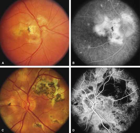
| Fig. 8 Right fundus (A) and late-phase fluorescein angiogram (B) of a 35-year-old woman with serpiginous choroidopathy. The visual acuity was 20/200. Left fundus (C) and fluorescein angiogram (D) of a 47-year-old man with advanced serpiginous choroidopathy, demonstrating several stages of lesions and subretinal gliosis. Visual acuity was 14/400. (A and B courtesy of Michael L. Klein, M.D., Portland, OR) |