|
|
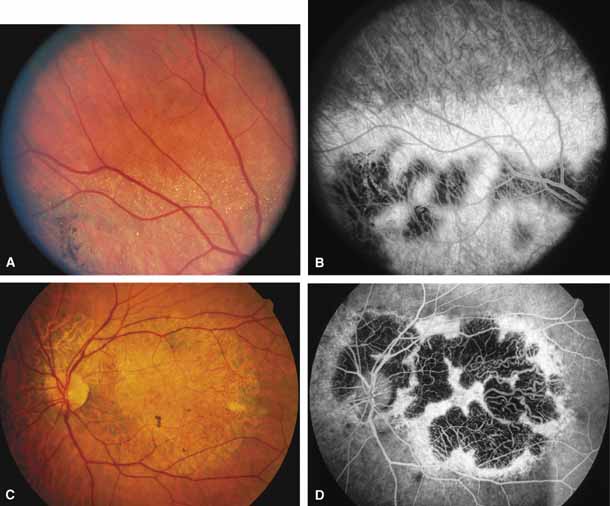
| Fig. 3 Right fundus (A) and fluorescein angiogram (B) of 52-year-old man with regional form of Bietti's crystalline corneal retinal dystrophy. Note retinal crystals adjacent to areas of dystrophy. Left fundus (C) and fluorescein angiogram (D) of 61-year-old brother of patient shown in A, showing marked regional atrophy of choroid and retina limited to the posterior pole and peripapillary regions. (A and B from Wilson DJ, Weleber RG, Klein ML, et al: Bietti's crystalline dystrophy: A clinicopathologic correlative study. Arch Ophthalmol 107:213–221, 1989; Copyright 1989, American Medical Association) |