|
|
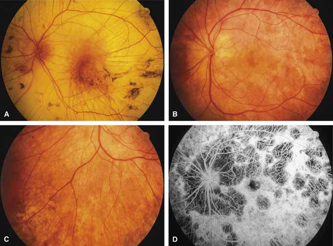
| Fig. 13 Left eye of a 53-year-old man with advanced choroideremia (A). Posterior fundus (B), inferior nasal retina (C), and fluorescein angiogram of posterior pole (D) of the left eye of a 30-year-old woman with carrier state for choroideremia, showing the characteristic mottled pigment epithelium and extensive patchy choroidal atrophy. At age 36, her visual acuity had fallen to 20/30 in each eye, and she has become significantly visually disabled by loss of pericentral visual field. She is the daughter of the patient shown in Figure A and the granddaughter of the carrier woman whose retinal histology was reported in reference 70. |