|
|
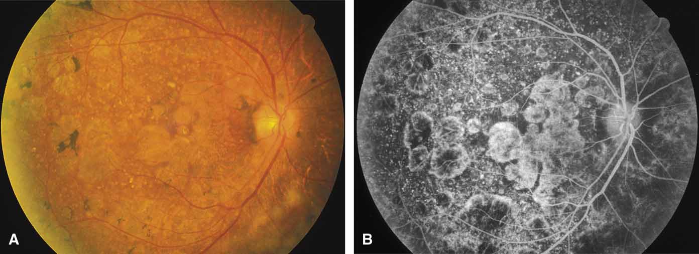
| Fig. 28 Right fundus (A) and fluorescein angiogram (B) of a 73-year-old woman with secondary syphilitic retinopathy, demonstrating patchy areas of atrophy of retinal pigment epithelium and underlying choriocapillaris. Visual acuity was 20/40. (From Weleber RG: Retinitis pigmentosa and allied disorders. In Ryan SJ [ed]: Retina, vol 1. Basic Science and Inherited Retinal Disease, 2nd ed. St. Louis: CV Mosby, 1994:335–420) |