|
|
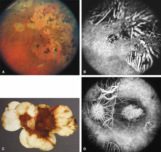
| Fig. 11 A. Transition zone between atrophic and more intact midperipheral retina in 37-year-old patient with B6-responsive gyrate atrophy. One allele is the Glu318Lys mutation of the ornithine aminotransferase gene, but the mutation affecting the other allele has yet to be defined (Inana G, Weleber RG, personal communication). B. Fluorescein angiogram of same region, showing pigment epithelial window defects and loss of choriocapillaris. C. Left fundus (C) and fluorescein angiogram (D) of advanced gyrate atrophy associated with macular atrophy in a 64-year-old woman homozygous for the most common Finnish mutation, the Leu402Pro mutation of exon 11 of the ornithine aminotransferase gene (Inana G, Weleber RG, unpublished finding). (B from Weleber RG, Kennaway NG: Gyrate atrophy of the choroid and retina. In Heckenlively JR [ed]: Retinitis Pigmentosa. Philadelphia: JB Lippincott, 1988:198–220. Images C and D courtesy of Robert Kalina, M.D., Seattle, WA) |