Chapter 23
Diseases of the Sclera and Episclera
PETER WATSON
Main Menu   Table Of Contents

Search

ANATOMIC CONSIDERATIONS
NORMAL ANTERIOR SEGMENT FLUORESCEIN ANGIOGRAPHY
CLINICAL EXAMINATION
EPISCLERITIS
SCLERITIS
CORNEAL CHANGES IN EPISCLERITIS AND SCLERITIS
ECTASIA AND STAPHYLOMA
CONGENITAL ANOMALIES
TRAUMA AND CHEMICAL INJURY
INFLAMMATION
DEGENERATIONS
TUMORS
REFERENCES

If the eye is considered as a much-modified ball and socket joint and the episclera as the synovial membrane, many of the conditions that afflict the sclera and episclera can be readily understood. The sclera consists of collagen, which resembles tendon, and much elastic tissue; therefore, the sclera suffers from the chronic, granulomatous, and destructive conditions that affect the joints and collagenous tissue elsewhere. Because the sclera is avascular, it is dependent on the vascular coats on either side, particularly the episclera, to provide a response to inflammation, so that scleral inflammation is almost always accompanied by an overlying episcleritis. Although the sclera is subject to degenerations and infections, these are insignificant when compared with the inflammatory changes of episcleritis and scleritis. Neoplasms of the sclera are unknown.
Back to Top
ANATOMIC CONSIDERATIONS
The function of the sclera is to provide a firm protective coat for the intraocular contents. This coat is resilient enough to allow for variations in the intraocular pressure yet firm enough to prevent severe distortion of the contents of the eye on movement or when pressed on by the muscles or external forces. To achieve this, the sclera is composed largely of collagen types I, III, and VIII and elastic tissue, the bundles of which are arranged to neutralize the external pressures imposed by the action of the muscles. These bundles vary in size from 10 to 15 μm in thickness and 100 to 150 μm in length, roughly parallel to the surface but interlacing among themselves.1 The somewhat irregular size and the criss-cross pattern account, in part, for the opaqueness of the sclera. The sclera is also fully hydrated at all times. If the water content is reduced to 40% by drying, the sclera becomes translucent. This phenomenon, which is often observed during detachment surgery, is probably produced by concentration of the proteoglycan, thus changing its refractive index to one that is near that of collagen. As will be seen, a similar change occurs after certain types of scleral disease in which the sclera becomes translucent as a result of changes in collagen and proteoglycan.

The shape of the sclera is determined by the inner layer of the optic cup, the retina, and the choroid. The sclera is derived from a condensation of mesectoderm (from the neural crest). The sclera develops outside the choroid late in development, starting anteriorly. The development of sclera and choroid is in turn induced by the retinal pigment epithelium. Abnormalities of development at this stage may lead to colobomas and in lesser instances to progressive myopia. Development of sclera over the posterior pole is not complete until the end of the fifth month of fetal life.2 From then on, the shape of the sclera is modified by the effect of the intraocular pressure. The elastic fibers in the sclera can be stretched beyond their limit of elasticity when not supported by a fully developed collagenous structure. The whole globe can therefore be distended until 3 years after birth; the lamina cribrosa is the only scleral structure that can be stretched after this age.

Although the sclera has a low metabolic activity, it requires some nutrition, which it derives from the underlying choroid and overlying episclera, the sclera being entirely permeable to water, glucose, and proteins. The sclera transmits blood vessels but retains a very scant supply for its own use. However, the sclera is supplied with nerves, particularly in the anterior segment near the muscles; damage to these nerves in destructive scleritis is undoubtedly the major cause of pain in this condition. The nerves also appear to be stimulated by distention of the sclera.

The episclera, or Tenon's capsule, provides for part of the nutrition of the sclera and for the cellular response to inflammation. In addition, the episclera also acts as a synovial membrane for smooth movement of the eye and, together with the muscular sheaths to which it is fused, provides a check on excessive movement. The episclera is a fibroelastic structure formed after the development of the sclera from a mesenchymal condensation of the areolar structures of the orbit, possibly in response to eye movement. A deep or visceral layer is closely applied to the sclera. The outer parietal layer combines with the muscle sheaths, fusing first with the visceral layer and then with conjunctiva near the limbus. The subconjunctival space is traversed by only a few fibers, which form very little barrier to edema; it is not a lymphatic space because it contains no endothelium. The parietal layer of Tenon's capsule is penetrated by the muscles, anterior ciliary artery, veins, nerves, and aqueous veins. The two layers of the episclera are thinly joined together by connecting fibers, and each layer is supplied with its own vascular network derived from the anterior ciliary arteries.

With the increasing use of anterior segment fluorescein angiography in the early detection of severe necrotizing disease of the sclera, it is necessary to have an understanding of the normal anatomy of the vasculature of the anterior segment of the eye.3–5 The blood supply to this region is enormous, being derived from the anterior ciliary arteries, but with extensive collateral arterial anastomoses to the posterior ciliary arteries at the root of the iris (Fig. 1). The anterior system is readily visible with the slit lamp and by anterior segment fluorescein angiography, especially if the eye is inflamed, and its recognition is of vital importance in the differentiation of episcleral and scleral conditions. The separation and displacement of these vascular layers give the most important clinical clues to the site and, hence, the severity of the inflammation. On slit lamp examination, three layers of vessels are readily visible. The conjunctival plexus, which is the most superficial layer of vessels, can be moved over the underlying structures. The superficial episcleral capillary plexus (Fig. 2) is a radially arranged series of vessels lying within the parietal layer of Tenon's capsule. The vessels in this layer anastomose at the limbus with the conjunctival vessels, with other members of the same plexus, and with the deep plexus. The deep episcleral capillary network (see Fig. 2) is closely applied to the sclera in the visceral layer of Tenon's capsule. The vessels anastomose freely with each other, forming a syncytium. The large vessels to and from the intrascleral plexus traverse the episclera near the insertions of the muscles. The conjunctival and superficial episcleral vessels can be blanched with 1:1000 epinephrine or 10% phenylephrine, but the deep vessels are affected slightly. This is of considerable assistance when attempting to differentiate deep and superficial inflammation.

Fig. 1. Anterior view montage of a cynomolgus monkey ocular casting with Tenon's and episcleral vessels removed. The anterior ciliary arteries (ACA) arborize at the limbus and interconnect via their lateral branches to form the episcleral circle. (CM, ciliary muscle capillary bed; CV, choroidal veins; EC, episcleral circle. (Original magnification, X20). (Morrison JC, van Buskirk EM: Anterior collateral circulation in the primate eye. Ophthalmology 90:707, 1983)

Fig. 2. The normal relationships of the capillary networks that can be seen with the slit lamp are a conjunctival (easily mobile) network, a superficial episcleral network in the parietal layer of Tenon's capsule, and a deep episcleral plexus closely applied to the sclera. These relationships are much more obvious in inflamed eyes (see Figs. 13, 14, and 30). (Watson PG, Hayreh S, Awdry P: Episcleritis and scleritis. Br J Ophthalmol 52(3):278– 279, 1968)

The direction of flow, the distinction between arteries and veins, and the integrity of the circulation can be determined only by the use of anterior segment fluorescein angiography.4,5

Back to Top
NORMAL ANTERIOR SEGMENT FLUORESCEIN ANGIOGRAPHY

ARTERIAL PHASE

Anterior Episcleral Arterial Circle

The anterior ciliary arteries run radially toward the limbus within Tenon's capsule, giving few, if any, branches until they reach the anterior part of the globe. Their positions are very variable, and they do not always follow the rectus muscles. They bifurcate 2 to 5 mm behind the limbus, and each division runs forward and circumferentially to anastomose with a branch from an adjacent artery. This results in an anterior episcleral arterial circle (Figs. 3 through 5). The divisions of the anterior ciliary arteries are typically superficial at their origins but run deeper at their anastomoses. They occasionally dip too deep to be seen in fluorescein angiograms (Fig. 6).

Fig. 3. Normal superior anterior segment angiography in a 35-year-old man. The main anterior ciliary arteries have filled, branching at or adjacent to the limbus to fill the limbal arcade, the recurrent branches of which are passing backward to fill the episcleral and conjunctival vessels. The episcleral circle here is superficial, and the anastomotic vessels are readily visible. There is a close resemblance to the vascular pattern in Figure 1, including the penetrating vessels adjacent to the limbus.

Fig. 4. Normal temporal angiogram of a 32-year-old woman. Fluorescein first enters the anterior ciliary artery above (as in Figure 3), filling the limbal arcade, and then dips deep into the sclera adjacent to the limbus; it can just be distinguished in the deep scleral tissue adjacent to the limbus. The posterior tarsal circulation can be seen filling at the same stage.

Fig. 5. Normal temporal angiogram in the same 32-year-old woman as in Figure 3 two seconds later. The superficial episcleral vessels are filling from a superficial branch of the anterior ciliary artery; the deep circle is now difficult to define but can be seen contributing to the iris vascular filling. The limbal arcade and conjunctiva near the limbus are filled from the superficial branches. Both the episcleral plexus and the conjunctival plexuses perfuse late, about 3 mm from the limbus. This watershed zone is important in the etiology of scleral disease.

Fig. 6. Normal left temporal angiogram of a 33-year-old man. In this angiogram the anterior conjunctival vessels and episcleral vessels are derived from the main ciliary artery trunk at the limbus, filling simultaneously with the iris vessels at a very early stage of the angiogram.

From the anterior episcleral arterial circle, four distinct circulations are supplied: episcleral, anterior conjunctival, limbal, and iris.

Episcleral Circulation

Immediately after their origin by bifurcation of the anterior ciliary arteries, the contributions to the anterior episcleral circle divide again to give recurrent branches that run posteriorly and subdivide to form a netlike episcleral plexus (see Fig. 5; Fig.7). The variability of the positioning of the anterior ciliary arteries inevitably leaves large areas of episclera far from such an arterial supply (see Figs. 4, 6, and 7). These areas receive other posterior branches from the episcleral circle. Where the circle runs deep within the sclera, such branches appear as isolated perforating vessels (see Fig. 6). They fill very shortly after the episcleral circle, and they also divide repeatedly as they run posteriorly.

Fig. 7. Normal left temporal angiogram of the same 33-year-old man as in Figure 6 eight seconds later. The episcleral circulation is being filled from a perforating vessel near the limbus derived from the deep circle. These eventually anastomose with vessels posteriorly and above. The conjunctival circulation is also late in filling. The main trunk of the artery can no longer be distinguished because of superficial leakage of dye.

Anterior Conjunctival Circulation

Throughout their superficial course, the arteries of the episcleral circle give off fine loops that run forward into the limbal reflection of the conjunctiva before curving back radially and dividing to form the lacework of the anterior conjunctival capillary plexus (see Fig. 2). The delicate column of blood within the anterior conjunctival loops may be punctuated by a string of individual erythrocytes, suggesting that the lumen is approximately 12 μm in diameter.

Anterior conjunctival loops may also arise from perforating posterior branches of the episcleral circle (see Fig. 6).

The anterior conjunctival circulation, supplied by the anterior ciliary arteries, always fills before the posterior conjunctival circulation, which is derived from the posterior tarsal vessels (see Figs. 4 and 5). The watershed zone between these sources can fill very late (see Fig. 5). However, anterior conjunctival loops do sometimes anastomose with arteries of similar caliber derived from the posterior tarsal circulation.

Limbal Arcades

Limbal arcades are supplied by anterior branches from the episcleral circle. Their origins are often shared with those of the anterior conjunctival loops, and, where the circle runs deep, they too are derived from the perforating posterior branches. They often fill very late during a normal angiogram (see Fig. 7).

The limbal capillary loops never leak fluorescein, even during high-dose angiograms, suggesting that their endothelial cells are united by tight junctions.

Iris Vessels

The first flush of fluorescein within the anterior episcleral arterial circle always coincides with filling of the radial arterioles of the iris. It may be implied from this that the iris receives a major supply from the anterior ciliary circulation (see Fig. 6). In some angiograms, the iris circulation appears to derive directly from the episcleral circle. This raises the possibility that the “major circle of the iris” and the episcleral arterial circle are less distinct entities than has hitherto been presumed.

VENOUS PHASE

Anterior ciliary veins accompany the arteries, but there is no well-organized venous ring corresponding to the anterior episcleral arterial circle. The posterior episcleral branches of the arterial circle are paralleled by centripetal venules, and looping anterior conjunctival venules are interspersed between the arterioles (see Figs. 4 and 5).

The posterior conjunctiva drains back into the tarsal circulation.

CAPILLARY PHASE

The episcleral capillary net is often difficult to discern below the more prominent conjunctival circulation. It is most clearly seen when the conjunctival circulation fills late for anatomic or pathologic reasons.

The anterior conjunctival capillary plexus forms an interlacing network between the anterior conjunctival arterioles. Perfusion of the watershed zone that separates the territories supplied by the anterior ciliary and posterior tarsal systems may be delayed by as much as 15 seconds after first flush (see Figs. 5 and 7). However, this region is often crossed by arteriolar anastomoses between the two circulations, and the destination of venous blood is irrespective of its origin from the anterior ciliary or posterior tarsal circulations.

Back to Top
CLINICAL EXAMINATION
Because of the collagenous nature of the sclera, diseases that affect it tend to be indolent, painful, and destructive, presenting as local manifestations of a generalized condition. Conditions involving the episclera tend to be acute, transient, and of little importance unless they indicate the presence of intercurrent disease. Therefore, it is of considerable importance to decide which tissue is involved from the onset of the disease so that the correct treatment may be given and the correct prognosis determined.

HISTORY

Careful taking of the patient's history sometimes reveals the cause. In general, the more rapid the onset, the more readily treatable the condition, and, consequently, the better the prognosis. Most scleral disease is bilateral and recurrent, and the history of suggestive attacks in the other eye should be sought. Alteration in visual acuity always indicates corneal or deep-seated disease.

Many eye diseases are relatively painless. However, pain is a prominent characteristic of scleral disease; it is often the pain, rather than the redness of the eye, that causes the patient to seek advice. The pain of deep-seated scleral disease is severe and boring in character. It often radiates to the forehead and brow and characteristically awakens the patient during the night. The pain in superficial conditions is localized to the eye.

On direct questioning, approximately one fourth of patients with episcleral and scleral disease will complain of lacrimation or photophobia. This is not a major symptom and is, surprisingly, not correlated with the presence of keratitis or any particular type of scleral condition.

Scleral disease can be a manifestation of disease of any system of the body; therefore, a routine inquiry should be made concerning the cardiovascular system for evidence of arteritis or hypertension; the respiratory system for evidence of tuberculosis or sarcoidosis; and the genitourinary system for evidence of renal tuberculosis or venereal disease. A very strong relationship exists between skeletal and scleral disease, and an attempt should be made to determine whether there is any suggestion of connective tissue disease (e.g., a history of general malaise, pains in multiple or single joints, pains in the back or in the neck, and the presence of morning stiffness). Skin disorders that accompany or precede the onset of scleral inflammation include herpes zoster, rosacea, psoriasis, and erythema nodosum or arteritis. No central nervous system disease appears to be associated with scleral disease. Many patients with episcleritis give a history of recent viral disease, hypersensitivity reactions, or contact with external irritants, particularly industrial solvents. A family history of atopy is occasionally found in patients with episcleritis.

Because most therapy for inflammatory scleral disease is administered systemically and is immunosuppressive in type, a history of gastric ulceration is of major importance because it may affect the type and extent of the therapy that can be given.

EYE EXAMINATION

Failure of vision is insidious, and the visual acuity must be measured at frequent intervals during the course of the disease. The external examination of the eye in daylight must never be omitted. This examination is essential to distinguish the deep discoloration, the increased transparency, and the area of maximum edema in deep scleral disease. No other method gives so much information; tungsten or fluorescent light is not as effective as daylight. Areas of deep inflammation and the extent of the progression of scleral disease have been seen quite often in daylight but have been invisible when examined with the slit lamp.

The object of slit lamp examination is to determine the depth and nature of scleral and episcleral conditions and the presence of corneal changes. The changes seen are drawn in the records. With the use of diffuse light with a neutral density filter, the vascular networks of both eyes are examined in detail to determine the layer in which the vessels show maximum congestion, the infiltration of episcleral tissues, and the edema of sclera, episclera, or subconjunctival space. Slit lamp examination is also used to ascertain the nature and depth of any corneal changes; the presence of scleral edema (for which it may be necessary to blanch the superficial tissues with epinephrine 1:1000 or phenylephrine 10%); the nature of any episcleral infiltration or mass; and the presence of cells in the anterior chamber or vitreous and posterior synechiae. The red-free (green) filter is extremely valuable in confirming the areas of maximum congestion and whether any areas are totally avascular. Because this is an important physical sign and is easily missed, examination in red-free light should be routinely performed. The green light brings the vessels into very sharp contrast with the background and enables the position of maximum inflammation to be determined with certainty. It also enables the paths and configurations of the vessels to be followed and will show lymphocytic infiltration of the episcleral tissue as yellow spots; this often indicates that the condition is more extensive than previously supposed (Fig. 8).

Fig. 8. Examination in red-free light. Blood vessels brought into sharp contrast reveal areas of lymphocytic infiltration in episcleral tissues, in this case due to herpes simplex virus.

Glaucoma often occurs secondary to scleral disease. Applanation readings should be undertaken at the first visit and subsequently during treatment, especially if corticosteroids are used.

Direct and indirect ophthalmoscopy are performed. This is particularly important in all cases of posterior scleritis or if cells are observed in the anterior or posterior chamber. Some patients with scleritis have granulomatous changes in the posterior segment that give rise to an exudative retinal detachment. Posterior scleritis and orbital disease such as pseudotumor, both of which involve the sclera and surrounding structures, will produce not only proptosis but also limitation of ocular movements. It is worth noting that these lesions can sometimes be completely symptom free even when the patient has developed a complete ophthalmoplegia (see Figs. 53 and 54).

Fig. 53. Proptosis of the right eye in a patient with both anterior and posterior scleritis.

Fig. 54. Retraction of the left lower lid occurs as a patient with posterior scleritis attempts elevation of the eye.

OTHER INVESTIGATIONS

Because so many patients with scleral disease have systemic disease, a thorough physical examination is essential. In the Scleritis Clinic at Moorfields Eye Hospital, this examination has been undertaken by a rheumatologist; this is a most satisfactory arrangement because the particular features that need thorough investigation are the joints, the skin, and the cardiovascular and respiratory systems.

The following routine investigations are performed:

  1. Hemoglobin
  2. White blood cell count and differential count
  3. Erythrocyte sedimentation rate
  4. If connective tissue disease is suspected, full immunologic investigations are undertaken, including levels of immunoglobulins and immunofluorescent studies for autoantibodies (including rheumatoid factor and antinuclear and anti-DNA antibodies); circulating immune complexes are searched for. If Wegener's granulomatosis or periarteritis nodosa are suspected, the anti-nuclear cytoplasmic antibody (ANCA) tests should be performed. The C-reactive protein is the best indicator of an active generalized inflammatory response.
  5. Serum uric acid
  6. Full serologic tests for syphilis

Radiologic investigations should include a chest roentgenogram and a roentgenogram of the sacroiliac joints. Physical examination may not reveal sacroiliitis of the rheumatoid type. Because this may be the only other systemic manifestation in scleral disease, this investigation should not be omitted. Roentgenograms of other joints are taken if a particular disease process such as gout, rheumatoid arthritis, or sarcoidosis is suspected.

Prick and patch testing of the skin has been universally unrewarding even when a known sensitizer has been found. Local challenge has been attempted, but the results are inconclusive.

Electroretinography and electro-oculography are of assistance only in the presence of cataract or severe necrotizing or posterior scleritis. There is sometimes a dramatic fall in the electric response at the onset of disease and an equally dramatic rise when the disease is suppressed, provided destructive changes have not occurred.

B-scan ultrasonography should never be omitted from the examination of patients with scleritis. Now that high-quality ultrasonography has become available, the extent and severity of the inflammation can be determined with great accuracy. Many patients who were formerly thought to have only anterior segment disease have been found to have extensive and sight-threatening posterior scleritis as well. It also has become known that many patients with posterior scleritis with few symptoms and signs have much more extensive disease than had previously been considered possible (Fig. 9). Anterior segment B-scan ultrasonography sometimes reveals extensive involvement of the deep scleral tissue around the ciliary body, indicating the need for urgent and intensive use of immunosuppressive therapy.

Fig. 9. B-scan ultrasonography in a patient with severe posterior scleritis. Note the thick sclera and the gap between scleral and episcleral tissue posteriorly.

B-scan ultrasonography has proved to be a much more valuable investigation than even high-resolution computed tomography (CT) scanning. However, if orbital extension of disease is thought to have occurred, then CT or magnetic resonance imaging (MRI) should be performed.

Back to Top
EPISCLERITIS
Episcleritis is almost always a benign inflammatory condition occurring in young adults, with a marked tendency to recur. The condition, which is frequently bilateral, may be divided on clinical grounds into simple episcleritis and nodular episcleritis.

ETIOLOGY

Thirty percent of patients with episcleritis had some associated general conditions,6–8 but the rest defied all attempts to discover an etiology. Although some patients had a strong family history of atopy, results of patch and prick testing were uniformly negative. Of those in whom an etiology was found, only 5% showed any association with collagen disease, 7% had an association with herpes zoster, and 3% each had an association with gout or syphilis; the rest had associated conditions such as erythema nodosum, Schönlein-Henoch purpura, erythema multiforme, contact with industrial solvents, or penicillin sensitivity, indicating an immune basis for the condition.

PATHOLOGY

Microscopic and electron microscopic studies of biopsy specimens from patients with simple and nodular episcleritis have been totally noncontributory in the attempt to discover the etiology of this condition. The inflamed area is packed with lymphocytes and a few other inflammatory cells, but there are no mast cells, plasma cells, or eosinophils.

CLINICAL MANIFESTATIONS

The onset is usually acute; the eye may become red and painful in as short a time as half an hour. The patient's main complaint is redness of the eye, which is often sectorial and may be accompanied by a feeling of hotness, pricking, and mild discomfort. There is no discharge, although the eye waters occasionally.

Pain may be absent, but the discomfort may be so severe that patients cannot pursue their normal occupation. The pain is localized to the eye, rarely radiating to the forehead and never producing the severe boring pain that is so commonly described in scleritis. In a severe attack the lids may become swollen, but this is a rare occurrence. If photophobia is present, an accompanying corneal condition should be suspected.

Simple and nodular episcleritis differ in their clinical courses, but in both the edema and infiltration are entirely within the episcleral tissues. The sclera is not involved. The maximum congestion is in the superficial episcleral network, with some slight congestion of the conjunctival vessels and deep episcleral vessels (Fig. 10). The intraocular structures are not involved in either variety, nor is the visual acuity affected. Anterior segment fluorescein angiography reveals a normal vascular pattern but a very rapid flow rate, with the whole transit of the dye being completed within 2 or 3 seconds (Figs. 11 and 12).

Fig. 10. Maximum congestion in the superficial vascular plexus in episcleritis. The conjunctival and deep episcleral networks are separated from the deep plexus by edema and infiltration in the episcleral tissue. (Watson PG, Hayreh S, Awdry P: Episcleritis and scleritis. Br J Ophthalmol 52(3):278–279, 1968)

Fig. 11. Anterior segment fluorescein angiogram of a 45-year-old woman with simple episcleritis. At first transit of dye, all the vessels are dilated and filling simultaneously. However, the vascular pattern is not disturbed.

Fig. 12. Angiogram of the same 45-year-old woman in Figure 11 one second later. Within 1 second, all the vessels are filled and there is even, venular filling except in the deep episcleral plexus. This is the rapid filling pattern seen in all forms of episcleritis and in diffuse anterior scleritis.

The redness of simple episcleritis may be intense, varying from a fiery-red or a brick-red discoloration to a mild red flush, but it does not have the bluish tinge that is seen in scleritis. The distribution is usually sectorial but can involve the whole anterior segment of the globe. The episcleral vessels are engorged but retain their normal radial position and architecture (Figs. 13 and 14; Color Plate 1A). In simple episcleritis, there is a diffuse edema of the episcleral tissues. These tissues are sometimes infiltrated with gray deposits that appear yellow in red-free light. Surprisingly, the eye is rarely tender to the touch.

Fig. 13. Infiltration of the episclera in which the superficial episcleral vessels show maximal congestion. Conjunctival vessels are slightly congested, as is the deep episcleral plexus, whose irregular criss-cross pattern can be seen deep to the radially arranged superficial episcleral plexus.

Fig. 14. Diffuse inflammation. Superficial vessels are maximally engorged and retain their radial pattern and architecture. (See Figures 27 and 34.) (Watson PG: Connective tissue disorders and the eye. In: Recent Advances in Ophthalmology, Vol 5, pp 214–277. London, Churchill-Livingstone, 1975)

Plate 1. A. Simple episcleritis. There is a diffuse dilatation of all the vessels, but the normal vascular pattern is not disturbed. The episcleral tissue is edematous but there is no swelling of the sclera. B. Nodular episcleritis. The episclera is edematous in the area of the nodule. The vessels over the nodule are dilated, but there is no displacement of the deep vessels, so the sclera is not edematous. C. Diffuse scleritis. The sclera is inflamed and swollen below the horizontal meridian, and the vessels are irregular and distorted. Above the horizontal, where there was previously inflammation, the color of the sclera has changed. This is not due to thinning of the sclera but rather to an increased transparency as a consequence of the alteration of the proteoglycan/collagen structure caused by recurrent inflammation. D. Sclerokeratitis in diffuse scleritis. Diffuse scleritis recurring at the same site often leads to opacification of the corner adjacent to it. This is caused by transudate from leaking vessels and sometimes leads to an immune ring in the cornea at this site. E. Necrotizing scleritis in Wegener's granulomatosis. There is an intense inflammatory response with granulomatous changes and necrosis of the sclera. The vessels are grossly distorted and the inflammation transgresses the limbus. The vessels that are entering the cornea leak profusely, and there is early erosion of the peripheral corneal tissue. F. Scleromalacia perforans. Quiet sequestration of the sclera in a 52-year-old woman with long-standing burned-out erosive articular rheumatoid arthritis. Note the complete lack of an inflammatory response even though this is at an active stage of the resorption of the tissue.

In contrast to simple episcleritis, the infiltration and edema of nodular episcleritis are localized to one part of the globe, forming a nodule and some surrounding congestion (Color Plate 1B). The nodule can be moved over the underlying sclera, which is not edematous. The scleral plexus of vessels can be distinguished deep to the nodule, lying flat on the sclera and slightly congested but otherwise normal in color and configuration (Figs. 15 and 16). Episcleral nodules may be single or multiple but do not undergo necrosis (see Fig. 16). After multiple attacks of nodular episcleritis in the same location, the superficial lamellae of the sclera show some alteration and become slightly more transparent in this one area.

Fig. 15. Episcleritis. In episcleritis, the vascular networks of the conjunctiva, episclera, and sclera are all congested. The edema is confined to the episcleral tissue so that the reflected light from the sclera shows no displacement. (Watson PG, Hayreh S, Awdry P: Episcleritis and scleritis. Br J Ophthalmol 52(3):278–279, 1968)

Fig. 16. Deep vessels, which are normal in configuration, can be traced beneath edematous nodules lying flat on the sclera. (Watson PG: Connective tissue disorders and the eye. In: Recent Advances in Ophthalmology, Vol 5, pp 214–277. London, Churchill-Livingstone, 1975)

TREATMENT

Even without therapy, simple episcleritis improves considerably within the first week and resolves within 3 weeks (Fig. 17). Provided the eye is not too uncomfortable, most patients can be persuaded to leave the eye untreated, because the condition will resolve spontaneously. However, if it is believed that some treatment is desirable, topical corticosteroids or locally applied nonsteroidal anti-inflammatory drugs (NSAIDs) may make the eye more comfortable and speed resolution slightly (see Fig. 17).9 Use of corticosteroid drops must be continued for several days after the inflammation has subsided to prevent the exacerbation of the condition that occurs if they are stopped suddenly. Prednisolone, betamethasone, or dexamethasone drops may be administered hourly until redness disappears, and then three times daily for 4 to 5 days. Under no circumstances should topical steroids be administered continually for more than a few weeks at a time because of the very real danger of inducing steroid glaucoma and cataract. If the condition fails to respond immediately, other treatment regimens should be sought. Ocular NSAIDs can be administered four times daily until redness disappears. Glaucoma and cataract have not been observed after prolonged use, but many patients become intolerant to the use of the ointment or complain of stinging and irritation.

Fig. 17. Improvement in signs and symptoms in a double-blind crossover trial of oxyphenbutazone (Tandearil) ointment, betamethasone (Betnasol) ointment, and placebo ointment in the treatment of episcleritis. Although active preparations improve episcleritis at a faster rate than the placebo, the condition resolves spontaneously within a month. (Watson PG, McKay DR, Clemmett RS et al: Treatment of episcleritis. Br J Ophthalmol 57: 866–870, 1973)

Whereas simple episcleritis resolves rapidly without therapy, the resolution of nodular episcleritis is much slower. Local therapy is consequently of much more value; the same regimen of treatment is followed.

In the few patients in whom episcleritis becomes indolent, or in whom recurrences are so numerous that the patient becomes incapacitated, it is reasonable to consider systemic therapy with NSAIDs such as flurbiprofen (Froben), 100 mg three times daily, which usually gives immediate and prolonged relief of symptoms and signs. It is important to note that not all of the NSAIDs work in this condition. Treatment may be terminated abruptly when the condition comes under control.

The complications of episcleritis are minor and are not responsible for any decrease in visual acuity.

COURSE AND PROGNOSIS

Whether treated or not, simple episcleritis will resolve in 10 to 21 days. It will usually reappear at irregular intervals and then eventually disappear. An accurate 12-month record kept by a patient who went without treatment is shown in Figure 18. He was free from any further attacks for 3 years. He then had four attacks in the next 3 months and has had none since. No etiologic or precipitating factor has been found.

Fig. 18. Record kept by a patient over a full year, during which time he had no treatment. Intensity of attack is indicated by height of peaks. After September, no further attacks occurred for 3 years.

In nodular episcleritis, the nodule initially increases rapidly in size, sometimes reaching the size of a split pea. Thereafter it gradually regresses over a variable period and eventually disappears, although this may take up to 2 months without treatment.

Recurrences occur also in nodular episcleritis, but the two varieties are not mutually exclusive (a simple episcleritis may recur as a nodular episcleritis and vice versa). However, episcleritis never develops into scleritis in the same attack, although it invariably accompanies scleritis. Of 180 patients initially diagnosed as having episcleritis, only 4 developed scleral involvement.10

Episcleritis is an entirely benign condition, although it may be a great nuisance to the patient. It may recur over a period of many years, but it rarely leaves any residual ocular changes except for some areas of scleral transparency or localized stromal keratitis in those patients who have had severe attacks of nodular disease occurring always at the same site. Of 180 patients analyzed,10 only 2% had a decrease in visual acuity of two lines or more within a year of the onset, and in every case this was from increasing involutional cataract.

Back to Top
SCLERITIS
Scleritis, unlike episcleritis, is a severe destructive disease, sometimes leading to the loss of an eye from deteriorating vision, severe pain, or even (occasionally) perforation of the globe. Such changes, when they occur, are rapid, so early diagnosis and effective treatment are essential. Scleral disease can be diagnosed when the patient is first seen if one remembers that whereas episcleritis rarely, if ever, involves the scleral tissue, in scleritis the episclera is always involved. Therefore, attention must be diverted to the sclera to detect the early changes of scleral edema or necrosis.

The onset of scleritis is usually gradual, building up over several days. By the time patients seek advice, the clinical types can be distinguished as anterior or posterior, or occasionally both. Anterior scleritis may be further subdivided into diffuse, nodular, or necrotizing. The last condition may present with signs of inflammation or with few or no signs of inflammation (scleromalacia perforans). Long-term follow-up of patients with scleral disease showed that only 8% of patients changed from one type of disease to another during the course of this disease, so although differentiation into these types does not usually indicate an etiology, it does have a direct bearing on the prognosis and the type of treatment to be used.10

Scleritis is most common in the fourth to sixth decades of life and occurs more frequently in women than men (8:5). Necrotizing scleritis occurs later than the other varieties, the mean age being 61 years. Scleritis is bilateral in 52% of patients. In half of these, the condition starts in both eyes simultaneously, with the rest becoming bilateral in 5 or more years.

ASSOCIATED SYSTEMIC DISORDERS

In a review of 1200 patients with scleritis who have attended the Scleritis Clinic at Moorfields Eye Hospital in London, an associated systemic disorder was found in all patients with scleromalacia perforans, in half of those with nodular and necrotizing disease, in a third of those with diffuse anterior scleritis, and in only 10% of those with posterior scleritis. Severe polyarticular rheumatoid arthritis and a case of porphyria accounted for the patients with scleromalacia perforans.

Forty percent of the patients with necrotizing scleritis had other connective tissue disorders, but, surprisingly, only 21% of the patients with diffuse anterior or nodular scleritis had rheumatoid arthritis or other connective tissue disorders. This percentage is much lower than that reported by other authors,7,11–13 but this may be because patients with the less severe scleral disease are referred to us only if the etiology is in doubt, thus biasing the results. Twelve percent of the patients with diffuse anterior and nodular scleritis had ankylosing spondylitis, and in a further 15% the scleritis followed an attack of herpes zoster ophthalmicus. A variety of other conditions, including syphilis, tuberculosis, gout, Reiter's disease, IgA nephropathy, and erythema nodosum, were thought to be definite etiologic factors because with appropriate treatment the eye changes disappeared. Fowler investigated a random selection of the patients with scleritis at Moorfields Eye Hospital and found only 7%, all of whom were young males, who did not have any other detectable physical abnormality.14 Forty percent of these patients had hypertension, which in some cases required treatment. It was thought that the hypertension could have been a manifestation of a generalized arteritis, but this was convincingly demonstrated in only 19% of these patients.

PATHOLOGY

The pathology of scleritis has received much attention in the past.6,8,15–19 Although certain inferences can be drawn from pathologic specimens of eyes removed because of pain, perforation, or mistaken diagnosis, these eyes have been severely damaged from advanced disease. Unfortunately, biopsies of scleral lesions have proved to be unsatisfactory, at best yielding material of limited diagnostic value and at worst leaving an area of exposed choroid that will not heal. Consequently, they should 
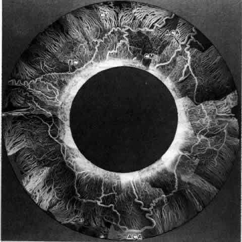
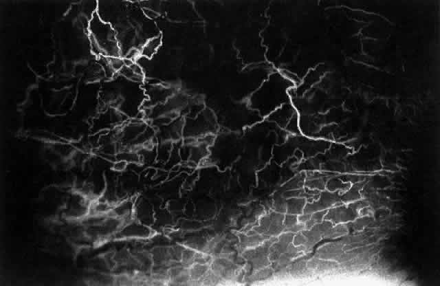
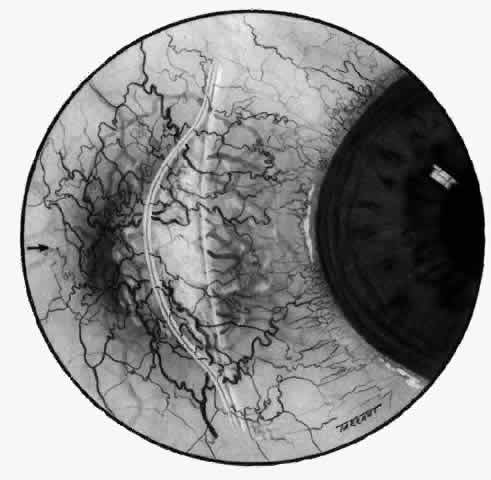
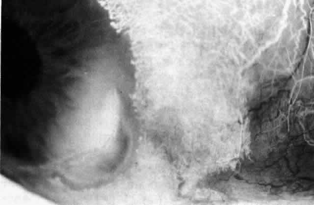
not be performed.

Scleritis usually affects the anterior segment of the eye, possibly because this is the area with the best blood supply, but with sluggish flow through the vessels (Fig. 19). The sclera is thickened and roughened in the affected area, which appears to be sharply demarcated from the rest of the sclera. However, tissue obtained at surgery during the course of grafting of areas adjacent to necrotic tissue shows marked pathologic changes.20,21 The area of affected sclera may be swollen, excavated, or frankly ulcerated with undermined edges covered with a thin layer of fibrous tissue. However, spontaneous perforation is extremely unusual and, where seen in pathologic specimens, has usually occurred at the time of removal 
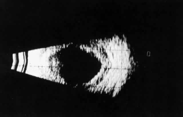
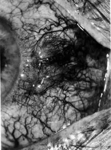
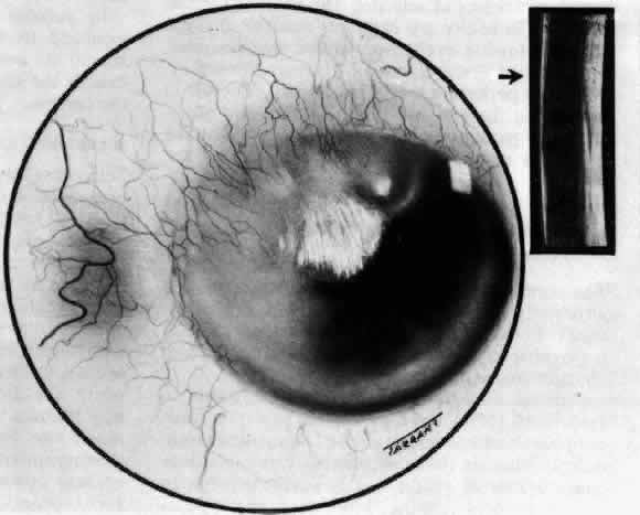
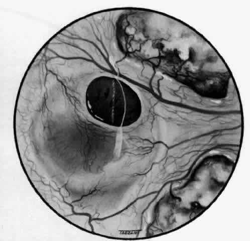
of the eye. A posterior scleritis often occurs as an extension of anterior disease; but, as in Figure 20, most of the inflammation (in some cases all of the inflammation) is in the posterior segment and the exudative detachments and subretinal granulomas 
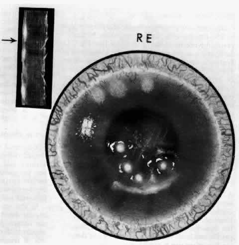
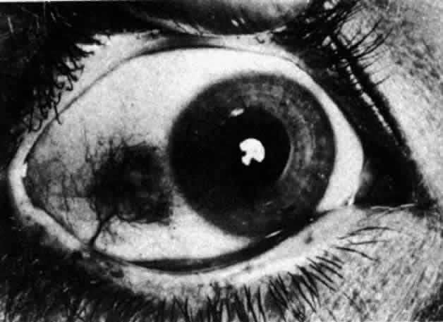
can be mistaken for malignant melanoma.

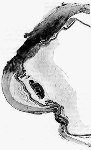
Fig. 19. Anterior necrotizing scleritis. The eye was removed because of loss of vision and intractable pain. No form of steroid was given to this patient because of a severe Pseudomonas infection of the chest. (Courtesy of Professor N. Ashton)

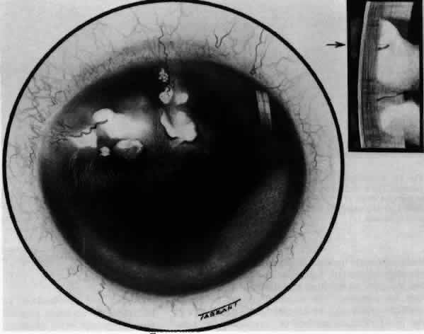
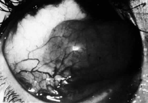
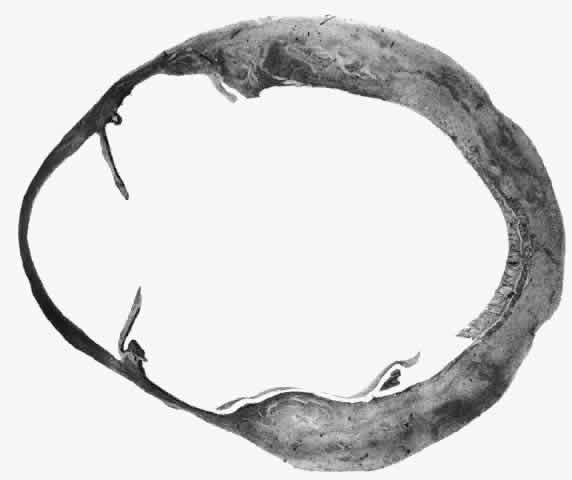
Fig. 20. Posterior scleritis. This eye was removed because of loss of vision and pain, mistakenly diagnosed as malignant melanoma. (Courtesy of Professor N. Ashton)

What is clinically represented solely by inflammation and edema is histopathologically a granulomatous lesion of the sclera, the center of which consists largely of plasma cells, lymphocytes, and mast cells (Figs. 21 through 23). Foster and colleagues have identified the cellular subsets and glycoproteins in both necrotizing and non-necrotizing scleritis.22 This shows an active T-cell inflammatory response with a high CD4/CD8 ratio and increased HLA/DR and CD14, indicating a macrophage-induced response that would lead to granuloma formation. Remote from the granuloma, the fibrocytes of the sclera become activated, the proteoglycan adjacent to them becomes altered, and the collagen fibrils of the sclera become unraveled (Figs. 23 and 24). These changes appear to take place prior to the invasion of the stroma by cells of the granuloma.20 The vessels in and around the necrotic area show medial necrosis and perivascular cuffing with lymphocytes, and endothelial swelling with microvascular occlusion. Ninety-six percent of the specimens examined by Foster and associates show a microangiopathy characterized by a neutrophil infiltrate in and around the vessel wall.22–23 This is most obvious at the center of the lesion where there may be occlusion of the vessel, thrombosis, or even aneurysm formation (Fig. 25). From these pathologic investigations, clinical observations, animal experiments, and the results of fluorescein angiography, it would appear that the scleral inflammation is initiated either by trauma (be it accidental or surgical)23–25 or by bacterial or viral infection. If circulating immune complexes are present because of the poor blood flow, they become precipitated in and around the vessel walls in the area of inflammation. In other patients, a persistence of tissue damage will lead to autoimmunization. Damage to the endothelial cells of the microvasculature leads to changes within the vessels detectable on angiography and to catabolic changes in the surrounding tissues. These changes, in turn, allow the granulomatous response that is seen in histopathologic sections, the first detectable change being in the scleral fibrocytes and the proteoglycan and collagen remote from the site of cellular infiltration.

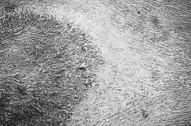
Fig. 21. Advancing edge of a granulomatous reaction. Scleral fibers are split and separated by edema and then disrupted when invaded by the granuloma

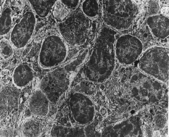
Fig. 22. Electron micrograph of an area of active scleritis showing the plasma cell infiltrate suggestive of an immune response. Note aggregated plasma cells, with the characteristic whorled rough endoplasmic reticulum, in the process of degeneration, releasing organelles and nuclear debris into the extracellular matrix. (Uranyl acetate and lead citrate. X3000) (Courtesy of Dr. R. Tripathi)

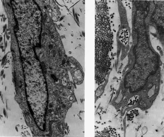
Fig. 23. Electron micrographs of scleral stroma at the periphery of an area of ulceration in a patient with necrotizing scleritis. The left shows an active fibroblastic cell, and the right shows collagen fibrils within intracellular vacuoles (V) in the fibroblastic cell. (Left X15,375; right X15,375) (Watson PG, Young RD: Changes at the periphery of a lesion necrotizing scleritis: Anterior segment fluorescein angiography correlated with electron microscopy. Br J Ophthalmol 68:781–789, 1984)

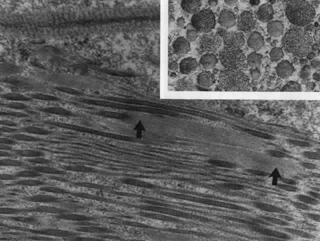
Fig. 24. Electron micrograph of scleral stroma at the periphery of an ulcer in necrotizing scleritis (same patient as in Figure 23) showing swelling and unraveling of collagen fibrils (arrows) in longitudinal section (X29,270) and in transverse section (inset, X44,000). Fibrils of all diameters are affected. (Watson PG, Young RD: Changes at the periphery of a lesion necrotizing scleritis: Anterior segment fluorescein angiography correlated with electron microscopy. Br J Ophthalmol 69:656–663, 1985)

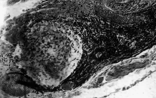
Fig. 25. Intense lymphocytic reaction and infiltration around a medium-sized arteriole and nerve.

CLINICAL MANIFESTATIONS

Lacrimation and photophobia are more common in scleritis than in episcleritis. However, they are not always clearly related to the severity of 
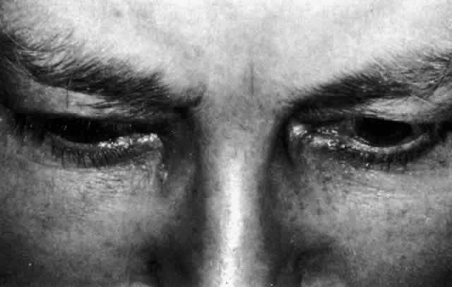
the scleritis or to the keratitis and uveitis that may accompany it.

The pain of scleritis is its most dominant feature and is the symptom that causes the patient to seek medical advice. The exception to this is scleromalacia perforans occurring in long-standing rheumatoid arthritis, which may be entirely pain free. Pain, when it occurs, may be localized to the eye, but in 66% of patients it is much more diffuse, radiating to the temple, the jaw, and the sinuses. It is boring in nature, severe enough to prevent sleep, accompanied by malaise, and only temporarily relieved by analgesics (Fig. 26). The pain is particularly severe in those patients suffering from progressive necrotizing scleritis with overlying inflammation; eyes have been removed for this reason alone. The pain can be a diagnostic problem, particularly in the early stages of posterior scleritis before the vision becomes affected. Patients with posterior scleritis are often referred to neurologists and others because of the severity of the headache or ophthalmoplegia. The pain is probably caused by distention of sensory nerve endings as a result of edema. In the necrotizing disease, the severity of the pain is increased by the destruction of the nerve 
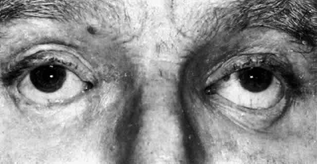
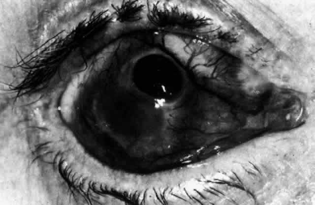
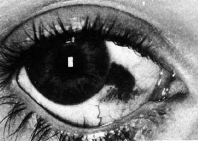
endings that takes place.

Fig. 26. Severe ptosis produced in a severe diffuse anterior scleritis. Pain radiated to temple and face and was severe enough to prevent sleep.

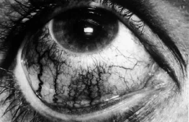
The inflammation of the eye is a prominent feature. The inflammation has a bluish-red hue in contrast to the brighter red of episcleritis and may be sectorial or diffuse. The severity of inflammation seems to depend on the amount of episcleral tissue present. Therefore, it is more prominent in younger people and is least prominent in those with rheumatoid arthritis in whom the episcleral tissue almost disappears.

Each of the various types of scleritis can be distinguished by its typical clinical appearance. Because the pathologic change is in the sclera, there is always edema and/or necrosis of that tissue. This gives rise to an overlying episcleral edema and to congestion that may be very severe and may need blanching with epinephrine 1:1000 or phenylephrine 10% to 
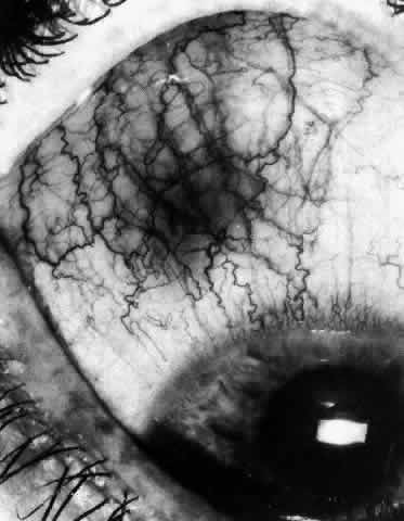
detect the underlying edema.

The sclera that is edematous is pushed forward, and the deep episcleral network is more congested than the superficial networks (Figs. 27 and 28). It is usually easy to ascertain by simple observation that the patient has scleritis and not episcleritis. However, it is not as easy to ascertain whether the patient has early necrotizing scleritis. It is in these patients that fluorescein angiography has considerable value, because the first changes are detectable in the ocular vasculature. Prompt and adequate treatment can prevent these changes from becoming irreversible.

Fig. 27. In scleritis, maximum congestion occurs in deep episcleral plexus, which is bowed forward by underlying scleral edema. Episcleral tissue is slightly infiltrated and superficial plexus is slightly congested (see Fig. 14). (Watson PG, Hayreh S, Awdry P: Episcleritis and scleritis. Br J Ophthalmol 52:278–279, 1968)

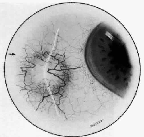
Fig. 28. Nodular scleritis. Both the anterior conjunctival slit and the deep scleral slit are displaced forward by the scleral edema. There is little separation between these two beams, indicating that all the edema is in the sclera and not in the overlying episclera. (Watson PG, Hayreh S, Awdry P: Episcleritis and scleritis. Br J Ophthalmol 52:278–279, 1968)

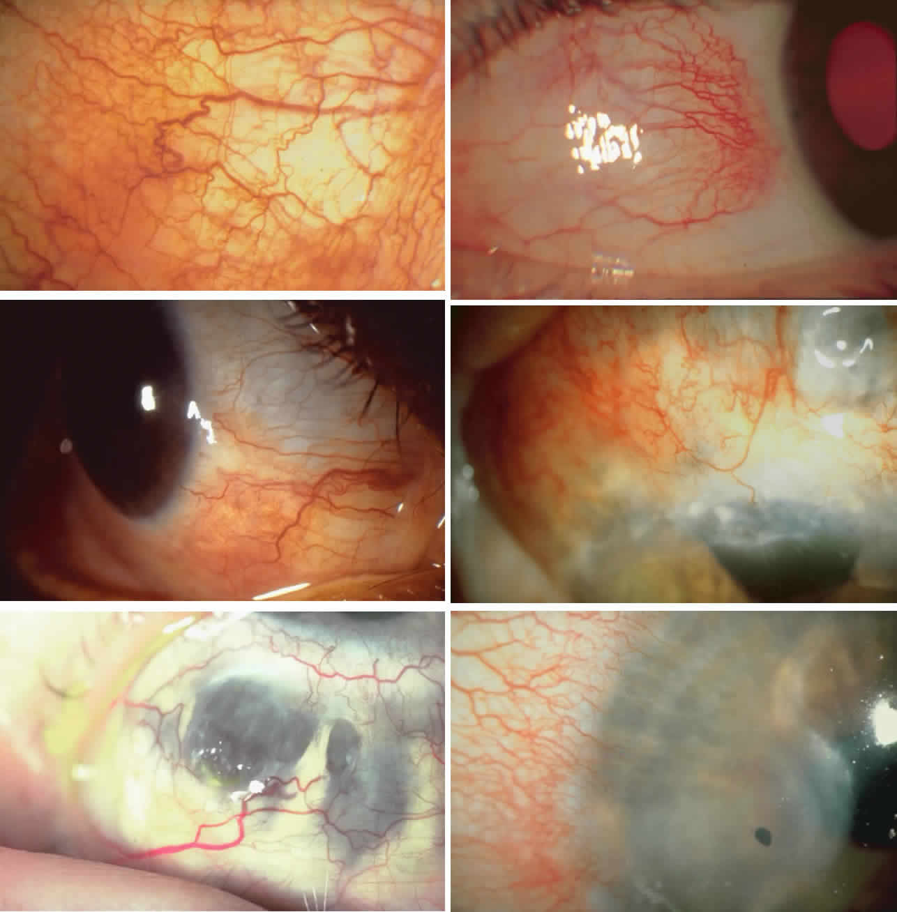
Diffuse Anterior Scleritis

Diffuse anterior scleritis is the most common and least severe type of scleritis. The inflammation is widespread, and it may involve either a small segment or the whole of the anterior segment, sometimes with such severe overlying inflammation as to justify the name “brawny” scleritis (Fig. 29). On slit lamp examination, the vascular pattern of both deep and superficial layers may be distorted, so that the normal radial pattern of the vessels is lost; large anastomotic channels develop, leading to beading and tortuosity of the remaining vessels (Figs. 30 and 31; Color Plate 1C).

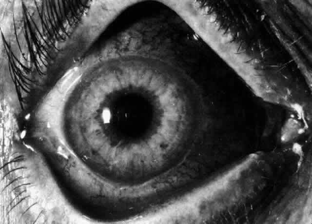
Fig. 29. Intense inflammation, edema, and conjunctival chemosis that accompany acute diffuse anterior scleritis.

Fig. 30. Diffuse anterior scleritis. During the acute attack, the vessels are dilated and distorted. New vessels or large vessels not normally seen have appeared adjacent to the limbus.

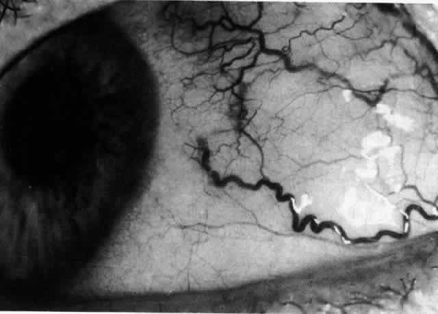
Fig. 31. Diffuse anterior scleritis after treatment. The dilated abnormal blood vessels remain even though no inflammation remains.

In this relatively benign form of scleral inflammation, the fluorescein angiogram reveals a rapid flow pattern in which the transit time of the dye is very rapid (as in episcleritis) (Figs. 32 and 33). Subtle changes occur in the capillary network, and abnormal leaking vessels appear after prolonged inflammation. These changes do not disappear after the inflammation subsides or is treated (see Fig. 31).

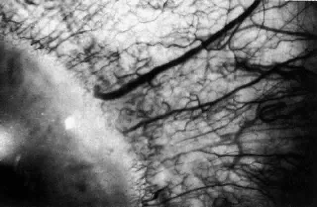
Fig. 32. Diffuse anterior scleritis associated with corneal edema. The limbal vessels are grossly dilated in association with generalized scleral edema. The cornea adjacent to these vessels is edematous.

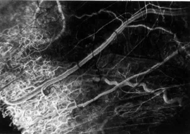
Fig. 33. Fluorescein angiogram of patient in Figure 32 four seconds after the appearance of the dye. This is a very rapid transit time. All the limbal capillaries are completely full, and all the major episcleral vessels contain fluorescein. Note that the very large vessel is a vein, and the narrow vessel below it is an artery. The deep vessels are distorted, and some are abnormal in configuration.

Nodular Anterior Scleritis

Although patients with nodular anterior scleritis resemble those with nodular episcleritis on cursory examination, detailed examination reveals marked differences. The nodule or nodules (they may be multiple) consist of scleral tissue that is immovable episclera is tightly adherent to the nodule, which is tender to the touch. Although the sclera sometimes becomes transparent below the nodule, it does not become necrotic, nor does the condition extend beyond the site of the nodule, as occurs in necrotizing scleral disease (Fig. 36).

(see Fig. 28; Figs. 34 and 35). The edematous

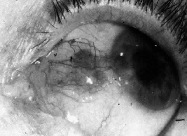
Fig. 36. Increased scleral transparency that occurred at the same site resulting from recurrent attacks of nodular scleritis after herpes zoster ophthalmicus.

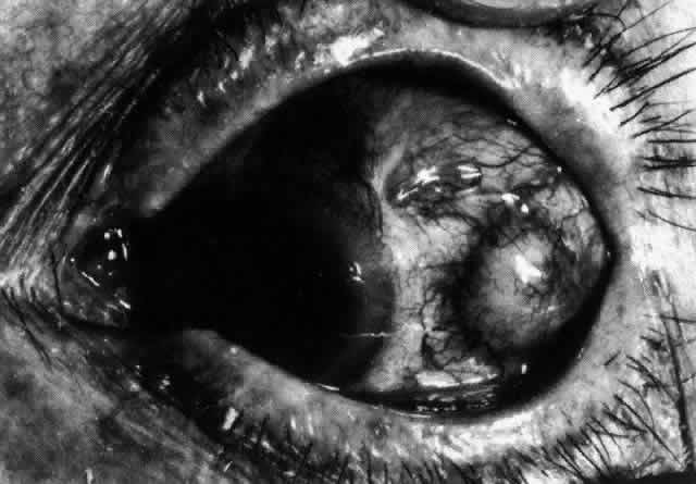
Fig. 34. Scleral edema has displaced all the vessel layers forward. Area surrounding the nodule is acutely inflamed.

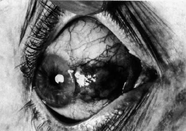
Fig. 35. Multiple scleral nodules. Surrounding inflammation is deep and intense. (Watson PG: Management of scleritis. In: Recent Advances in Ophthalmology, Vol 5. London, Churchill-Livingstone, 1975)

The angiogram is similar to that of diffuse anterior scleritis (i.e., there is a rapid filling pattern and deep scleral leakage of dye).26

Necrotizing Anterior Scleritis with Inflammation

Patients with necrotizing anterior scleritis with inflammation not only suffer extremes of discomfort but are in serious danger of losing an eye. Therefore, it is of great importance that the condition be detected early and treated adequately. (It is of equal importance that those varieties of scleral inflammation that are not destructive to the eye should not be treated with drugs that are themselves dangerous.) Accurate 
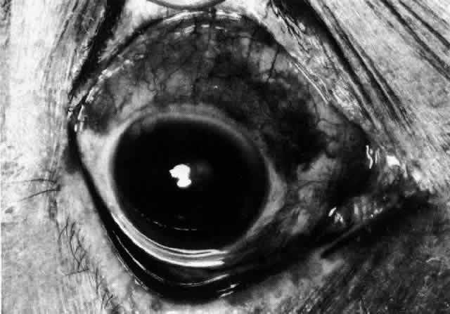
diagnosis is the key.

Necrotizing scleritis accompanied by inflammation is always painful, waking the patient at night, increasing in intensity day by day, and leading to severe distress. The sclera is swollen, and the overlying inflammation is localized to the center of a lesion or to either end of an 
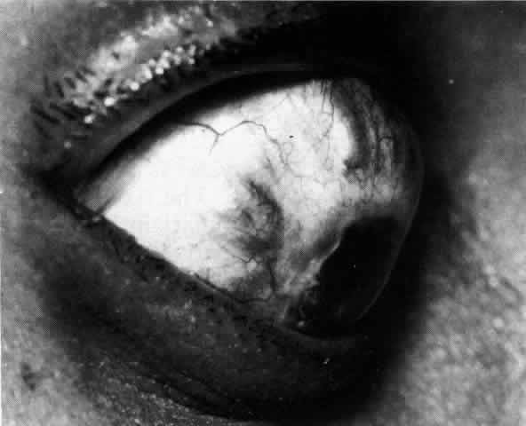
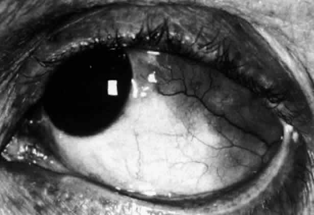
extending lesion (Fig. 37; Color Plate 1D). After inflammation, the sclera becomes transparent so that the underlying choroidal pigment becomes visible when viewed in daylight (Fig. 38). These areas may be invisible with the slit lamp. The area of inflammation extends outward around the globe from the original site of inflammation, often joining with other areas of scleritis that have subsequently appeared. If the inflammation is not suppressed, the process will 
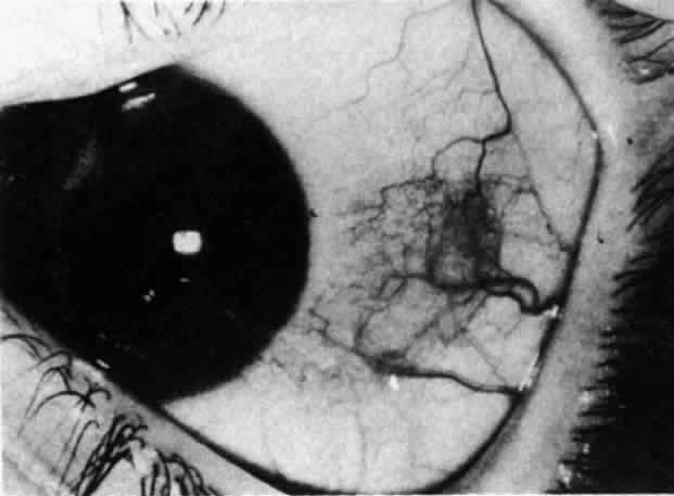
progress around the globe until the whole anterior segment is involved (Fig. 39).

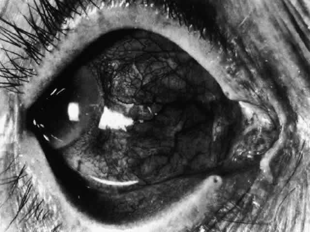
Fig. 37. Necrotizing anterior scleritis. Early stage in which there is diffuse, intense, scleral congestion in one segment of the globe, and anomalies of the vascular pattern. (Watson PG: Management of scleritis. In: Recent Advances in Ophthalmology, Vol 5, pp 77–87. London, Churchill-Livingstone, 1975)

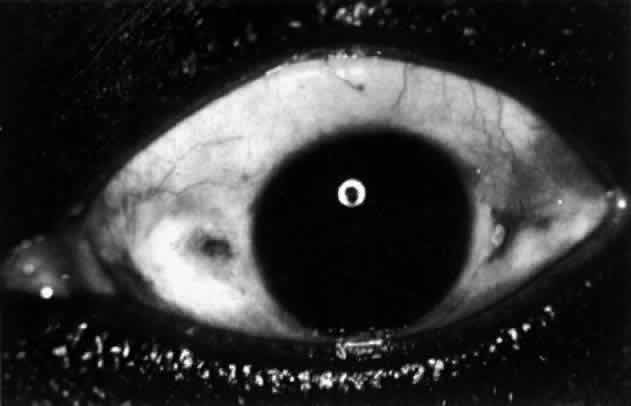
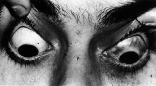
Fig. 38. Diffuse circumferential increased scleral pigmentation in left eye of a young patient after treatment of a necrotizing anterior scleritis. Subtle color changes of this type can be differentiated from similar changes of increased transparency only if the patient is examined in daylight.

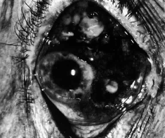
Fig. 39. Necrosis occurs in areas behind the advancing edge, which is at 4 o'clock. The sclera at 6 o'clock is so far not affected.

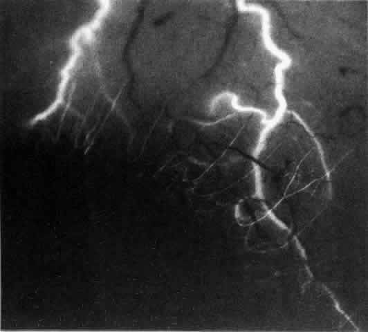
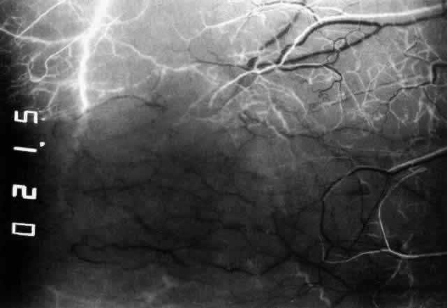
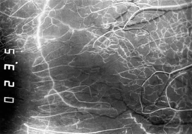
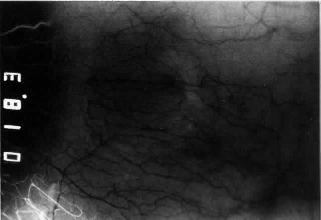
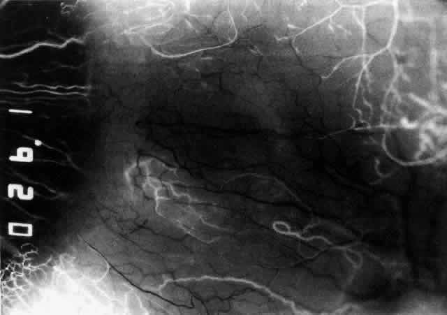
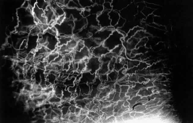
The characteristic features of necrotizing scleritis on fluorescein angiography are hypoperfusion and, eventually, nonperfusion of the vascular networks (Figs. 40 through 43).26 The initial changes are on the venous side of the capillary network; the transit time of the dye increases even if the eye is red and congested. If the disease process persists or has been present for a long time, thrombosis and permanent vaso-occlusive changes occur. These vessels (or the occluded capillary network) are bypassed by the opening of anastomotic channels. New vessels in a granuloma give rise to deep intrascleral 
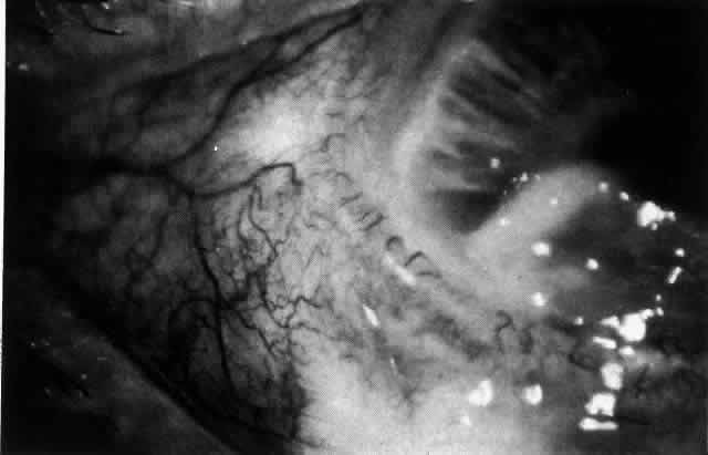
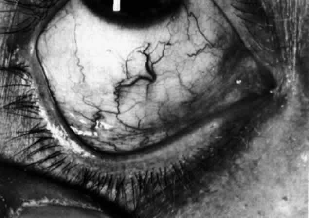
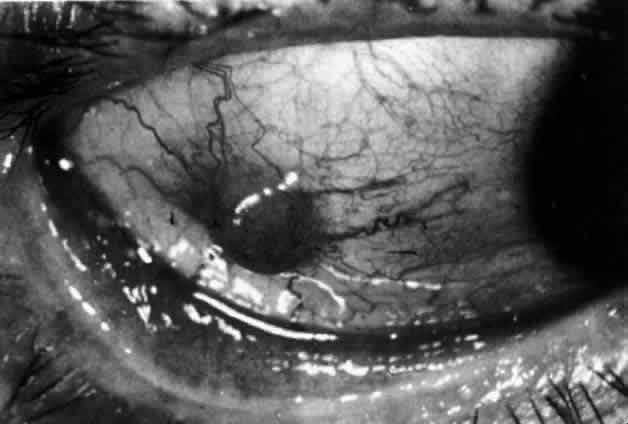
leakage of dye (see Fig. 43). Conjunctival and episcleral involvement by the destructive change is late but is always preceded by vaso-occlusive changes that can sometimes be detected with use of the red-free light on the slit lamp (Figs. 44 and 45).

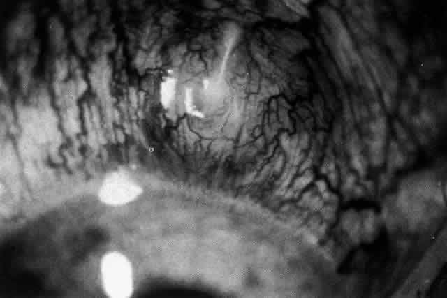
Fig. 40. Early necrotizing scleritis. There is characteristic yellow discoloration of the sclera underlying the conjunctiva at a point of necrosis. In this instance a small filament of tissue has penetrated the conjunctiva.

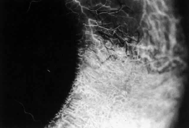
Fig. 41. Late stage of fluorescein angiogram adjacent to the site of necrosis in the same patient as in Figure 40. Although the eye is uniformly congested, the area near the necrosis shows vascular shutdown, whereas the rest of the conjunctiva and episclera is normally perfused.

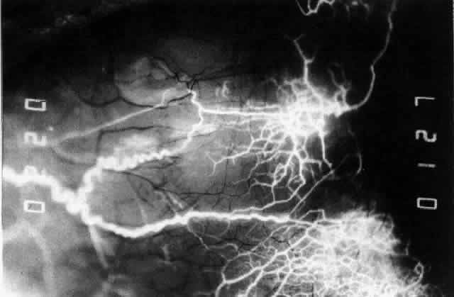
Fig. 42. Late arterial phase of fluorescein angiogram in a patient with necrotizing scleritis. All the vessels except the main trunk and the vessels around the limbal perforating vessels are occluded and remain unperfused throughout the angiogram.

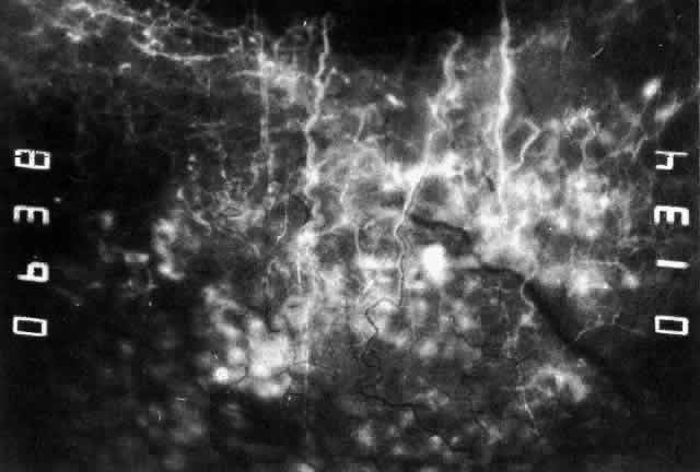
Fig. 43. Late venous phase of angiogram of a patient with necrotizing scleritis showing late deep leakage from vessels on the surface of the sclera and leakage of the capillary network at the limbus and the vessels draining it, together with poor or absent perfusion of the remaining vessels.

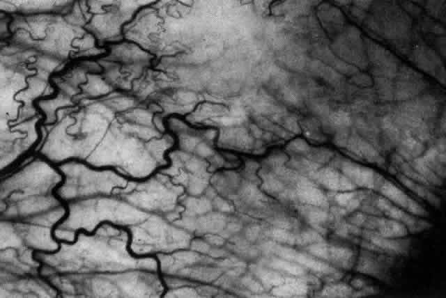
Fig. 44. Necrotizing scleritis. An avascular patch is seen in red-free light. If left untreated, this will progress to the situation found in Figure 45.

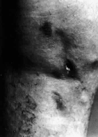
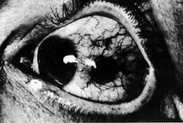
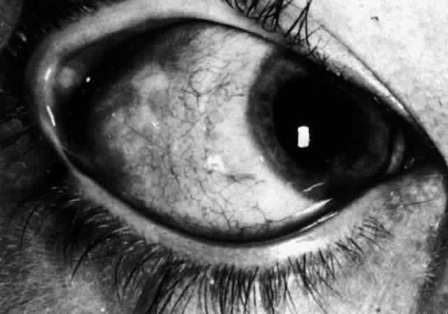
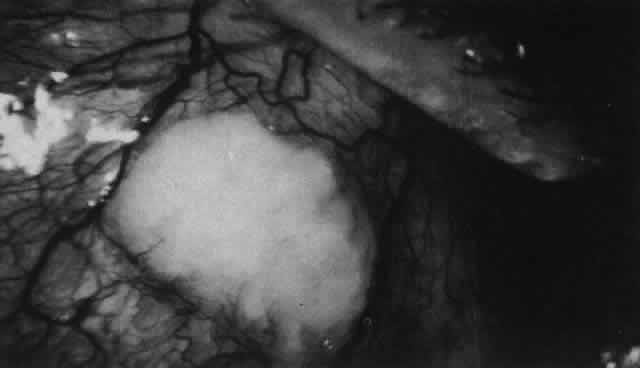
Fig. 45. Necrotizing scleritis. An area of necrosis is evident in the eye of this patient with localized Wegener's granulomatosis. The conjunctiva adjacent to the white necrotic tissue becomes adherent to the underlying episclera.

Uveitis, lens changes, glaucoma and other serious complications such as central vein occlusion do not seem to occur until the disease process 
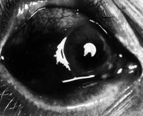
affects the whole circumference of the eye.27 Sometimes scleral thinning, as well as increased transparency, occurs, but unless the intraocular pressure rises above 40 mm Hg, staphylomas are extremely rare. As the disease is brought under control, the necrotic areas are absorbed or sequestered, leaving an ectasia with the underlying uvea exposed or covered with a thin film of conjunctiva or episclera (Fig. 46). If the defect is small, new collagen will cover it (Figs. 47 and 48). If the defect is large or if it is thought to have been the source of a persisting antigenic stimulus, the necrotic tissue may need to be excised and then covered by scleral grafts. However, this procedure is usually performed for aesthetic reasons rather than because the vision is endangered. The underlying disease process is not affected by the presence of a scleral graft, which has to be covered by conjunctiva and, preferably, episclera if it is to survive. Surgery must never be undertaken until the disease process has been suppressed. Temporary gluing may be used in perforated eyes until this has been achieved.

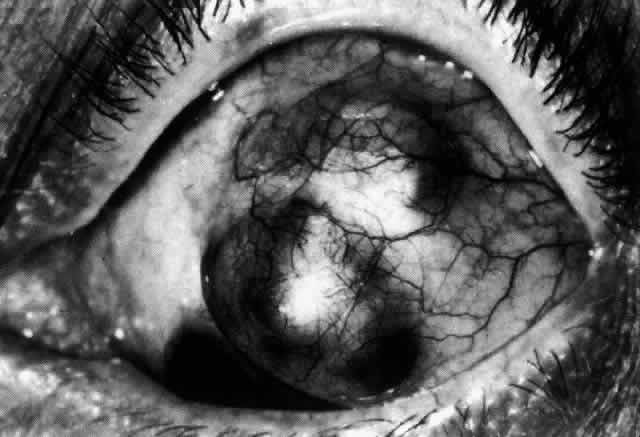
Fig. 46. Localized anterior staphyloma after necrotizing anterior scleritis and seconday glaucoma, during the acute phase of which the intraocular pressure rose to 50 mm Hg.

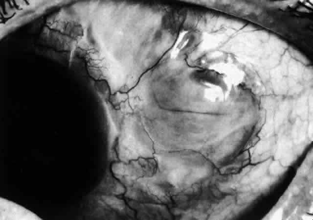
Fig. 47. Healing of a large scleral defect. Sclera is flat, without staphyloma formation. Newly formed fibers are thin and radially arranged but adequate to support a normal intraocular pressure. (Courtesy of Mr. HE Hobbs)

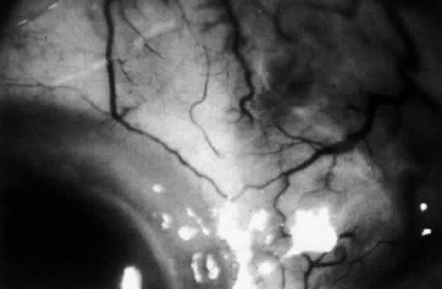
Fig. 48. Inactive necrotizing scleritis. The vessels have remained abnormal. The sclera has been absorbed, leaving the deep, blue-gray choroid visible beneath a very thin layer of conjunctiva. The adjacent cornea shows pitting and lipid deposition because of the impaired venous drainage in the adjacent episclera and conjunctiva.

Necrotizing Anterior ScleritisdWithout Adjacent Inflammationd(Scleromalacia Perforans)

Necrotizing anterior scleritis without adjacent inflammation appears to be a well-defined condition with little relation in clinical features to necrotizing scleral disease, even though the pathology is similar and the final result is the same. Scleromalacia perforans is characterized by the almost total lack of any symptoms. It occurs almost exclusively in patients with long-standing polyarticular rheumatoid arthritis, the 
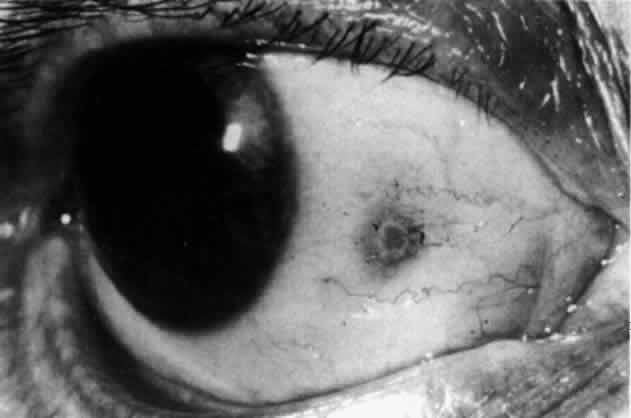
majority of whom are female (Figs. 49 and 50; Color Plate 1E).

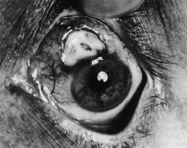
Fig. 49. A white necrotic plaque developing in an area of sclera with practically no surrounding inflammation in a 60-year-old woman who had had Crohn's disease for 17 years.

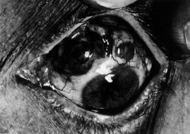
Fig. 50. Scleromalacia perforans after treatment. The very thin sclera is covered by conjunctiva only and a few remaining large blood vessels. (Courtesy of Mr. HE Hobbs)

The anterior sclera loses its covering of episclera and develops an area of yellow-white necrotic slough over many months; this eventually separates or is absorbed, leaving the underlying choroid covered by either conjunctiva or nothing at all. As with necrotizing disease, the choroid does not bulge into this ectatic area; but unlike necrotizing disease, spontaneous healing of even small perforations is very limited once the necrotic tissue has been removed (see Fig. 50).

Fluorescein angiography is not helpful, except to indicate areas of vascular closure in an otherwise extremely thin, atrophic episcleral tissue.4 The formation of a sequestrum appears to be caused by arteriolar closure as opposed to the venular disease seen in the other forms of necrotizing scleritis.

Posterior Scleritis

Because the posterior sclera is invisible, the diagnosis of posterior scleritis is made only if the anterior sclera is also involved or some other sign or symptom leads one to suspect it. Posterior scleritis is much more common than previously suspected, as recent clinical and pathologic studies have shown.19,28,29 There are two distinct forms of posterior scleritis. The first is usually associated with an anterior scleritis. This granulomatous disorder, like its anterior counterpart, can be diffuse, nodular, or necrotizing in character and is associated with the connective tissue diseases. The second form occurs in young patients of all races who are 9 to 40 years of age. It is always diffuse in character but is not associated with any systemic disorder. Both forms may cause uveitis if the inflammation affects the ciliary body, and in both forms the patient may develop exudative retinal detachments, choroidal folds, and swelling of the disc (Figs. 51 and 52). The granulomatous type may also involve the structures outside the globe, causing proptosis (Fig. 53), limitation of ocular muscle movement, and, uniquely, retraction of the lower lid on attempted elevation of the eye (Fig. 54). Diagnosis is with B-scan ultrasonography.

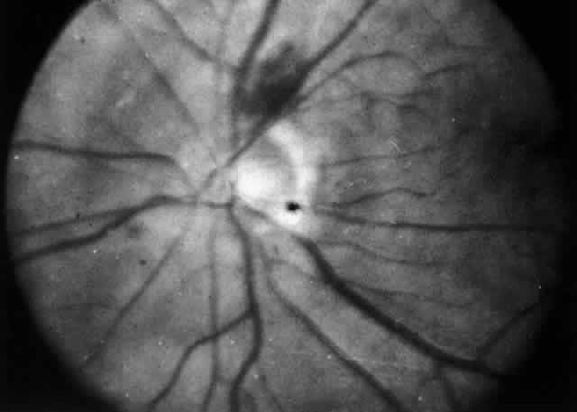
Fig. 51. Swelling of the optic nerve head and hemorrhage near the disc in a patient with posterior scleritis. The poor quality of the photograph is partly due to vitreous haze that accompanied the inflammation.

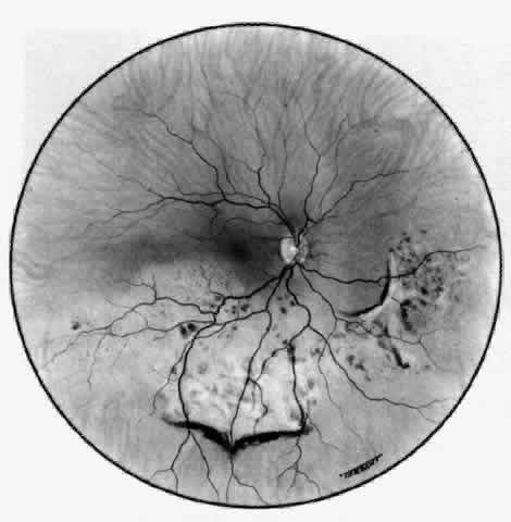
Fig. 52. Fundus appearance after resolution of exudative detachment in patient with severe posterior scleritis. Macula was affected and vision much impaired. (Watson PG: Management of scleritis. In: Recent Advances in Ophthalmology, Vol 5. London, Churchill-Livingstone, 1975)

TREATMENT

Scleritis is almost always accompanied by very severe pain that prevents sleep. A response to treatment is heralded by a dramatic relief of pain even though the condition might appear to be getting worse (Figs. 55 through 59). Treatment may be modified with confidence once the pain has disappeared.

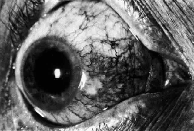
Fig. 55. Necrotizing scleritis 2 days after onset of severe pain in eye and temple. The eye was red, the sclera edematous, and the overlying episclera congested. A small paralimbal avascular area was noted. The patient was treated with 400 mg oxyphenbutazone for 7 days. (Watson PG: Contemporary Ophthalmology. Baltimore, Williams & Wilkins, 1972

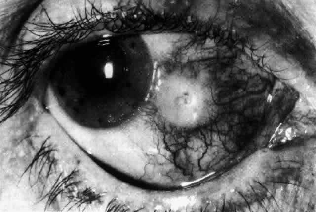
Fig. 56. Appearance of same eye as seen in Figure 55 one week later. The pain persisted, and the avascular patch was much enlarged with a necrotic center. Treatment was not altered, but the patient was admitted to the hospital. (Watson PG, Hayreh S, Awdry P: Episcleritis and scleritis. Br J Ophthalmol 52(3): 278–279, 1968)

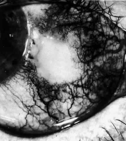
Fig. 57. Appearance of the same eye as seen in Figure 56 five days later. The area has increased in size, and the necrotic center is beginning to separate. Treatment was changed to 100 mg prednisolone daily. Within 24 hours, the severe ocular and facial pain had disappeared, but the appearance of the eye did not change.

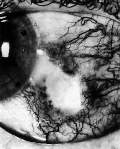
Fig. 58. Appearance of the same eye as in Figure 57. After 5 days' treatment with a high dosage of steroids, new vessels can be seen growing into the necrotic area both superficially and deep. The steroids were reduced to a maintenance level of 20 mg and continued for 6 weeks. (Watson PG: Contemporary Ophthalmology. Baltimore, Williams & Wilkins, 1972)

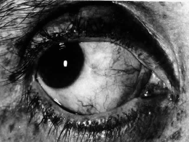
Fig. 59. Same eye as seen in Figures 55 through 58. Four months after the onset of disease, the ulcerated area has completely healed and filled in with new collagen material, which has assumed the usual radial pattern of scleral fibers. (Watson PE: Contemporary Ophthalmology. Baltimore, Williams & Wilkins, 1972)

Local Corticosteroids

Local steroid therapy increases the patient's comfort, but it is not effective in suppressing scleral inflammation. It is occasionally justified to use local steroid therapy alone when the inflammation is mild, the pain is slight, and corneal involvement is present, or very occasionally between attacks in the more severe forms of the disease to prevent remission. However, local steroids should be used only sparingly, if at all, in scleral disease because of the high chance of developing steroid-induced glaucoma or cataract.

Systemic Therapy

NONSTEROIDAL ANTI-INFLAMMATORY AGENTS.

Nonsteroidal anti-inflammatory agents are effective in suppressing the inflammatory response in the majority of patients with diffuse and nodular scleritis, especially if they exhibit a high flow pattern on fluorescein angiography. Dosage levels need to be high initially and, as a consequence, care must be taken to monitor the patients to ensure that no toxic side effects occur. Treatment must be continued until the inflammation subsides, after which it can be stopped abruptly.

In assessing the effect of treatment, pain, tenderness, episcleral and scleral injection, and corneal and intraocular involvement should be used as parameters of activity of the disease. In a series of double-blind controlled trials, the effects of different anti-inflammatory and immunosuppressive agents have been compared. The suggested routines of treatment are based on the results of these trials. Unfortunately, not all of the nonsteroidal anti-inflammatory agents are effective in controlling scleral inflammation. The current practice is to use flurbiprofen (Froben), 100 mg three times daily, for at least 1 week in all patients who present with scleritis of whatever type, provided there is no evidence of vascular closure or scleral destruction on slit lamp examination. The response, if it is going to occur, is immediate, with the pain disappearing within 48 hours. Within a week, the results of investigations are known, including those of the angiographic films if they have been done. If there is a poor response to flurbiprofen and the angiogram shows a high flow pattern, the drug is changed to another nonsteroidal anti-inflammatory agent, because there is an individual susceptibility among the patients. Only if there is no response in the progression of the disease or if there is evidence of vascular closure are systemic steroids or other immunosuppressive drugs used.

SYSTEMIC STEROID THERAPY.

If the scleritis is severe or necrotizing or if areas of vascular closure are detected with slit lamp examination or fluorescein angiography, then the use of systemic steroids is mandatory (see Figs. 40 through 44). Prednisone and prednisolone are most commonly used.

The principle of treatment with systemic steroids is that a sufficient amount must be given to suppress the condition; once this has occurred, the dosage may be rapidly reduced to a maintenance level, which may have to be continued until a natural remission occurs, or the steroid may be replaced by a nonsteroidal anti-inflammatory agent. Provided sufficient amounts are given and the patient can tolerate them, systemic steroids will control scleritis. The problem is deciding what dosage is appropriate. The following scheme has been found to be effective. If the angiogram shows early vascular shutdown and treatment with flurbiprofen has not been effective, oral prednisolone, 60 or 80 mg, is given for 2 days and is reduced over 1 week to 20 mg. The dose of prednisolone is then reduced by 2.5 mg every other day until the pain recurs or signs of inflammation begin to recur. This maintenance dose is continued for about 1 month, and then the dose is reduced by 1-mg steps. This final phase may be aided by the addition of a nonsteroidal anti-inflammatory drug. Pain relief is by far the most sensitive indicator of control of the disease.

If this course of treatment is not effective, intravenous pulse therapy of high doses of methylprednisolone, with or without the use of immunosuppressive 
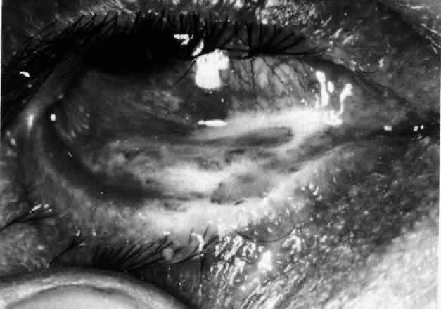
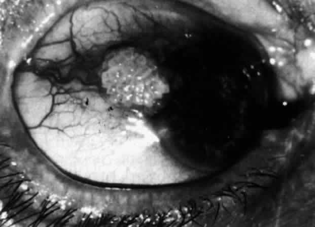
therapy, should be considered (Fig. 60).

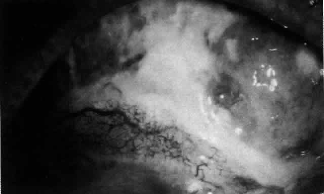
Fig. 60. Severe necrotizing scleritis. The surface of the necrotic sclera is covered with a thick, sticky, white mucus. The presence of such mucus is a sensitive indicator that the disease is active. This patient required two pulses of 1 g of methylprednisolone and 500 mg intravenous cyclophosphamide (5 days apart) to bring the condition under control. Thereafter, systemic low-dosage steroid (5 mg daily) and 50 mg of cyclophosphamide maintained control. The first sign of poor control was the reappearance of the mucus.

Pulsed Therapy With Methylprednisolone.

In very severe necrotizing scleritis or posterior scleritis when the vision is being threatened, it is important to suppress the inflammatory reaction completely. This is achieved by the use of intravenous methylprednisolone. The patient must be carefully monitored because this therapy inhibits all cellular transfer mechanisms, including the heart, and is therefore potentially dangerous. Methylprednisolone is administered by intravenous infusion as 500 mg or 1 g given over a period of 1 hour. The clinical response is observed. If this is unsatisfactory, then the dose may be repeated at 1- to 3-day intervals for three doses. It may be necessary to add other immunosuppressives to this regimen.

Immunosuppressive Therapy

The commonly used immunosuppression drugs used in the control of scleral disease are cyclophosphamide, cyclosporine, azathioprine, methotrexate or chlorambucil. Cyclophosphamide is the drug of choice in patients with systemic vasculitis, periarteritis nodosa, and Wegener's granulomatosis if they are not of child-bearing age. In severe cases of these disorders, cyclophosphamide is given with methylprednisolone as a bolus of 500 mg. A high fluid intake is essential to prevent hemorrhagic cystitis. Cyclophosphamide is then given orally at a dose of 50 mg three times per day. This treatment has a profound effect on the lymphocyte count, which must be monitored at weekly intervals for the first few months of treatment. Azathioprine can also be used as an adjunct to steroid therapy in those patients who require an unacceptably high maintenance dose of steroid (i.e., 15 mg prednisolone or more) to control their disease and in those with known immune complex disease (Fig. 61). Cyclosporine is helpful sometimes. Considering that pathologically there is always evidence of T-cell activation, it might be expected that cyclosporine would be the drug of choice in these diseases. Experience shows that this is not the case (in sharp contrast to the treatment of uveitis). Cyclosporine should therefore remain a second-line treatment and should be reserved for recalcitrant cases. It is strongly recommended that ophthalmologists always work in close cooperation with internists or other physicians when undertaking immunosuppressive therapy.

Fig. 61. The course of treatment in a patient who had lost the sight in one eye from posterior scleritis and who had developed a severe posterior scleritis in the other eye. She had extremely high circulating immune complexes, but eliminating these alone was not sufficient to control the scleral disease. Control was maintained for 3 years after the episode, using 7.5 mg prednisolone and 50 mg cyclophosphamide daily. Two exacerbations required treatment with short 7-day courses of oral prednisolone. (Watson PG: The nature and treatment of scleral inflammation [Doyne Memorial Lecture]. Trans Ophthalmol Soc UK 102:257–281, 1982)

SUBCONJUNCTIVAL AND ORBITAL FLOOR STEROIDS.

Treatment with subconjunctival steroids is contraindicated in scleritis. Perforation can occur at the site of subconjunctival injections (Fig. 62). Depot steroids should not be used because the particulate matter may induce or perpetuate the inflammatory reaction in the sclera. Orbital floor steroids are occasionally helpful in patients with necrotizing disease who are unable to take systemic steroids. The effects unfortunately tend to be transient, and the injections often need to be repeated at 7- to 10-day intervals. In this situation, intravenous pulse therapy should be considered as an alternative.

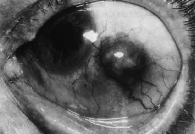
Fig. 62. Extreme scleral thinning in nasal and inferior quadrants surrounding a deposit of hyodrocortisone given for suppression of scleral disease. All sclera in the area later disappeared, and the eye threatened to perforate.

Surgery

Surgical treatment for defects in the sclera is rarely necessary. Adequate medical therapy allows the base of all small scleral defects to be covered by newly formed collagen, rendering them safe from perforation (see Fig. 59).

However, very large defects may have to be covered with sclera or cornea. Provided these grafts can be covered by conjunctiva, they usually remain in place, apparently viable. Scleral grafts ensure the comfort of the patient but do not prevent progressive necrotizing disease in the host sclera or even the graft (Figs. 63 and 64). Scleral replacement should be performed with the use of cornea rather than scleral tissue. Sclera rapidly resorbs, whereas corneal tissue is attacked only if there is a recurrence of the original disease.

Fig. 63. Severe necrotizing scleritis.

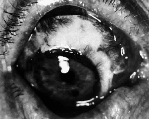
Fig. 64. Scleral graft in eye shown in Figure 63. Three months later the scleritis was still being treated with steroids. New vessels invaded the graft, which later took on the appearance of normal sclera.

Keratolysis or progressive peripheral corneal thinning sometimes requires lamellar corneal grafting (Figs. 65 and 66). Penetrating keratoplasty should be avoided if possible. The endothelium always remains normal in sclerokeratitis, and because of the proximity of the grafts to the limbus, larger penetrating grafts do less well. Under no circumstances should surgery be attempted until the systemic disease and ocular inflammation are brought under control. If necessary, cyanacrylate glue can be used to seal a perforation until immunosuppression is achieved. Methylprednisolone 500 mg should be given during the operation and, if required, after surgery.

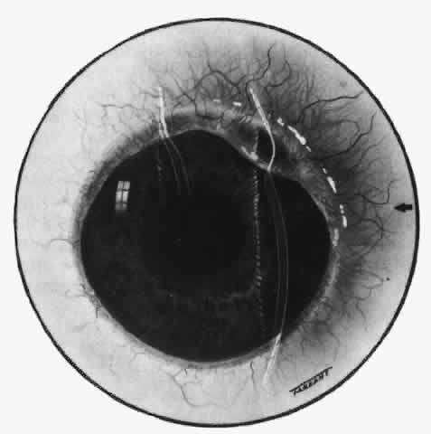
Fig. 65. Sclerokeratitis. Terrien-like ulcer at site of long-standing scleritis. The eye began to expand in this area after 6 years. (Watson PG: Contemporary Ophthalmology. Baltimore, Williams & Wilkins, 1972)

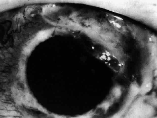
Fig. 66. The affected area of cornea seen in Figure 65 covered by a corneal graft. The scleritis has since settled and no longer requires treament.

COMPLICATIONS

Complications occur late in the disease and vary with the severity of inflammation. They occur most frequently in posterior scleritis and in severe necrotizing disease, particularly when the condition has become circumferential and when the inflammation is so severe as to produce secondary intraocular inflammation.

Visual Acuity

The object of early diagnosis and treatment is to prevent a decrease in visual acuity. The treatment must not produce iatrogenic changes that cause decreased acuity.

Over a 3-year period, approximately 27% of the patients who develop this disease will experience a decrease in visual acuity of two or more lines, which can be the result of cataracts and keratitis developing in patients with severe diffuse anterior scleritis. However, over a 25-year period, only 3% have lost useful vision.

Increased Scleral Transparency and Thinning

Alteration in the collagen and ground substance results in increased scleral transparency. Scleral thinning occurred, particularly in necrotizing disease or scleromalacia perforans. Of these patients, 22% showed increased scleral transparency after the first attack; however, only 6% developed a scleral defect. If scleral defects are small, they will refill with new collagen after treatment; but if they are very large, they may have to be covered with a graft (see Figs. 63 and 64).

Uveitis

Although roughly 35% of patients with scleral disease show some evidence of cellular activity in either the anterior or the posterior segment, a severe uveitis with a marked flare and heavy cellular response is very unusual. If it does occur, it is a serious sign, and intensive treatment must be instituted at once with systemic steroids. In posterior scleritis, if the granuloma is behind the equator, there may be little or no intravitreal cellular reaction, even though there is a visible granuloma and a retinal detachment. Scleritis occurring between the pars plana and the equator affects the ciliary body, so some inflammatory response occurs. Unless patients with this form of inflammation are treated rapidly, the intraocular pressure sometimes rises disastrously. Most patients with posterior scleritis have high intraocular pressures at some stage in the disease.

As the scleral disease is brought under control, the uveitis resolves, leaving anterior and posterior synechiae unless care is taken to prevent them. The inflammation of the pars plana sometimes leads to massive pigment migration at the retinal periphery, leaving a reaction rather like a diathermy or cryotherapy reaction in retinal detachment surgery (see Fig. 52).

Glaucoma

The intraocular pressure may become raised at any stage of the disease because of an acute congestion of the outflow channels,27 raised episcleral venous pressure, angle closure, or a steroid-induced rise. Therefore, it is important that the intraocular pressure be monitored; 13.5% of all patients with nodular or necrotizing scleritis had a pressure rise, albeit transient, during the course of the disease. Permanent field changes occurred in 5%. Patients with posterior scleritis are particularly prone to develop rises of intraocular pressure.

The treatment of the glaucoma is the treatment of the scleritis. Once the scleritis is controlled, the pressure will fall to normal. While the eye is inflamed, particularly if there is a limbitis, acetazolamide should be used to control the intraocular pressure. Should the pressure remain high after the attack, topical timolol can often help to control the intraocular pressure. If control fails, trabeculectomy can be performed successfully in an area of normal sclera and conjunctiva.

Cataract

Involutional changes that are already present will be increased by the presence of a severe inflammation. However, there is no doubt that the transparency of the lens can be affected directly in patients who have had previously normal lenses and who have developed severe necrotizing scleral disease.

If a cataract advances to the extent that it requires removal, the extraction can be performed with use of a corneal section in spite of the presence of scleritis. Healing is a little delayed in some cases, but no operative or postoperative complications have occurred.

Cataract extraction and, for that matter, any other surgical procedure can precipitate scleral inflammation in a patient who is predisposed, usually because of circulating immune complex disease. These patients usually have necrotizing scleritis and require vigorous therapy (Fig. 67).23–25,30–33

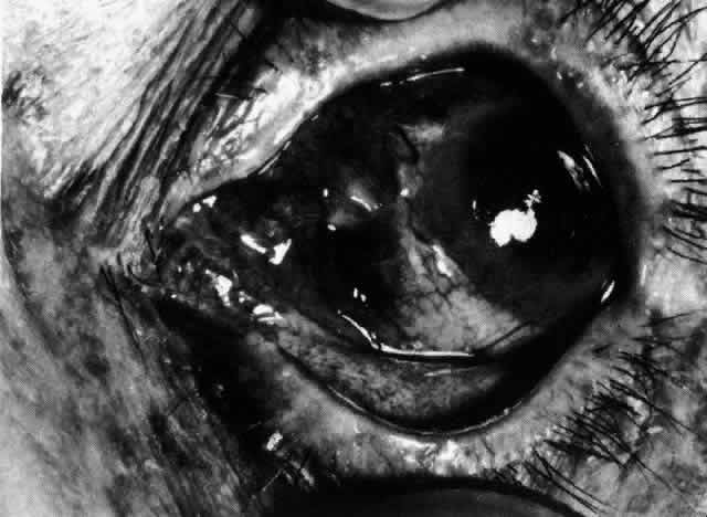
Fig. 67. Severe necrotizing scleritis that started in the wound edges 3 weeks after cataract extraction. The patient had no previous history of eye disease other than involutional cataract. Ill-advised subconjunctival depot steroid led to tissue loss at 8 o'clock. (Watson PG: Management of scleritis. In: Recent Advances in Ophthalmology, Vol 5. London, Churchill-Livingstone, 1975)

Retinal Detachment

Exudative retinal detachment occurs in patients who have posterior scleritis, and it may, indeed, be the only sign in a very painful eye. The detachment is poorly mobile. A pale gray granuloma can be seen extending from the choroid beneath the retina and is accompanied by a poorly mobile serous detachment that may become total. The scleral granuloma sometimes leaves a permanent, inward indentation of the retina and a subretinal mass, although this does not always occur. An increasing hypermetropia has also been noted; it is of rapid onset (over a period of 1 week) and is caused by the diffuse scleral edema in the early stages of the disease before the detachment of the retina occurs.

The exudative detachment usually resolves completely with treatment of the scleritis. However, if the inflammatory changes have affected the macular area, vision will be severely and permanently affected. After resolution, the retina shows a diffuse, heavy pigmentation of the affected area with a “high-water mark” at the edge (see Fig. 52). Patchy changes outside this area do not seem to occur. Surgery is not indicated.

Optic Nerve Swelling

Granulomatous processes inside the muscle cone or affecting the optic nerve sheaths may be accompanied by edema of the optic nerve (see Fig. 51). Although it is not possible to make a diagnosis of posterior scleritis on the basis of this sign alone, should there be severe pain, proptosis, limitation of movement, and a retinal detachment, a presumptive diagnosis is permissible; however, it can be confirmed only if the anterior sclera becomes involved later in the disease. B-scan ultrasonography is very helpful in defining granulomas involving the sclera and the optic nerve. Swelling of the disc in patients who have presented with anterior scleritis is unusual, but it has occurred in patients in whom it was known that the process had advanced to involve the posterior segment.

Back to Top
CORNEAL CHANGES IN EPISCLERITIS AND SCLERITIS
The corneal changes that accompany scleritis are absolutely characteristic, occurring in 37% of patients. Between attacks, when the eye is white, it is possible to say with certainty from the corneal changes that a patient has had scleritis.

Corneal changes of a similar type occur in episcleritis (14%), but they are less severe, always peripheral, and never a source of permanent visual defect, whereas those of scleritis can sometimes cause a loss of vision that is severe enough to require corneal grafting. More than one type of corneal involvement may occur in the same patient, and the classification of such involvement is presented in the following list:

  1. Diffuse scleritis
    1. Acute stromal keratitis
    2. Sclerosing keratitis
    3. Limbal guttering

  2. Nodular scleritis
    1. Localized stromal keratitis
    2. Localized sclerosing keratitis

  3. Necrotizing scleritis
    1. Sclerosing keratitis
    2. Keratolysis

ACUTE STROMAL KERATITIS

The corneal signs may arise simultaneously with the scleritis or occur later in the disease. An acute keratitis will often produce a limbal flush, which has occasionally been confused with a scleral inflammation. In lupus erythematosus, the corneal lesions may appear first, to be followed within 2 to 3 days by a diffuse scleritis.

In the acute stage of the disease, the vision rapidly deteriorates because of a diffuse clouding of the cornea. Superficial and midstromal opacities appear both centrally and around the limbus (Fig. 68). If treatment is not instituted at this stage, vascularization will follow and the opacities will become permanent. However, if treatment is successful, the edema and the opacities can disappear completely.

Fig. 68. Acute stromal keratitis in a patient with acute anterior scleritis. The cornea is edematous and there is an associated anterior uveitis. (Watson PG, Hayreh S, Awdry P: Episcleritis and scleritis. Br J Ophthalmol 52(4):368–369, 1968)

In nodular scleritis, the corneal changes are localized to the area of the inflammation, and should the condition recur, the opacity returns in exactly the same area, often extending farther into the cornea (Color Plate 1F).

SCLEROSING KERATITIS

Sclerosing keratitis is the most common corneal complication of scleral disease. Depending on the type of scleritis, the corneal changes may be diffuse, annular, or localized.

The first change is a diffuse striate appearance affecting the full depth of the cornea that becomes slightly thickened and appears gray. These changes move toward the center of the cornea with surprising speed unless the condition is vigorously treated. Behind the advancing edge, spotty white opacities may develop in the cornea that may become “crystalline” in appearance, looking much like floss candy (Fig. 69). Occasionally, “precipitin rings” may appear around the white corneal opacities. Vascularization, when it occurs, appears to be entirely passive. The vessels show no active budding and follow a long way behind the advancing edge. The vascularization is usually superficial but can occur at any level.

Fig. 69. Sclerokeratitis. A diffuse peripheral change affects the whole corneal stroma at the site of the scleritis. Behind the advancing edge, the corneal lamellae take on a crystalline appearance like floss candy. A “precipitin ring” has formed around one opacity. (Watson PG: Contemporary Ophthalmology. Baltimore, Williams & Wilkins, 1972)

The cornea may become quite white and look like porcelain, but there remains a comparatively clear zone near the limbus. In recurrent nodular scleritis, the changes remain localized to one area.

The change in the corneal stroma results in defective nutrition, so that, if the condition persists, lipid becomes deposited adjacent to the outer edge of the lesion, leaving a yellow opacity in the midstroma (Fig. 70).

Fig. 70. Lipid deposition in an area of sclerokeratitis. Fine floss candy opacities surround this area and appear below the diffuse corneal haze that characterizes the early stages of the condition. (Watson PG, Hayreh S, Awdry P: Episcleritis and scleritis. Br J Ophthalmol 52(4):368–369, 1968)

LIMBAL GUTTERING

Limbal guttering may follow active scleral disease but is not characteristic of scleritis, and when guttering occurs without scleral inflammation in long-standing rheumatoid arthritis, the cornea looks as if it is covered by a corneal contact lens; the center is much thicker than the periphery.

When limbal guttering follows a scleritis, lipid deposition is the rule. Occasionally, a Terrien-like ulcer can appear at the site of a prolonged scleritis, or the deep corneal layers may break, causing aqueous leakage into or through the cornea (see Figs. 65 and 66).

When there is a severe vascular shutdown at the limbus or a severe necrotizing disease of this region, peripheral ulcerative lesions very similar to those seen in Mooren's ulceration may develop. These ulcers are infiltrated and swollen on the central margin, and there is also a marked infiltration at the advancing edge of the peripheral ulcer. The ulcer may be sloping or undermined. The epithelium almost always remains intact, and the base may be vascularized. The limbal arcades adjacent to the advancing edge are disrupted, and the new vessels that arise from the limbal arcades leak into corneal tissue.

KERATOLYSIS

Keratolysis occurs in very severe necrotizing disease or scleromalacia perforans in which the superficial layers of the previously clear cornea melt away, sometimes over areas of several millimeters (Figs. 71 through 74). In very severe cases, descemetoceles form, and although spontaneous rupture is unusual, the cornea is so thin that it can easily be ruptured by minor trauma, in which case the eye may well be lost (see Fig. 73). Treatment is urgent. If medical treatment can be instituted at once, the process will stop and some repair of the cornea will result (see Fig. 74). However, once descemetocele formation has occurred, surgery is the only treatment. Lamellar keratoplasty retains the integrity of the host endothelium and is effective for removing the necrotic tissue. Although multiple grafts may be required, this procedure is the most effective method of controlling the condition. Graft sutures will not hold unless the accompanying scleritis is adequately controlled; therefore, systemic therapy should not be discontinued at the time of surgery.

Fig. 71. Keratolysis associated with severe necrotizing scleritis. The early loss of scleral tissue was at 7 o'clock. There is a gutter from 12 o'clock to 9 o'clock, and the cornea is infiltrated and necrotic. This soft area extends into clinically normal cornea.

Fig. 72. Late fluorescein angiogram in a situation similar to that shown in Figure 71. The conjunctiva has filled in all areas, but the deep vessels are nonperfused in the region of the gutter.

Fig. 73. Keratolysis. Severe necrotizing scleritis with large preferential vessels coursing over the limbal area. The superficial layers of the cornea have melted away, leaving a descemetocele. (Watson PG, Hayreh S, Awdry P: Episcleritis and scleritis. Br J Ophthalmol 52(4):348–349, 1968)

Fig. 74. Eye of the same patient as seen in Figure 73 after 10 days of treatment. The scleral areas of necrosis are filling in, but the descemetocele is still present. This was later covered with a lamellar corneal graft. (Watson PG: Management of scleritis. In: Recent Advances in Ophthalmology, Vol 5. London, Churchill-Livingstone, 1975)

SURGICALLY INDUCED NECROTIZING SCLERITIS (SINS)

Scleritis can occur after any type of ocular surgery in susceptible individuals, particularly those with intermittent systemic connective tissue disease.23–25,31–33 The scleritis is almost always of the necrotizing variety and is often extremely resistant to treatment. It can follow cataract, trabeculectomy, retinal detachment, strabismus, or any type of ocular surgery. The longest interval between the surgery and the scleritis developing at the site of the original surgery has been 24 years. Although the scleritis usually occurs adjacent to the site of surgery, it can, particularly in detachment and strabismus patients, occur at the site of the original surgery. Because this response to a surgical stimulus occurs at a site remote from the surgery, it suggests that the resident tissue macrophage has become the antigen-presenting cell that stimulates the inflammatory response.

COURSE AND PROGNOSIS

The corneal changes that occur in scleritis presumably have the same etiology as the scleral condition and, fortunately, respond to the same treatment. If an acute stromal keratitis is treated vigorously and early permanent changes can be avoided, the disruption of the corneal lamellae in sclerosing keratitis may partly resolve and become less obvious, but some permanent scarring remains. Corneal defects in keratolysis sometimes fill in with steroid therapy but more often leave a thin cornea that is permanently scarred and requires corneal grafting (see Figs. 65 and 66).

Although the majority of patients with necrotizing scleral disease have some other systemic disorder, it is not always clear why the eye becomes involved, nor is it clear whether the scleral inflammation is due to the presence of a particular organism or to immune reactions induced by these organisms. For example, in scleritis associated with herpes zoster, no virus has been demonstrated in the sclera at the time of the primary infection. Nevertheless, these patients have developed a typical scleritis indistinguishable both clinically and cytologically from any other form of noninfectious scleritis.

Scleritis can also be induced by accidental or surgical trauma in susceptible individuals. All forms of scleritis, if left untreated, will leave some scleral defect--increased transparency from changes in collagen and ground substance, actual thinning, or even ectasia. In necrotizing scleritis, the condition is progressive and exceptionally painful and leads to destruction of the eye through the complications it produces.

Decreased visual acuity is not confined to severe posterior or necrotizing disease, as might be expected, but is common in the diffuse and nodular varieties of scleritis. The reasons for this decrease in acuity are cataract, macular changes (often following uveitis), optic disc changes, and, in posterior disease, retinal changes. However, early and persistent treatment is very effective in preserving the eye and vision, but careful monitoring of patients is essential to ensure that the minimum drug therapy is used to control the disorder.

Back to Top
ECTASIA AND STAPHYLOMA
If the sclera gives way without the involvement of uveal tissue, this is known as an ectasia (see Fig. 49). If the uvea is included in the ectasia, this is then termed a staphyloma (see Fig. 47; Fig. 75). In scleritis, the inflammation starts superficially and becomes deep, producing an ectasia. However, only if the intraocular pressure rises above 40 mm Hg does the uvea prolapse into the defect with staphyloma formation. This is a very rare occurrence. Staphylomas following inflammation are strictly localized to the site of inflammation. Staphylomas more commonly form from within outward, stretching and attenuating the normal sclera either because of congenital weakness of its structure or because of prolonged periods of high intraocular pressure associated with intraocular inflammation (see Fig. 75).

Fig. 75. Staphyloma developing at site of necrotizing scleritis after intraocular pressure rose to 50 mm Hg over a 6-week period.

An anterior staphyloma has an abrupt sharp margin with no surrounding inflammation. The scleral thinning is irregular; the numerous ridges on the surface are made more obvious by the dark underlying choroid. Glaucoma is frequently associated with anterior or intercalary staphylomas, and detachment of the retina with equatorial and posterior staphylomas.

Back to Top
CONGENITAL ANOMALIES
Pigmentation of the sclera may be either diffuse or localized to a single discrete patch, and although it is more common in blacks or Asians, it is still seen in whites. Strictly, the pigmentation is not congenital because it appears shortly after birth from migration of chromatophores through the sclera or via the perivascular channels in the anterior sclera. It is not unusual to note a cuff of pigment around the exit of the vessels from the sclera in blacks as a normal phenomenon, and this is particularly noticeable near the limbus. The episclera can also be diffusely pigmented, probably from the downward extension of the pigment cells of the basal epithelium of the conjunctiva (Fig. 76). Very rarely, pigmentation of sclera in white races may indicate an intraocular melanoma, although not necessarily representing an extraocular extension of the malignant disease (Fig. 77).

Fig. 76. Diffuse scleral pigmentation occurring in a white patient who was without any other ocular disease.

Fig. 77. Dilated deep episcleral vessels overlying a malignant melanoma of the choroid. Diffuse scleral pigmentation is seen at exit of main vessel from sclera.

Translucency of the sclera can occur through thinning, dehydration, and reorientation of the collagen structure of the cornea. A diffuse uniform bluish tinge is common in white newborns and is a feature of young children's eyes. The blue color is almost universal in premature infants when the underlying pigment can be seen through the translucent sclera. Blue sclerae persisting beyond the first few months of infancy are pathologic and may indicate either stretching of the anterior segment of the eye, as in congenital glaucoma, or van der Hoeve's syndrome, a condition known to all physicians and seen by few. It is characterized by blue sclerae, fragilitas ossium, deafness, and other mesodermal defects in the skull, teeth, and eye. Changes in the supporting tissues of the joints lead to their subluxation and dislocation. An autosomal dominant, regular inheritance is characteristic: 50% of the members of a family are affected. The etiology, presumed to be an enzyme defect, is unknown, and, consequently, there is no effective treatment. The blue sclerae are due to congenital thinning of the sclera and cornea with an increase in the mucopolysaccharide ground substance, indicating that the collagen tissue has never reached its adult form.

Encroachment of sclera on the cornea is so common, particularly at the upper limbus, that it is difficult to know what is normal and what is to be regarded as abnormal, particularly when searching for pathologic signs in chlamydial infections or vernal conjunctivitis. Gross degrees are not uncommon but fortunately rarely affect vision or require active treatment. The defect is most commonly encountered in microphthalmic eyes or eyes containing other congenital defects, in particular anterior chamber cleavage syndromes or posterior colobomas. Very rarely, the cornea encroaches on the sclera, particularly in Axenfeld's syndrome, giving the appearance of megalocornea or buphthalmos. The intraocular pressure must always be measured in these patients.

A complete failure of condensation of mesoderm in the region of a coloboma of the choroid is rare and probably not compatible with the formation of a functional eye. However, areas of ectasia of the sclera have been seen, particularly around the disc, for these patients the normal disc can be seen at the bottom of a deep, sharp-edged pit or posterior staphyloma. Malformation of the sclera at the posterior pole of the eye may be the reason for the increased axial length found in congenital myopia.

Cartilage is found only in grossly abnormal eyes. This is surprising considering the nature of the sclera and the fact that cartilaginous ocular coats occur in lower animals.

Back to Top
TRAUMA AND CHEMICAL INJURY
The healing of scleral wounds is slow and dependent on the vascular tissue superficial and deep to it for the production of the scar tissue. The ease with which the edges of scleral wounds can be separated even after many years is well known to all ophthalmic surgeons. The sclera, being avascular, cannot heal by primary intention but is repaired from fibrous tissue derived from episclera and choroid. The fibrous scar unites the edges of the sclera; the fibers become reoriented to lie in the same direction as normal sclera. It is important, therefore, that scleral wounds be sutured early and securely and covered by conjunctiva and episclera. The most common source of injury is spectacle or windshield glass, and the rupture is often much larger than it appears at first sight. All such injuries should be examined under anesthesia with the conjunctiva being opened widely to determine the full extent of the laceration.

All detachment surgeons have reason to know how poorly sclera heals; wounds often open years after surgery or open easily when surgery is repeated. Traumatic wounds, therefore, require careful and exact apposition, reposing or excising uveal tissue between the scleral edges. If strong chemicals, whether acid or alkali, come into direct contact with the sclera, they may destroy the collagen, leading to death of the tissue (Fig. 78). However, the scleral tissue rarely sequestrates but remains to form a matrix on which the fibrous scar forms. Being freely permeable, the underlying choroid is subject to more damage, and the overlying episclera may be totally destroyed. If the exposed sclera is covered by viable conjunctiva or mucous membrane, the sclera may retain sufficient integrity for some scar formation to occur. It is impossible to suture into the friable tissue, and the graft must be anchored elsewhere. Severely damaged sclera can be removed and replaced by donor tissue, which again acts as a matrix for scar tissue. However, an eye so severely damaged as to require this procedure usually becomes phthisical.

Fig. 78. Necrosis of conjunctiva and sclera produced by concentrated sulfuric acid. The scleral tissue sequestrated but healed without further complication.

Back to Top
INFLAMMATION
The nonspecific granulomatous inflammations that make up most cases of episcleritis and scleritis have been dealt with in detail. In the review of 1200 patients with scleral disease seen at Moorfields Eye Hospital, a cause for the scleritis was found in 56% of the patients: connective tissue disease accounted for 30% of these, and some other specific cause was found in the others. Often, there is nothing in the clinical presentation that gives a clue to the etiology, which has to be sought by history taking, general examination, or laboratory investigation.

INFECTIVE

Exogenous

Abscess formation must be differentiated from the necrotic areas seen in necrotizing scleritis. An abscess rapidly affects the overlying conjunctiva and episclera, discharging on the surface. When the infecting agent is pyogenic, the surrounding inflammation is intense and is always accompanied by intraocular inflammation (Fig. 79). Mycotic infections and inflammation produced by the caterpillar or plant hairs in ophthalmia nodosa are much more indolent and in the early stages are indistinguishable from a nodular scleritis. However, a history of injury can usually be elicited and the caterpillar hairs readily seen in the cornea if this is the cause (Fig. 80). Treatment with steroids makes the condition worsen rapidly, and should this happen in an apparent nodular scleritis, an infective agent should be suspected. Occasionally, however, and especially in patients who have circulating immune complexes, such an infection will trigger an autoimmune necrotizing scleritis that is independent of the initiating organism and requires steroids for its control.

Fig. 79. Scleral abscess (Staphylococcus aureus). The intensity of inflammation is more severe than that found in scleral disease and is accompanied by fever, pain, and swelling of the preauricular gland.(See also Figures 29, 34, 35, 37, and 39.)

Fig. 80. Scleral granuloma caused by the hairs from the caterpillar of the fox moth (Macrothylacia rubi). Additional hairs were seen in the cornea and anterior chamber. (Watson PG, Sevel D: Ophthalmia nodosa. Br J Ophthalmol 50(4):209, 1966)

Infection around implants used in detachment surgery and stitch abscesses following squint and other scleral surgery are not uncommon, and it is surprising how rarely complications follow. As soon as the offending irritant has been removed, the sclera readily heals without apparent tissue defect. If the irritation is prolonged, a progressive necrotizing scleritis can result. Scleritis from this cause is very persistent and very difficult to treat.

Endogenous

PYOGENIC.

Endogenous pyogenic infections are a rarity, presumably because of the poor blood supply of the sclera. The sclera is so resistant to the pyogenic infection inside the eye that the contents can be eviscerated in patients with panophthalmitis, leaving no infected areas. When, however, a pyogenic infection does affect the anterior sclera, a discharging abscess forms, accompanied by severe intraocular inflammation and occasionally hypopyon. Infection of the posterior sclera is far more difficult to diagnose. The inflammation and pain are intense, the vision invariably reduced, and the eye proptosed, sometimes giving the impression of an orbital tumor. However, very frequently the patient is feverish, the preauricular gland is enlarged and tender, and the lids are swollen. This is a situation that is never found in granulomatous posterior scleritis. Early diagnosis is essential if effective treatment is to be instituted. Provided any abscess is opened and the appropriate antibiotic is given, the results are good. Patients with scleral abscesses that have perforated the wall of the globe with loss of some of the intraocular contents have been known to recover full vision.34

Other organisms known to cause scleral abscesses are Pseudomonas aeruginosa, the streptococci, and Proteus. Fungal infections with Aspergillus in particular are not uncommon, and Acanthamoeba can invade the sclera in a severe infection. In all these instances there is difficulty in deciding whether this is a localized abscess or a severe noninfective scleritis because it is rare for there to be any purulent discharge. It is important to know that infections of the sclera can and do occur.

SPECIFIC GRANULOMATOUS CONDITIONS.

Whereas pyogenic infections are uncommon, the nonpyogenic granulomatous conditions are common enough to be looked for routinely, particularly because their presentation is no different from that of nonspecific granulomatous scleritis. Although there may be other signs of these infections in the uveal tract, this is by no means universal. Both syphilis and tuberculosis can present with a scleritis. None of the patients we have seen have had any other ocular signs or general symptoms (Fig. 81). The patients with syphilis presented with a diffuse anterior scleritis, and those with tuberculosis presented with a nodular scleritis (see Fig. 34). Caseation is unusual, but perforation is said to occur.35,36 Treatment is with the appropriate chemotherapy and antibiotic, followed by an intensive local steroid regimen to suppress the local inflammatory response.

Fig. 81. Tertiary syphilis. A gummatous ulcer of the skin of the leg in a patient who presented with diffuse anterior scleritis.

VIRAL. HERPES SIMPLEX.

Herpes simplex sclerokeratitis or episcleritis is uncommon, although some patients with recurrent episcleritis will often find that the episcleral inflammation occurs at the same time as the skin eruptions (see Fig. 8). It is difficult to isolate the virus from the conjunctiva or episclera at the time, although Foster and colleagues have isolated the virus and have detected viral antigen in the sclera in patients with keratoscleritis.8

Herpes Zoster.

The pattern of disease in herpes zoster infection is both interesting and instructive. In the stage before the eruption, the patient sometimes presents with intense pain in the eye and episcleritis. The diagnosis soon becomes apparent; vesicles can form on the conjunctiva, and these can involve the episclera, but more commonly a nodular episcleritis occurs that resolves in 3 to 4 weeks with no residual signs. After a variable period of 1 to 4 months, the patient sometimes presents again with a typical nodular scleritis that may be necrotizing (see Fig. 35). This nodule is very resistant to treatment and may take months to resolve, frequently leaving an area of thin sclera at the site of the nodule. Recurrences in the same site are not infrequent. These observations indicate that the autoimmune scleritis has been triggered by the virus and that the recurrent inflammation is thereafter independent of the presence of the virus.

NONSPECIFIC GRANULOMATOUS CONDITIONS.

The endogenous varieties of nonspecific granulomatous episcleritis and scleritis have been dealt with in detail, but it must be remembered that the same physical signs can be produced by an exogenous cause. Exposure to certain industrial solvents produces an instant reddening of the eye and episcleritis in some patients. The sclera may become involved in an orbital granulomatous process or may be involved in the granulomas of sympathetic ophthalmia or severe posterior uveitis, particularly in Vogt-Koyanagi-Harada syndrome.

METABOLIC

Gout

Gout is a defect of purine metabolism causing a rise in serum uric acid and deposition of sodium urate in the tissues. Episcleritis or scleritis may be the first or only clinical manifestation of gout. Of 15 patients discovered to have a persistently raised serum uric acid on routine testing, 9 had other clinical signs suggestive of gout. Of the others, 2 have developed symptoms that responded to specific therapy within 3 years of their presenting with an episcleritis or scleritis. The episcleritis or scleritis is not in any way diagnostic of the condition, but occasionally what appear to be very fine crystals can be detected in the episclera close to the vessels. These crystals are longer and thinner than the fine globules often seen beside the vessels overlying the rectus muscles. Because no biopsy specimen of these crystals has yet been obtained, their significance remains “sub judice,” but if they are seen in the presence of an episcleritis, a serum uric acid should be performed and, if raised, should be repeated. Gouty tophi have been described by McWilliams37 and Wood38 but are exceptionally rare.

Cystinosis

This defect of amino acid metabolism gives rise to deposition of cystine in the tissues. Although cystine crystals can be seen in the episclera, superficial scleral tissues, and cornea, they do not give rise to any inflammatory changes.

Alkaptonuria

Alkaptonuria is a defect of homogentisic acid metabolism giving rise to melanin deposition in the tissues. The pigmentation of the sclera that occurs in this condition is patchy but involves the areas exposed to light so that the dark areas of the sclera have a triangular pattern with a base at the limbus on the inner and outer sides. The pigmentation increases with age.

Porphyria

This defect of blood and bile pigment metabolism gives rise to secretion of porphyrin or porphobilinogen. Two distinct types exist: a photosensitive type occurring in childhood and an acute intermittent form presenting in the second or third decade. A form of scleromalacia perforans is described39 in which areas of superficial sclera become eroded, leaving the appearance of a scleral hyaline plaque over a large area with calcareous degeneration in the adjacent area (Fig. 82). The destruction is rarely sufficient to cause visual defect. The only patient in the correct age group who might have fit this description did not have porphyria or any other metabolic defect.

Fig. 82. Erosion of the superficial sclera appearing much like a hyaline plaque in a 30-year-old South African with congenital porphyria. (Courtesy of Dr. W. Douglas)

Xanthomatosis

In xanthomatosis, defects of fat metabolism lead to the deposition of lipids in the tissues. Yellow-white deposits are not infrequently seen in the episcleral tissues of normal individuals with no abnormality of their lipid metabolism, but scleral deposits are most unusual, and, if seen, a full serum lipid investigation should be undertaken. Scleral deposits have been seen in patients with essential hypercholesterolemic xanthomatosis, xanthoma dissemination, Hand-Schüller-Christian disease, and Letterer-Siwe disease.

Hypercalcemia

This defect is due to the imbalance of calcium and phosphorous metabolism giving rise to excess deposition of calcium in the tissues. Although the major changes occur in the cornea, calcium is also deposited in scleral tissue as an extension of the band keratopathy usually seen in hyperparathyroidism, sarcoidosis, hypervitaminosis D, and Labrador keratopathy.40 The appearances are of white translucent plaques that seem to separate the scleral lamellae. Histologically, these areas of calcification can be seen in all layers of the sclera, particularly near the ciliary body.

Back to Top
DEGENERATIONS
Lipid and calcareous degenerations are largely responsible for the “rheumy” eye of old age. Not only is there a reduction of greasy components and volume of the tear flow, but there is also a loss of episcleral tissue. These factors lead to a lack of luster of the conjunctiva, but even more subtly there is a change in the lipid content of the sclera. Lipid becomes deposited between the fibers (particularly those of the deep layers in middle age), producing a yellowish tinge. This color change deepens with advancing age, and white flecks appear among the yellow as calcium in its turn becomes deposited.

Calcareous degeneration may also follow severe inflammation, particularly if this has occurred near the recti muscles. Amyloid degeneration has also been described following severe scleral inflammation.39

SENILE HYALINE PLAQUE

Senile hyaline plaques are a common but rarely noticed scleral change. They are almost always found between the insertion of the lateral or medial rectus muscles and the limbus. The sclera becomes translucent and slightly depressed over a small area; the overlying episcleral tissue remains normal. The periphery of the hyaline ring is often hard and white and calcareous (Fig. 83). These plaques are caused by the tug of the recti muscle insertion on the scleral fibers; however, they never cause any trouble and require no treatment.

Fig. 83. Scleral hyaline plaque. This is a common anomaly in the sclera in which a hyaline ring is surrounded by an area of increased translucency of the sclera. No treatment is required.

SPONTANEOUS INTERCALARY PERFORATION

A spontaneous intercalary perforation describes a small well-defined hole that appears in the sclera of young individuals without any signs surrounding the inflammation. Initially, the hole grows slightly in size and then remains stationary. Sometimes fluid is driven through the hole subconjunctivally and choroidal tissue becomes incarcerated in it. A small vessel appears to run through the defect to anastomose with the posterior ciliary circulation. These small communicating vessels are not uncommonly seen in normal individuals, sometimes running in the same channels as the aqueous veins. It is at these sites that spontaneous intercalary perforations occur. The perforations are entirely benign and nonprogressive expansions of the normal perforating channels. They require no treatment.

Back to Top
TUMORS
Tumors of the sclera do not occur. However, the sclera and the overlying episclera can be involved by neoplastic changes within and outside the eye. When tumors occur in conjunctiva and involve episclera, they form part of the differential diagnosis of episcleritis and scleritis. The technique for determining the nature and the site of the inflammation in scleral and episcleral tissues has been dealt with in detail, but nowhere is it more important to ascertain the exact site than where a tumor is suspected.

The sclera forms a barrier for the spread of intraocular tumors, but the vascular and nervous intrascleral canals form a channel through which tumor cells can extend into the episcleral tissues. In certain instances, particularly in intraocular malignant melanomas, myelomas, and sarcoidosis, a vascular response is excited in the deep episcleral tissues with dilatation, tortuosity, and newly formed vessels (see Fig. 77). Deposits in episcleral tissue and tumors that arise from episclera often cause difficulty in diagnosis.

The localized epithelial tumors, such as the intraepithelial epithelioma (Bowen's disease), present little difficulty because the deep tissues are rarely involved and the tumor ulcerates onto the surface (Fig. 84). The pigmented tumors also do not cause any great difficulty in diagnosis, but occasionally a nonpigmented nevus of the episclera of conjunctiva occurs. In the initial stages, these form a diffuse infiltrated lesion of the episclera usually very close to the limbus. As time passes, new vessels pass to the lesion, which may remain the same size or grow slowly (Fig. 85). Should malignant change occur within the lesion, the surrounding tissues will become inflamed and the feeding vessels greatly enlarged (Fig. 86). In necrotizing nodular scleritis, the large vessels adjacent to the inflammation enlarge but do not pass into the lesion itself, so the engorged vessels appear to skirt the nodule (see Figs. 27 and 28).

Fig. 84. Bowen's disease. Intraepithelial epithelioma at the limbus in a 75-year-old man. This patient was originally diagnosed as having an episcleral nodule at the limbus.

Fig. 85. Nonpigmented nevus involving the episcleral tissues. This lesion had been present for 15 years and has not altered in 4 years. The vessels are larger leading to and from the lesion, but their size has not altered.

Fig. 86. Malignant change in a subconjunctival nevus that occurred over a 6-month period. The mass became deeply pigmented and the vessels became engorged and altered so as to radiate from the mass.

In the early stages of the vascular tumors, all that can be seen is a dilatation of some of the vessels of the deep episcleral network (Fig. 87). In some congenital varieties, these anomalies can be extensive, but, as in the skin hemangiomas, they regress with age. However, from time to time, angiomas arise within the episclera; they resemble an amelanotic melanoma and sometimes cannot be distinguished except by biopsy (Fig. 88). Here again, the new vessels that form in the tumor itself and in response to its presence appear to radiate from the mass rather than skirt it, as in the inflammatory conditions.

Fig. 87. Spontaneous hemorrhage from a small hemangioma of the episcleral vessels in a 12-year-old boy.

Fig. 88. Episcleral angioma. This lesion in a 24-year-old woman had been present since puberty and had not altered in size. From time to time it became very injected and was removed for cosmetic reasons. Histology confirmed the diagnosis.

Lymphomatous infiltrates of the episclera present a real diagnostic problem. These patients may present with an apparent episcleritis. However, several features are valuable in helping to distinguish the reticulosis from the inflammation change (Fig. 89):

Fig. 89. Lymphomatous infiltration of the episcleral tissue. The color was a dull red and the edges ill defined. The infiltrative lesion could be easily identified with the red-free light on the slit lamp.

  1. Color. The color of the lymphoma is a dullish red in contrast to the brick red of the episcleritis and the blue red of the scleritis. The color differences are particularly noticeable in daylight.
  2. Edges. No posterior defined limit can be found for a lymphoma. Almost all patients with episcleritis and scleritis have a readily definable margin to the inflammation.
  3. Infiltration. Lymphomatous infiltration can be seen in the episcleral tissue when the patient is examined with the slit lamp by use of red-free light in which the infiltrated area shows up yellow.
  4. The size of the area grows with time rather than regressing spontaneously.

Secondary carcinomatous spread into the episcleral tissue is rare but does occur. In young patients, a dermolipoma can be confused with an episcleral infiltrate of a more serious nature (Fig. 90). The child in Figure 91 was originally referred with a nodular episcleritis, but within 2 weeks the mass, which proved to be a rhabdomyosarcoma, had doubled in size. The orbit was exenterated immediately after radiation therapy; the child is still well and free from recurrence 12 years later.

Fig. 90. Dermolipoma that appeared to be entirely confined to the episclera, causing doubt as to its etiology.

Fig. 91. Rhabdomyosarcoma that had taken only 2 weeks to grow and was originally thought to be an episcleral nodule.Color

Back to Top
REFERENCES

1. Yomai Y, Ushiki T: The three-dimensional organization of collagen fibrils in the human cornea and sclera. Invest Ophthalmol Vis Sci 32:2244, 1991

2. Ozanics F, Rayborn M, Saun D: Some aspects of corneal and scleral differentiation of the primate. Exp Eye Res 22:305, 1976

3. Morrison JC, Van Buskirk M: Anterior collateral circulation in the primate eye. Ophthalmology 90:707, 1983

4. Meyer P, Watson PG: Low dose fluorescein angiography of the conjunctiva and episclera. Br J Ophthalmol 71:2, 1987

5. Meyer PA: Pattern of flow in episcleral vessels studied by low dose fluorescein video angiography. Eye 2:533, 1988

6. Watson PG, Hazleman BL: The Sclera and Systemic Disorders, pp 155–207. London, Philadelphia, WB Saunders, 1976

7. Watson PG, Hayreh SS: Scleritis and episcleritis. Br J Ophthalmol 60:163, 1976

8. Foster S, Sainz de la Maza M: The Sclera, pp 100–101. New York, Springer-Verlag, 1993

9. Lyons CJ, Hakin KN, Watson PG: Topical flurbiprofen: An effective treatment for episcleritis? Eye 4:521, 1990

10. Tuft SJ, Watson PG: Progression of scleral disease. Ophthalmology 98:467, 1991

11. McGavin DDM, Williamson J, Forrester JV et al: Episcleritis and scleritis: A study of their clinical manifestations and association with rheumatoid arthritis. Br J Ophthalmol 60:192, 1976

12. Lyne AJ, Pitkeathley DA: Episcleritis and scleritis: Association with connective tissue disease. Arch Ophthalmol 80:171, 1968

13. Sevel D: Necrogranulomatous scleritis: Clinical and histological features. Am J Ophthalmol 64:1125, 1967

14. Fowler PD: Rheumatic disease in patients suffering from scleral disease. Ann Rheum Dis 29:195, 1970

15. Kostenitsch J: Uber einen Fall von Skleritis; pathologische-anatomische Untersuchung. Arch Augenheilkd 28:27, 1894

16. Verhoeff FH, King MJ: Scleromalacia perforans: A report of a case in which the eye was examined microscopically. Arch Ophthalmol 20:1013, 1938

17. Ashton N, Hobbs HE: Effect of cortisone on rheumatoid nodules of the sclera (scleromalacia perforans). Br J Ophthalmol 36:373, 1952

18. Ferry AP: Histopathology of rheumatoid episcleral nodules: An extra-articular manifestation of rheumatoid arthritis. Arch Ophthalmol 82:77, 1969

19. Fraunfelder FT, Watson PG: Evaluation of eyes enucleated for scleritis. Br J Ophthalmol 60:227, 1976

20. Watson PG, Young RD: Changes at the periphery of a lesion in necrotising scleritis: Anterior segment fluorescein angiography correlated with electron microscopy. Br J Ophthalmol 69:656, 1985

21. Sainz de la Maza M, Tourber J, Foster CS: Scleral grafting for necrotizing scleritis. Ophthalmology 96:306, 1989

22. Fong LP, Sainz de la Maza M, Rice BA, Foster CS: Immunopathology of scleritis. Ophthalmology 98:472, 1991

23. Rao NA, Marak GE, Hydayat AA: Necrotizing scleritis. A clinicopathologic study of 41 cases. Ophthalmology 98:472, 1991

24. Lyne AJ, Lloyd-Jones D: Necrotising scleritis after ocular surgery. Trans Ophthalmol Soc UK 99:146, 1979

25. Sainz de la Maza M, Foster CS: Necrotizing scleritis after ocular surgery: A clinicopathological study. Ophthalmology 98:1720, 1991

26. Watson PG, Bovey E: Anterior segment fluorescein angiography in the diagnosis of scleral inflammation. Ophthalmology 92:1, 1985

27. Wilhelmus K, Grierson I, Watson PG: Histopathologic and clinical associations of scleritis and glaucoma. Am J Ophthalmol 91:697, 1981

28. Calthorpe CM, Watson PG, McCartney ACE: Posterior scleritis: A clinical and histological survey. Eye 2:267, 1988

29. Benson WE: Posterior scleritis. Surv Ophthalmol 32:297, 1988

30. Bloomfield SE, Becker CG, Christian CL et al: Bilateral necrotising scleritis with marginal corneal ulceration after cataract surgery in a patient with vasculitis. Br J Ophthalmol 64:170, 1980

31. O'Donaghue E, Lightman S, Tuft S, Watson PG: Surgically induced necrotising sclerokeratitis (S.I.N.S.): Precipitating factors and response to treatment. Br J Ophthalmol 76:17, 1992

32. Scott JA, Clearkin LG: Surgically induced diffuse scleritis following cataract surgery. Eye 8:292, 1994

33. Salamon SM, Mondino BJ, Zaidman GW: Peripheral corneal ulcers, conjunctival ulcers, and scleritis after cataract surgery. Am J Ophthalmol 93:334, 1982

34. Hudson AC: Staphylococcal abscess of the sclerotic. Proc R Soc Med 17:148, 1934

35. Duke-Elder S: System of Ophthalmology, Vol 8. Diseases of the Outer Eye: Conjunctiva, Cornea, and Sclera. St. Louis, CV Mosby, 1965

36. Bonnet P: Scleritis symptomatic of miliary granulomas in the choroid. Bull Soc Ophtalmol Fr 2:144, 1952

37. McWilliams JR: Ocular findings in gout. Am J Ophthalmol 35:1778, 1952

38. Wood DJ: Inflammatory disease in the eye caused by gout. Br J Ophthalmol 20:510, 1936

39. Coats G: Diseases of the sclera. Trans Ophthalmol Soc UK 35:257, 1915

40. Freedman A: Labrador keratopathy. Arch Ophthalmol 74:198, 1965

Back to Top