|
|
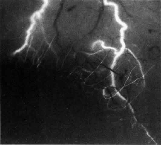
| Fig. 3. Normal superior anterior segment angiography in a 35-year-old man. The main anterior ciliary arteries have filled, branching at or adjacent to the limbus to fill the limbal arcade, the recurrent branches of which are passing backward to fill the episcleral and conjunctival vessels. The episcleral circle here is superficial, and the anastomotic vessels are readily visible. There is a close resemblance to the vascular pattern in Figure 1, including the penetrating vessels adjacent to the limbus. |