|
|
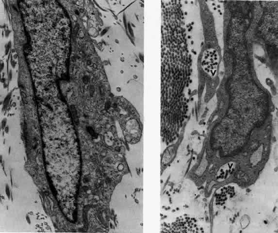
| Fig. 23. Electron micrographs of scleral stroma at the periphery of an area of ulceration in a patient with necrotizing scleritis. The left shows an active fibroblastic cell, and the right shows collagen fibrils within intracellular vacuoles (V) in the fibroblastic cell. (Left X15,375; right X15,375) (Watson PG, Young RD: Changes at the periphery of a lesion necrotizing scleritis: Anterior segment fluorescein angiography correlated with electron microscopy. Br J Ophthalmol 68:781–789, 1984) |