|
|
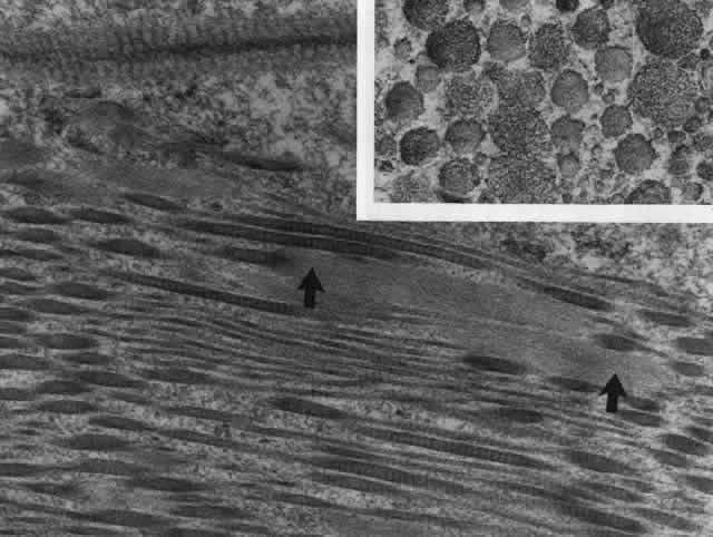
| Fig. 24. Electron micrograph of scleral stroma at the periphery of an ulcer in necrotizing scleritis (same patient as in Figure 23) showing swelling and unraveling of collagen fibrils (arrows) in longitudinal section (X29,270) and in transverse section (inset, X44,000). Fibrils of all diameters are affected. (Watson PG, Young RD: Changes at the periphery of a lesion necrotizing scleritis: Anterior segment fluorescein angiography correlated with electron microscopy. Br J Ophthalmol 69:656–663, 1985) |