|
|
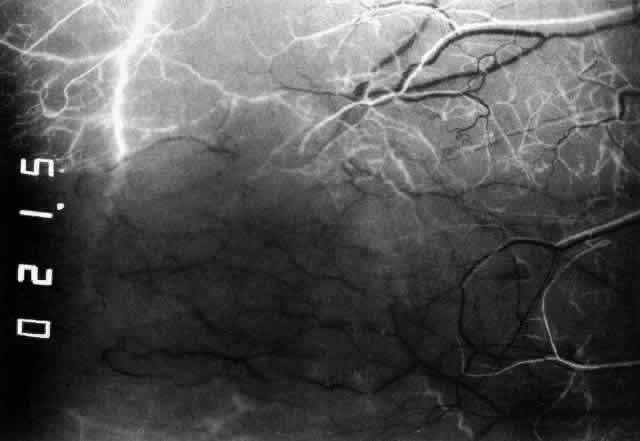
| Fig. 4. Normal temporal angiogram of a 32-year-old woman. Fluorescein first enters the anterior ciliary artery above (as in Figure 3), filling the limbal arcade, and then dips deep into the sclera adjacent to the limbus; it can just be distinguished in the deep scleral tissue adjacent to the limbus. The posterior tarsal circulation can be seen filling at the same stage. |