Chapter 30
Diabetic Retinopathy
BRETT J. ROSENBLATT and WILLIAM E. BENSON
Main Menu   Table Of Contents

Search

CLINICAL FINDINGS
RISK FACTORS OF DIABETIC RETINOPATHY
PATHOGENESIS
PSYCHOPHYSICAL AND ELECTROPHYSIOLOGIC ABNORMALITIES
DIFFERENTIAL DIAGNOSIS
TREATMENT
OTHER OCULAR COMPLICATIONS OF DIABETES MELLITUS
REFERENCES

CLINICAL FINDINGS
Diabetic retinopathy is usually divided into nonproliferative diabetic retinopathy (NPDR) and proliferative diabetic retinopathy (PDR).

EARLY NONPROLIFERATIVE DIABETIC RETINOPATHY

Although microaneurysms are the first ophthalmoscopically detectable change in diabetic retinopathy, the earliest abnormalities seen histopathologically are thickening of the capillary basement membrane1,2 and pericyte dropout.3,4 Pericytes are mesothelial cells that surround and support the retinal capillary endothelial cells. Normally there is one pericyte per endothelial cell. In people with diabetes, however, the pericytes die off and are decreased in number (Fig. 1). Their absence weakens the capillaries and permits thin-walled dilatations, called microaneurysms, to develop. Later, endothelial cells proliferate and lay down layers of basement membrane material. Fibrin may accumulate within the microaneurysm along with erythrocytes, and the lumen of the microaneurysm may become occluded (Fig. 2). Initially, most microaneurysms are on the venous side of the capillaries, but later they are seen on the arterial side as well. Clinically, they appear as small red dots (Fig. 3). Despite the multiple layers of basement membrane, microaneurysms are permeable to water and large molecules, allowing the transudation of fluid and lipid into the retina.

Fig. 1 A. Trypsin digest preparation of early background retinopathy. Normal retinal capillaries, with one pericyte (closed arrows) per endothelial cell (open arrows). B. Retinal capillary of a patient with diabetes with necrotic pericytes (arrows). (Courtesy of Dr. Myron Yanoff)

Fig. 2 Trypsin digest preparation of early background retinopathy. Early microaneurysm (closed arrow), aneurysm with endothelial proliferation (open arrow), and aneurysm occluded with fibrin (curved arrow). (Courtesy of Dr. Myron Yanoff)

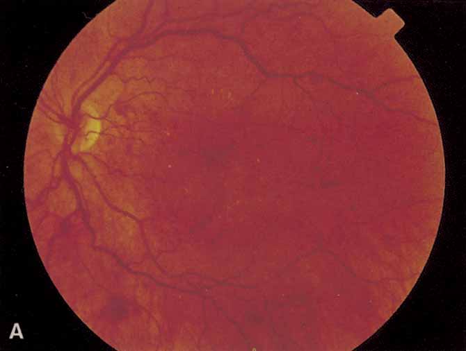
Fig. 3 A. Diabetic retinopathy with multiple microaneurysms, dot hemorrhages, and early neovascularization of the optic disc (NVD). A small blot hemorrhage is seen inferiorly. B. Continued. Midphase of the fluorescein angiogram. Patent microaneurysms are seen as hyperfluorescent dots. Note that most microaneurysms cannot be seen ophthalmoscopically. There is some enlargement of the foveal avascular zone because of some occluded capillaries. Temporally there is a larger zone of capillary nonperfusion. The NVD is beginning to leak. C. Late phase of the fluorescein angiogram showing diffuse leakage of fluorescein into the macula.

It is often difficult to distinguish a small dot hemorrhage from a microaneurysm by ophthalmoscopy alone. On fluorescein angiography patent microaneurysms will fill with dye quickly and then leak,5 unlike a small dot hemorrhage that will block fluorescence (see Fig. 3). However, angiography cannot distinguish a hemorrhage from a microaneurysm filled with clotted blood. Because fluorescein passes easily though them, we usually see many more microaneurysms on fluorescein angiography than are apparent on examination.6

When the wall of a capillary or microaneurysm is thin, it may rupture, giving rise to an intraretinal hemorrhage. If the hemorrhage is deep (i.e., in the inner nuclear layer or outer plexiform layer), it usually has a round or oval shape (“dot or blot”) (see Fig. 3). Superficial (nerve fiber layer) hemorrhages, on the other hand, become flame- or splinter-shaped indistinguishable from that seen in hypertensive retinopathy. Although people with diabetes with normal blood pressure may have multiple splinter hemorrhages, they should nevertheless have their blood pressure checked because a frequent complication of diabetes is systemic hypertension.

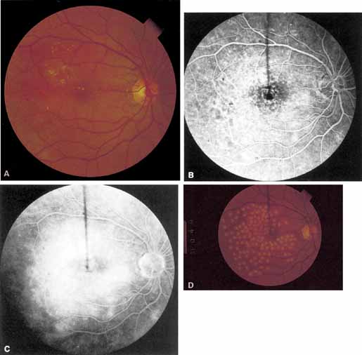
Macular edema (retinal thickening) is an important manifestation of NPDR because it is the leading cause of legal blindness in patients with diabetes. The intercellular fluid comes from both leaking microaneurysms and diffuse capillary leakage. It separates retinal cells, causing multiple intraretinal interfaces which scatter light, decreasing the retina's normal translucency and blurring the normal retinal pigment epithelial and choroidal background pattern (Figs. 3, 4, and 5). Clinically, macular edema is detected by biomicroscopy with a contact lens or by a 60- or 80-diopter hand-held lens. Optical coherence tomography (OCT) is a new diagnostic tool that accurately defines retinal thickness and cross-sectional anatomy (see Fig. 5). In severe cases of edema, the pockets of fluid in the outer plexiform layer are large enough to be seen. This is called cystoid macular edema (CME) (Fig. 6). Usually CME is seen in eyes with other signs of severe NPDR such as numerous hemorrhages or hard exudates, but in rare cases, generalized diffuse leakage from the entire capillary network can result in CME with few other signs of diabetic retinopathy.7

Fig. 4 A Exudates surround an area of hypoperfused retina. Note soft exudate superiorly. The macular edema thickens the retina and obscures the normal choroidal appearance. B. In the center of the hard exudates the fluorescein angiogram shows capillary non perfusion surrounded by microaneurysms. C. The late phase of the angiogram shows leakage into the retina.

Fig. 5 A. Circinate retinopathy inferotemporal to the center of the macula. B. The midphase of the fluorescein angiogram shows a cluster of microaneurysms in the center of the circinate ring. C. The late phase of the angiogram shows leakage of fluorescein. D. Optical coherence tomogram centered on the fovea of an eye with diabetic macular edema. The area of marked retina thickening contains numerous hyporeflective cystoid spaces (fine arrow). The outer retina is limited by the hyperreflective pigment epithelial band (thick arrow).

Fig. 6 A. Background diabetic retinopathy. B. The midphase of the fluorescein angiogram shows multiple microaneurysms. C. Late phase of the angiogram shows cystoid macular edema.

If the leakage of fluid is severe enough, lipid may accumulate in the retina (see Figs. 4 and 5). Again, the outer plexiform layer is first to be affected. In some cases, lipid is scattered through the macula, in others, it accumulates in a ring around a group of leaking microaneurysms, or around areas of capillary nonperfusion (see Figs. 4 and 5). This pattern is called circinate retinopathy.

In addition to the retinal vascular abnormalities, the choriocapillaris may be involved in NPDR. Initially there is a thickening of the basement membrane.8 Later, a periodic acid-Schiff (PAS)–positive material accumulates, which impinges on and may occlude the lumen of the choroidal capillaries in the posterior pole.4

ADVANCED NONPROLIFERATIVE DIABETIC RETINOPATHY

In advanced NPDR, signs of increasing retinal hypoxia appear, including multiple retinal hemorrhages, cotton-wool spots (Fig. 7), venous beading and loops (Figs. 7 and 8), intraretinal microvascular abnormalities (IRMA) (see Figs. 7 and 8), and large areas of capillary nonperfusion.

Fig. 7 A. Severe nonproliferative retinopathy with venous dilatation and beading, soft exudates, and intraretinal microvascular abnormalities B. The midphase of the angiogram shows the intraretinal microvascular abnormalities (IRMA) and severe capillary nonperfusion.

Fig. 8 Venous loop (large arrow) and intraretinal microvascular abnormalities (IRMA; small arrow).

Cotton-wool spots, also referred to as soft exudates, are actually nerve fiber layer infarctions. They are white, fluffy-appearing lesions in the nerve fiber layer that result from occlusion of precapillary arterioles. Fluorescein angiography confirms the lack of capillary perfusion. Microaneurysms frequently surround older cotton-wool spots as well as larger areas of capillary nonperfusion.

Venous beading (see Fig. 7) and venous loops (see Fig. 8) indicates sluggish retinal circulation and are nearly always adjacent to extensive areas of capillary nonperfusion. Focal vitreous traction is thought to contribute to their formation.9 Capillaries next to areas of nonperfusion that dilate and function as collaterals are referred to as IRMA. They are frequently difficult to differentiate from surface retinal neovascularization. Fluorescein, however, does not leak from IRMA but leaks profusely from neovascularization (see Fig. 7).

The Early Treatment Diabetic Retinopathy Study (ETDRS)10 found that IRMA, multiple retinal hemorrhages, venous beading and loops, widespread capillary nonperfusion, and widespread leakage on fluorescein angiography are all significant risk factors for the development of proliferative retinopathy. Interestingly, cotton-wool spots, in the absence of the other findings, are not. Approximately 50% of patients with severe NPDR progress to proliferative retinopathy with high-risk characteristics (discussed later) within 1 year.11

PROLIFERATIVE DIABETIC RETINOPATHY

Proliferative vessels usually arise from veins and often begin as a collection of fine vessels. When they arise on or within 1 disc diameter of the optic disc they are referred to as neovascularization of the disc (NVD) (Fig. 9). When they arise further than 1 disc diameter away, they are called neovascularization elsewhere (NVE) (Fig. 10). NVE nearly always grows toward and into zones of retinal capillary nonperfusion, but capillary nonperfusion is nearly always more widespread in eyes with NVD than it is in NVE.12 Interestingly, it is seen more often in patients younger than 40 compared to older patients with diabetes.13

Fig. 9 Advanced neovascularization of the disc.

Fig. 10 Neovascularization elsewhere (NVE).

Once the stimulus for growth of new vessels is present the vessels grow along the path of least resistance. The absence of the internal limiting membrane over the optic disc could explain the proclivity of new vessel growth at that location. Neovascularization grows readily along connective tissue scaffolding such as the posterior hyaloidal face (Fig. 11).

Fig. 11 Autopsy eye. Neovascular stalk adherent to and growing on the posterior cortical vitreous which has partially detached. (Courtesy of Dr. Myron Yanoff)

The new vessels, initially naked, usually progress through a stage of further proliferation with associated connective tissue formation. As PDR progresses, the fibrous component becomes more prominent. Fibrotic tissue can be vascular or avascular. The fibrovascular variety is usually found in association with vessels extending into the vitreous cavity or with abnormal new vessels on the surface of the retina or disc. The avascular variety usually results from organization or thickening of the posterior hyaloid face.

Posterior vitreous detachment in diabetics is characterized by a slow, overall shrinkage of the entire formed vitreous rather than by the formation of cavities caused by vitreous destruction.14 Davis15 has stressed the role of the contracting vitreous in the production of vitreous hemorrhage, retinal breaks, and retinal detachment. Neovascular vessels do not “grow” forward into the vitreous cavity; they are pulled into it by the contracting vitreous to which they are adherent (see Fig. 11). Vitrectomized eyes rarely develop new areas of neovascularization and existent neovascularization tends to regress. If severe enough, posterior vitreous detachment may result in retinoschisis, retinal detachment, and retinal break formation. In eyes fortunate enough not to develop these complications, the neovascularization may burn out, leading to atrophy of the new vessels.

An additional complication of contracting vitreous is traction involving the optic nerve that causes stria of the macula or even macular heterotopia (Fig. 13)20; both may cause decreased visual acuity.

Fig. 13 A. Macula prior to dragging. Arrows have been placed for reference. B. Two years later. Note the foveola and inferior vessels have been dragged superonasally.

Although the macular edema, exudates, and capillary occlusions seen in NPDR often cause legal blindness, affected patients usually maintain at least ambulatory vision. PDR, on the other hand, often results in severe vitreous hemorrhage or retinal detachment with hand-movements vision or worse. It has long been assumed that sudden vitreous contractions tear the fragile new vessels, causing vitreous hemorrhage. However, 62% to 83% of diabetic vitreous hemorrhages occur during sleep,16,17 possibly because of an increase in blood pressure secondary to early morning hypoglycemia or to rapid eye movement (REM) sleep. Because so few hemorrhages occur during exercise, we do not restrict the activity of patients with proliferative retinopathy. The location of the hemorrhage is important in predicting whether it will clear on its own. If blood is behind the posterior vitreous face it is likely to settle to the bottom of the eye and be absorbed. However, when hemorrhage breaks into formed vitreous it is less likely to clear spontaneously.

A large superficial hemorrhage may separate the internal limiting membrane from the rest of the retina. Such hemorrhages usually are round or oval but may also be boat-shaped (Fig. 12). The blood may remain confined between the internal limiting membrane and the underlying retina for weeks or months before breaking into the vitreous. Subinternal limiting membrane hemorrhages were formerly thought to lie between the internal limiting membrane and the cortical vitreous and were called subhyaloid or preretinal hemorrhages. It is now felt that true subhyaloid hemorrhages are probably quite rare. Tight subinternal limiting membrane hemorrhages are dangerous beause they may progress to traction retinal detachment.18,19

Fig. 12 Subinternal limiting membrane hemorrhages.

Two types of diabetic retinal detachments occur, those that are caused by traction alone (nonrhegmatogenous) (Fig. 14), and those caused by retinal break formation (rhegmatogenous) (Figs. 15 and 16). Characteristics of nonrhegmatogenous (traction) detachment in PDR include the following: (1) the detached retina is usually confined to the posterior fundus and infrequently extends more than two-thirds of the distancd to the equator, (2) it has a taut and shiny surface, (3) it is concave toward the pupil, and (4) there is no shifting of subretinal fluid.

Fig. 14 Traction retinal detachment. The detached retina has a smooth noncorrugated appearance and is convex toward the pupil.

Fig. 15 Combined traction/rhegmatogenous retinal detachment. The detached retina has a corrugated appearance and is concave toward the pupil.

Fig. 16 Round hole near fibrous proliferation.

Vitreous traction may also cause focal areas of retinoschisis that may be difficult to distinguish from full-thickness traction retinal detachment. In retinoschisis the elevated layer is thinner and more translucent (Fig. 17).

Fig. 17 Traction retinoschisis nasal to the disc (Stereo). There is a small inner wall hole along the vessel that leaves the disc at the 2 o'clock position.

In combined traction/rhegmatogenous detachment, the borders of the elevated retina usually extend to the ora serrata; the retinal surface is dull and grayish and undulates because of retinal mobility caused by shifting subretinal fluid. The causative retinal breaks are usually found in the posterior pole near areas of fibrovascular proliferation. They are oval in shape and appear to be partly the result of tangential traction from the proliferative tissue as well as vitreous traction. Determining the location of retinal holes may be complicated by many factors, particularly poor dilation of the pupil, lens opacity, increased vitreous turbidity, vitreous hemorrhage, intraretinal hemorrhage, and obscuration of the breaks by overlying proliferative tissue. Often they are only located during vitrectomy surgery.

Back to Top
RISK FACTORS OF DIABETIC RETINOPATHY

DURATION OF DIABETES

The best predictor of diabetic retinopathy is the duration of the disease.21–28 Patients who have had type 1 or insulin-dependent diabetes mellitus (IDDM) for 5 years or less rarely show any evidence of diabetic retinopathy.27,29 However, 27% of those who have had diabetes for 5 to 10 years and 71% to 90% of those who have had diabetes for longer than 10 years have diabetic retinopathy.30 After 20 to 30 years, the incidence rises to 95% and approximately one-third to one-half of these patients have PDR.29

Determining the role of duratioo of diabetes as a predictor of retinopathy in type 2 or noninsulin dependent diabetes mellitus (NIDDM) is more difficult because of the uncertainty of the onset in many patients. In some, the diagnosis of diabetes is made only after retinopathy is discovered. With these limitations, the best studies are from Wisconsin and Israel. Yanko and co-workers30 found that the prevalence of nonproliferative retinopathy 11 to 13 years after the onset of type 2 diabetes was 23%. After 16 or more years, it was 60%. Eleven or more years after the onset, 3% of the patients had proliferative retinopathy. Klein found that 10 years after the diagnosis of type 2 diabetes, 67% of patients had retinopathy and 10% had PDR. The risk was lowest in patients who did not require insulin.31

AGE OF THE PATIENT

Although the duration of diabetes is the most important determinant of retinopathy, the years that a patient has diabetes before the onset of puberty do not count against him or her. In other words, the risk of retinopathy is roughly the same in two 25 year-old patients, one of whom developed type 1 at the age of 6 and the other of whom developed it at age 12 or 13.21,25,28

SYSTEMIC FACTORS

Control of Blood Glucose

The decades-old controversy regarding whether or not intensive metabolic control prevents the development or progression of retinopathy was finally laid to rest by the Diabetes Control and Complications Trial (DCCT) 31,32 and the United Kingdom Prospective Diabetes Study (UKPDS).33 In the DCCT, patients who closely monitored their blood glucose and who were treated with insulin at least three times per day by injection or by insulin pump were compared to patients treated with conventional therapy. The intensive-treatment group had a 76% reduction in the rate of development of any retinopathy and an 80% reduction in progression of established retinopathy. Ophthalmologists must be aware that after institution of strict control, there is often an initial worsening of preproliferative retinopathy.36–38 Fortunately, after 2 years of control, the strict-control groups had the same or less retinopathy than groups treated conventionally.39 The UKPDS was a randomized, controlled clinical trial involving newly diagnosed type 2 diabetics. Patients were randomly assigned to intensive glycemic control with sulfonylurea agents or insulin or to conventional control with diet. After 12 years of follow-up, progression of diabetic retinopathy in the intensive-control group was reduced by 21%.34

The benefits of rigorous control of blood glucose do not extend to all eyes with advanced retinopathy. Even patients who are made normoglycemic by pancreatic transplantation continue to show progression.40

Renal Disease

Most patients with renal disease, as evidenced by proteinuria, elevated blood urea nitrogen (BUN), and elevated blood creatinine, also have retinopathy.28,41–43 Even patients with microalbuminuria are at high risk of having retinopathy.44–48 On the other hand, only 35% of patients with symptomatic retinopathy have proteinuria, elevated BUN or elevated creatinine.49

Systemic Hypertension

The literature indicates that elevated systolic blood pressure is a moderate risk factor for diabetic retinopathy.28,42,43,50 Other studies have found, however, that when patients with nephropathy are excluded, blood pressure is not a strong risk factor.51–53

The Hypertension in Diabetes Study (HDS), part of the UKPDS, evaluated the effect blood pressure control on the progression of diabetic retinopathy. Patients who were kept under strict control of blood pressure (< 150/85 mm Hg) had a 34% risk reduction in microvascular changes compared to the conventional blood pressure control group (< 180/105).54 For every 10-mm Hg reduction in systolic blood pressure there was an associated 13% reduction of microvascular end points. Lisinopril, an angiotensin-converting enzyme inhibitor, has been shown in another study to decrease by 50% the progression of NPDR in normotensive type 1 diabetics; suggesting a benefit for some antihypertensives independent of there blood pressure lowering effect.55

Pregnancy

Women with diabetes who begin a pregnancy with no retinopathy have a 10% to 26% risk of developing some NPDR.56–58 Those who have NPDR at the onset of pregnancy and those who have low hemoglobin or systemic hypertension tend to show accelerated progression, with increased hemorrhages, cotton-wool spots and macular edema.58–60 Fortunately, there is usually some regression of NPDR after delivery.61–63

Women who begin pregnancy with NPDR have a 22% to 40% incidence of progression to PDR.58,61,64 Those with untreated PDR at the onset frequently do poorly unless they are treated with aggressive panretinal photocoagulation (PRP),65,66 but those with previously treated PDR usually do well.

There is no doubt that women who maintain good metabolic control during pregnancy have fewer spontaneous abortions and fewer children with birth defects.67 Therefore, obstetricians strive for strict control. However, women who begin pregnancy with poorly controlled diabetes who are suddenly brought under strict control frequently have severe deterioration of their retinopathy and do not always recover after delivery.56,59,62,68 It is probably best to gradually bring the blood glucose under control.

Race

Retinopathy is more likely to be present in blacks than in whites.69 Moreover, blacks have a higher rate of severe macular edema and of blindness possibly because of a higher incidence of systemic hypertension.70 Contrary to these findings, the study by Arfkin and co-workers71 'suggested that blacks had a slightly slower rate of progression than whites.

Cigarette Smoking

Cigarette smoking, because it increases blood carbon monoxide, platelet aggregation, and causes vasoconstriction, might be expected to accelerate diabetic retinopathy but a large study recently found no effect.72

Serum Lipids

Elevated serum cholesterol is a strong predictor for the rate of visual loss. Patients with both elevated cholesterol and low-density lipoprotein (LDL) cholesterol are much more likely to have vision loss associated with hard exudates in the macula.73

Back to Top
PATHOGENESIS
The final metabolic pathway which causes diabetic retinopathy is unknown. There are several theories.

ALDOSE REDUCTASE

Aldose reductase is an enzyme that converts sugars, when present in high concentration, into alcohols. For example, glucose is converted to sorbitol (and later to fructose by polyol dehydrogenase) and galactose is converted to dulcitol. Because sorbitol, dulcitol, and fructose cannot easily diffuse out of cells, their intracellular concentration increases. Osmotic forces draw water into the cells resulting in electrolyte imbalance. The resultant damage to lens epithelial cells, which have a high concentration of aldose reductase, is responsible for the cataracts seen in children with galactosemia and in animals with experimental diabetes mellitus. Because aldose reductase is also found in high concentration in retinal pericytes and Schwann cells, some investigators suggest that diabetic retinopathy and neuropathy may be caused by aldose reductase-mediated damage.74,75 Strong support for this theory is that aldose reductase inhibitors inhibit cataract formation,76 permeability to small molecules,77 capillary basement membrane thickening,78,79 and pericyte loss.80 Furthermore, they improve nerve conduction velocity,81,82 decrease pain from peripheral neuropathy,83 decrease proteinuria,84 and decrease vascular permeability.77,85

However, clinical trials have thus far failed to show a reduction in the incidence of diabetic retinopathy or of neuropathy by aldose reductase inhibitors,87,88 possibly because an effective aldose reductase inhibitor with few systemic side effects has yet to be developed.89

VASOPROLIFERATIVE FACTORS

In 1954, Michaelson90 first proposed that hypoxic retina produces a “vasoproliferative factor” that diffuses to nearby blood vessels inducing neovascularization. There is some clinical evidence for such a substance. First, neoplasms produce a diffusible substance, tumor angiogenic factor, which causes blood vessels from adjacent normal tissues to grow toward and into the tumor, thereby providing the tumor with oxygen and other nutrients. Second, in many conditions (e.g., branch retinal vein occlusion, sickle cell disease, Eales' disease, and retinopathy of prematurity), ischemic areas of retina are adjacent to or near to areas of neovascularization that tends to grow into the hypoxic area.91–93 Third, the development of neovascularization of the optic disc and iris, both of which can be reversed by PRP, argues for a diffusible factor. Finally, several laboratory studies have found substances that induce neovascularization. Experimental evidence suggests that a diffusible factor exists.94 Vascular endothelial growth factor (VEGF), which inhibits the growth of retinal endothelial cells in vitro, has been implicated in diabetic retinopathy. VEGF is found in the vitreous of patients with diabetic retinopathy and decreases after PRP.95–97 Furthermore, experimental intravitreal injections of VEGF produce retinal ischemia and microangiopathy in primates.98 Studies are underway evaluating the use of anti-VEGF compounds in patients with diabetic retinopathy.

GROWTH HORMONE

After Poulson99 noted reversal of florid diabetic retinopathy in a woman who had postpartum hemorrhagic necrosis of the pituitary gland (Simmonds' syndrome), growth hormone was suspected to play a causative or at least an important supportive role in the development and progression of diabetic vascular complications. In the 1950's and 1960's surgical pituitary ablation was considered by some to be an effective treatment for diabetic retinopathy, but was hotly debated. The success of PRP ended the argument. More recently, growth hormone deficiency was found to be somewhat protective against retinopathy.100

PLATELETS AND BLOOD VISCOSITY

Several lines of evidence strongly suggest that platelet abnormalities in diabetics may contribute to retinopathy.101 There are three steps in platelet coagulation: initial adhesion, secretion, and further aggregation. Adhesion refers to the propensity of platelets, aided by von Willebrand factor (factor VIII) to stick to basement membrane, damaged endothelial cells, and collagen.102 It has been shown that the platelets in diabetic patients are “stickier” than platelets of patients without diabetes.103

Once some platelets adhere to the basement membrane or to damaged cell walls, they secrete prostaglandins which cause other platelets to adhere to them (aggregation) (Fig. 18). This is initiated shortly after adhesion when phospholipase releases arachadonic acid frol an arachadonic acid–phospholipid ester in the platelet's cell membrane. Arachadonic acid is then convdrted through several prostaglandin intermediaries to another prostaglandin, thromboxane A2, which is one of the most potent vasoconstricting and platelet aggregating agents known. As a byproduct of these events, adenosine diphosphate (ADP), another platelet aggregating agent, is released. Diabetic platelets are especially sensitive to thromboxane and to other aggregating agents such as epinephrine.103,104 It has been postulated that abnormal platelet adhesion and aggregation causes focal capillary occlusion and focal areas of ischemia in the retina, which in turn contribute to the development of diabetic retinopathy.101 However, it should be mentioned that PDR has been reported in patients with severe platelet dysfunction.105

Fig. 18 Platelet adhesion and aggregation.

OTHER HEMATOLOGIC ABNORMALITIES

Other hematologic abnormalities seen in diabetics are increased blood viscosity,106 increased erythrocyte aggregation,107 and decreased erythrocyte deformacility,108 all of which may contribute to sluggish circulation and endothelial damage.

Back to Top
PSYCHOPHYSICAL AND ELECTROPHYSIOLOGIC ABNORMALITIES

PSYCHOPHYSICAL ABNORMALITIES

One of the early symptoms of diabetic retinopathy is poor night vision (dark adaptation) and poor recovery from bright lights (photostress).113–116 Also, patients with diabetes, even those without retinopathy, are more likely to have abnormal color vision than are those without diabetes matched for age.117–120 Blue–yellow discrimination is affected earlier and more severely than is red–green discrimination. As retinopathy advances, color vision deteriorates. Contrast sensitivity may be abnormal in patients without diabetic retinopathy at a time that Snellen visual acuity is normal.121–123 Ocular hypertension worsens both color vision and contrast sensitivity.124

ELECTROPHYSIOLOGIC ABNORMALITIES

One of the earliest electrophysiologic aboormalities seen in patients with diabetes without ophthalmoscopically visible retinopathy is diminution of the amplitude of the oscillatory potentials (OP's) of the electroretinogram at a time when both the a- and b-waves are normal.125–129 This abnormality probably reflects ischemia in the inner nuclear layer of the retina. Diminished OP's are a good predictor of progression of retinopathy.130 As the severity of diabetic retinopathy increases, the amplitude of the b-wave decreases.

Back to Top
DIFFERENTIAL DIAGNOSIS
Conditions that have features similar to diabetic retinopathy are radiation retinopathy, hypertensive retinopathy, retinal venous obstruction, the ocular ischemic syndrome, anemia, leukemia, Coats' disease, retinal telangiectasia, and sickle cell retinopathy.131
Back to Top
TREATMENT

REDUCTION OF SYSTEMIC RISK FACTORS

Medical Therapy

ASPIRIN.

Aspirin blocks the conversion of arachadonic acid to prostaglandins, thereby inhibiting platelet secretion and aggregation (see Fig. 18). Because of its success in decreasing the incidence of transient ischemic attacks (TIAs), clinicians theorized that it might retard the progression of diabetic retinopathy. The Early Treatment Diabetic Retinopathy Study found that 650 mg of aspirin daily did not influence the progression of retinopathy, did not affect visual acuity, and did not influence the incidence of vitreous hemorrhages.109 On the other hand, another group found that 990 mg of aspirin daily alone or combined with 225 mg of dipyridamole (Persantine) significantly slowed the annual appearance of new microaneurysms, but the study was not carried out long enough to demonstrate any clinical significance.110

TICLOPIDINE.

Ticlopidine (Ticlid) inhibits ADP-induced platelet aggregation. Similar to aspirin, the effect is permanent for the life of a labeled platelet. It has been shown to decrease the risk of stroke in patients with TIAs. One short-term study showed statistically significant reduction in the development of diabetic retinopathy.111

PENTOXIFYLLINE.

Pentoxifylline (Trental) increases retinal capillary blood flow velocity probably by improving erythrocyte and leukocyte flexibility. It decreases blood viscosity. To date, however, a clinical benefit has not been shown for diabetic retinopathy.112

Panretinal Photocoagulation

When well-focused, intense light is absorbed by pigmented cells (such as erythrocytes or pigment epithelial cells), it is converted to heat, coagulating the cells and surrounding tissues. The first photocoagulator, the xenon arc, produced an intense light that was successful in obliterating neovascularization that was either on the surface of the retina or only slightly elevated (Fig. 19). Clinicians thought that eyes treated with xenon arc photocoagulation had good regression of retinopathy and fewer vitreous hemorrhages than would have been expected had they not been treated. However, xenon arc photocoagulation had severe limitations. First, the heat generated was often insufficient to obliterate highly elevated neovascularization. Second, neovascularization arising from the optic nerve could not be directly treated because the intense light beam damaged the optic nerve. Finally, in many cases, all of the neovascularization was initially obliterated, but new areas of neovascularization later developed. For these reasons, the long-term results of photocoagulation were considered by many observers to be no better than no treatment at all.

Fig. 19 A. Neovascularization immediately after xenon-arc photocoagulation. B. One year later. The neovascularization elsewhere (NVE) has been obliterated.

To prevent new areas of neovascularization, several ophthalmologists began to photocoagulate not only neovascularization but all intraretinal hemorrhages as well (“shoot the red”), on the grounds that they represented areas of hypoxia that could later develop into proliferative retinopathy. They soon noted that the cases that had the most intraretinal hemorrhages and therefore received the most initial photocoagulation frequently had the best long-term results, with permanent regression of neovascularization (Fig. 20). In such cases, the optic disc often became pale, indicating optic atrophy.

Fig. 20 A. Neovascularization of the disc (NVD) and a small vitreous hemorrhage. Panretinal photocoagulation was given. B. Two months later, the NVD has completely regressed.

At about the same time, Aiello and co-workers,132,133 and others134–137 noted that patients with unilateral high myopia, extensive chorioretinal scarring, glaucoma, and optic nerve atrophy frequently had markedly asymmetrical retinopathy. The prior retinal or optic nerve damage seemed to protect that eye from diabetic retinopathy (Fig. 21). These investigators initiated the concept of PRP (Fig. 22). They theorized that photocoagulation burns scattered throughout the retina would decrease the retina's need for oxygen and thereby prevent neovascularization from developing or might even cause regression of existent neovascularization. Early studies showing the benefits of PRP were criticized on statistical and other grounds and were not widely accepted.138,139

Fig. 21 A. Right eye of a patient with neovascularization of the disc (NVD), numerous retinal hemorrhages, soft and hard exudates. B. Left eye of the same patient. Note pale optic disc from previous ischemic optic neuropathy and minimal diabetic retinopathy.

Fig. 22 Panretinal photocoagulation.

It remained for a national collaborative study initiated by Davis and sponsored by the National Institutes of Health (The Diabetic Retinopathy Study (DRS)) to prove that both xenon-arc and argon-laser PRP significantly decrease the likelihood that an eye with high-risk characteristics will progress to severe visual loss.140–142 High-risk characteristics are defined as eyes with (1) NVD greater than one-fourth to one-third disc area, (2) any NVD and vitreous hemorrhage, or (3) NVE greater than one-half disc area and vitreous or preretinal hemorrhage (see Fig. 20). The main findings of the DRS are summarized in Table 1.


TABLE 1. Results of the DRS at Three Years


VA less than 5/200With PRPWithout PRP
NVD <1/2 DD with VH4.3%25.6%
NVD > 1/2 DD w/o VH8.5%26.2%
NVD > 1/2 DD with VH20.1%36.9%
NVE > 1/2 DD with VH7.2%29.7%

DRS, Diabetic Retinopathy Study; DD, disc diameters; VA, visual acuity; VH, vitreous hemorrhage.


After PRP, retinal circulation is definitely improved. There is a better regulatory response to hypoxia and decreased blood flow.143,144 The exact mechanism by which PRP works remains unknown. Some investigators believe that PRP decreases production of vasoproliferative factors first by eliminating some of the hypoxic retina or by stimulating the release of anthangiogenic factors from the retinal pigment epithelium.145 An alternative hypothesis is that vessel dilatation caused by chronic hypoxia is the direct stimulus for endothelial cell proliferation and neovascularization and that PRP works by thinning the retina. Vasodilatation is reduced by increased diffusion of oxygen from the choroid.146–149 Another possibility is that PRP decreases choroidal circulation in the midperiphery, which in turn shunts blood flow centrally, (“reverse choroidal steal”) decreasing the stimulus for NVD.150 Finally, others suggest that PRP leads to an increase in vasoinhibitors either by stimulating the retinal pigment epithelium to produce inhibitors of vasoproliferation145,151 or by causing a breakdown of the blood–retinal barrier so that serum vasoinhibitors can diffuse into the vitreous.152

The goal of PRP is to arrest or to cause regression of the proliferating new vessels. The currently recommended therapy is 1600 to 2000 burns, 500 μm in diameter delivered through a wide-field or three-mirrored lens.153 The burns should be intense enough to whiten the overlying retina. This usually requires a power of 200 to 600 mW and duration of 0.1 seconds. The usual PRP decreases the electroretinogram by 40% to 70%154,155 and destroys approximately 14% of the total retinal area.155 Most ophthalmologists use the argon blue–green or green laser, but a large clinical trial has shown that krypton red is equally effective.156

The number of burns necessary to achieve these goals has not been established. Some retinal specialists feel that there is no upper limit to the total number of burns and that treatment should be continued until regression occurs.157–160 However, it remains to be proven that the eye benefits from thousands of burns. In fact, the only prospective, controlled study found that eyes that received supplementary treatment had no difference in reduction in risk factors or better visual acuity than did eyes that received only a standard PRP.161

Once the DRS had proved that PRP was effective in preventing severe visual loss in patients with high-risk characteristics, the question arose as to whether treatment of mild PDR or severe NPDR would lower still further the risk of blindness from diabetic retinopathy. The ETDRS found that PRP significantly retards the development of high-risk characteristics in eyes with severe NPDR and macular edema.11 After 7 years of follow-up, 25% of eyes that received PRP developed high-risk characteristics compared to 75% of eyes in which PRP was deferred until high-risk characteristics developed. Nevertheless, the ETDRS concluded that treatment of PPDR and of PDR short of high-risk characteristics was not indicated. First, after 7 years of follow-up, 25% of eyes assigned to deferral of PRP never developed high-risk characteristics. Second, when patients are closely monitored and PRP is given as soon as high-risk characteristics develop severe visual loss can be prevented. After 7 years of follow-up, 4.0% of eyes that did not receive PRP until high-risk characteristics developed had a visual acuity of 5/200 or less compared to 2.5% of eyes assigned to immediate PRP. The difference was neither clinically nor statistically significant. So, if all eyes with severe NPDR received PRP, many would be treated unnecessarily. Third, PRP has significant complications. PRP often causes decreased visual acuity by increasing macular edema162–164 or by causing macular pucker. Fortunately, the edema frequently regresses spontaneously over 6 months. The visual field is usually moderately decreased.113 Color vision and dark adaptation, which are often already impaired, are also worsened by PRP.114–116 Finally, PDR is associated with an increased risk of myocardial infarction and increased mortality.165 Many patients will die before they develop complications of the PDR.

Peripheral Retinal Cryotherapy

Peripheral retinal cryotherapy (PRC) is used to treat eyes with high-risk characteristics and with media too hazy for PRP. Reported benefits include resorption of vitreous hemorrhages and regression of NVD, NVE, and NVI.166–172 The main complication is the development or acceleration of traction retinal detachment in 25% to 38% of eyes.170,173 Therefore, this treatment should be avoided in patients with known traction retinal detachment and all patients must be carefully monitored. An alternative treatment in eyes with hazy media is transscleral diode laser photocoagulation.

Treatment of Macular Edema

Patz and co-workers174 were the first to show that focal argon laser photocoagulation decreases or stabilizes macular edema. Later, the ETDRS confirmed their results. The ETDRS defined clinically significant macular edema (CSME) as (1) retinal thickening involving the center of the macula, (2) hard exudates within 400 μm of the center of the macula (if they are associated with retinal thickening), or (3) an area of macular edema greater than one disc area which is within one disc diameter of the center of the macula. The treatment strategy was to treat all leaking microaneurysms farther than 500 μm from the center of the macula (Fig. 23) and to place a grid of 100 to 200 μ burns in areas of diffuse capillary leakage and in areas of capillary nonperfusion. After 3 years of follow-up, 15% of eyes with eyes with CSME had doubling of the visual angle as opposed to 32% of nontreated control eyes.175 Recent subgroup analysis showed that treatment could be deferred in eyes in which the center of the fovea is not thickened as long as hard exudates are not threatening the center. However, such eyes must be closely observed.175

Fig. 23 A. Fifty-two-year-old man with clinically significant macular edema and a partial circinate ring of hard exudates. B. Midphase of the fluorescein angiogram shows a cluster of microaneurysms in the center of the are a edema. C. Late phase shows severe leakage. D. Several months after treatment, the edema is no longer present and the visual acuity is 20/25.

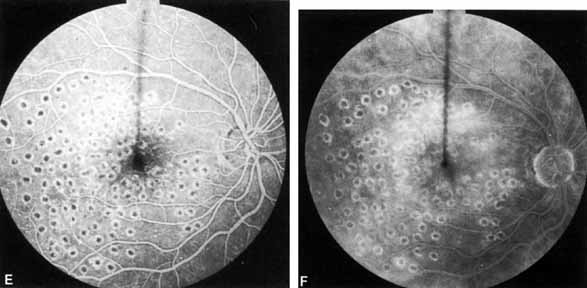
The ETDRS also showed that PRP is not part of the treatment strategy of CSME. Eyes that received PRP along with their focal treatment were much more likely to have an immediate decrease in visual acuity than were eyes that received focal treatment alone.11 An alternative treatment to the ETDRS strategy is a grid treatment (Fig. 24).176

Fig. 24 A. Thirty-five-year-old woman with diffuse macular edema and a visual acuity of 20/60. B. The midphase of the angiogram shows diffuse macular edema C. The late phase shows severe leakage and cystoid macular edema D. Grid treatment. E. The midphase of the angiogram done 4 months later shows minimal leakage. F. The late phase also shows minimal leakage. The visual acuity is 20/25.

Patients with macular edema who have the best prognosis for improved vision have circinate retinopathy of recent duration or focal, well-defined leaking areas and good capillary perfusion surrounding the avascular zone of the retina. Patients with an especially poor prognosis have dense lipid exudate in the center of the fovea (Fig. 25). Other poor prognostic signs include diffuse edema with multiple leaking areas, capillary closure around the fovea (Fig. 26), increased blood pressure, and cystoid macular edema.174,177 Nevertheless, the ETDRS found that even eyes with these adverse findings benefited from treatment compared to control eyes.175 The use of intravitreal steroids is also receiving attention for its potential role in the treatment of persistent diabetic edema. Small uncontrolled series have demonstrated dramatic reduction of macula thickness with associated improvement in visual function.178,179 Although encouraging, intravitreal corticosteroids are associated with frequent elevated intraocular pressure and occasionally endophthalmitis.180,181

Fig. 25 Circinate retinopathy with large hard exudates plaque in the center of the macula.

Fig. 26 Ischemic diabetic maculopathy. Notice large central areas of capillary nonperfusion surrounded by microaneurysms.

Vitrectomy techniques are also increasingly being considered for diabetic edema management. Early reports found up to 90% of eyes undergoing vitrectomy for the treatment of edema associated with a taut or thickened posterior hyaloid had visual improvement.182,183 Recent larger series confirm the efficacy of vitrectomy for eyes with abnormal hyaloid–macula interface.184,185 The success in this subgroup of patients is predicted by the hypothesis that tangential traction exerted by attached vitreous contributes to macular edema.186

More modest results have been reported with vitrectomy for the treatment of eyes with attached hyaloid that were not taut or thickened. Published series report approximately one-half of the eyes improve by only a line of acuity.187 Interestingly macular edema resolved in most of these eyes. Otani and colleagues188 reviewed seven patients with symmetric diabetic edema without an abnormally taut hyaloid who had one eye randomized to vitrectomy. They used OCT to demonstrate an average decrease in macular thickness of 353 μm postoperatively compared to a 60 μm average decrease in thickness in the fellow eyes.

In summary, DRS and the ETDRS conclusively proved that timely laser photocoagulation of diabetic retinopathy can reduce severe visual loss by 95%.189 Such treatment makes sense not only from the humanitarian point of view but is extremely cost-effective as well, saving approximately $250 to $500 million per year by keeping patients off disability and welfare.190–192 Nevertheless, only half of Americans with diabetes, especially the poor and minority population, fail to receive an annual dilated eye examination.193–194 The American Diabetes Association recommends that patients with type 1 diabetes should be screened annually beginning 5 years after the onset of the disease and patients with type 2 should be screened immediately and then annually thereafter.195 Alternatives methods of screening when ophthalmologists are not available include the use of primary care physicians or digital photography with remote image interpretation. Although primary care physicians commonly fail to detect significant retinopathy with direct ophthalmoscopy, training significantly improves their ability.86,196

Vitrectomy in Patients with Diabetes

Regarding this topic, the reader should also consult Chapter 56, Volume 6 for details.202 Vitrectomy, introduced by Robert Machemer, plays a vital role in the management of severe complications of diabetic retinopathy. The major indications are non-clearing vitreous hemorrhage, traction retinal detachment, and combined traction/rhegmatogenous retinal detachment. Less common indications are macular edema with a thickened and taut posterior hyaloid,203,204 macular heterotopia, and tight preretinal macular hemorrhage,18,19

To evaluate whether early vitrectomy (in the absence of vitreous hemorrhage) might improve the visual prognosis by eliminating the possibility of later traction macular detachment, the Diabetic Retinopathy Vitrectomy Study (DRVS) randomized 370 eyes with florid neovascularization and visual acuity of 10/200 or better to either early vitrectomy or to observation.205 After 4 years of follow-up, approximately 50% of both groups had 20/60 or better and approximately 20% of each group had light perception or worse. Thus, the results indicate that such patients probably do not benefit from early vitrectomy. They should be observed closely so that vitrectomy, when indicated, can promptly be undertaken.

If a patient has a vitreous hemorrhage severe enough to cause a visual acuity of 5/200 or less, the chances of visual recovery within 1 year are only approximately 17%.206 The DRVS randomized patients who had a visual acuity of 5/200 or less for more than 6 months into two groups: those who received an immediate vitrectomy and those for whom vitrectomy was deferred for an additional 6 months.207 Fifteen percent of those who had a deferred vitrectomy had a final visual acuity of 20/40 or better as opposed to 25% of those who had an immediate vitrectomy. In patients with type 1 diabetes, 12% of those who had a deferred vitrectomy had a final visual acuity of 20/40 or better as opposed to 36% of those who had an immediate vitrectomy. The reason for this discrepancy was thought to be excessive growth of fibrovascular proliferation during the waiting period. For this reason, the DRVS concluded that strong consideration should be given to immediate vitrectomy, especially in type 1 diabetics. (In type 2 diabetics, the final visual results were similar.) In most cases, vitrectomy should be deferred for approximately 6 months or longer if the retina is attached to give the patient a chance for spontaneous clearing. Some patients will never need the surgery, but more importantly, 25% of the patients in the DRVS who received an immediate vitrectomy had a final visual acuity of NLP. Patients with bilateral visual loss because of vitreous hemorrhage, with chronically recurring hemorrhage, with no history of PRP, and with known traction retinal detachment close to the macula are offered surgery sooner. If surgery is deferred, ultrasonography should be performed at regular intervals to make sure that traction retinal detachment is not developing behind the hemorrhage. The goals of surgery are to release all anterior–posterior vitreous traction and to perform a complete PRP to reduce the incidence of recurrent hemorrhage. Furthermore, the results of vitrectomy for nonclearing vitreous hemorrhage are excellent (Table 2).


TABLE 2. Overal Visual Results


 BensonThompson208DRVS207
20/20–20/40292425
20/50–20/100292429
20/120–20/30016225
20/400–CF8127
HM-LP121310
NLP6625

DRVS, Diabetic Retinopathy Vitrectomy Study. HM-LP, ·; NLP, ·.


In patients who have recurrent vitreous hemorrhage after vitrectomy, a simple outpatient air/liquid exchange may restore vision without the need for a repeat vitrectomy.209

Traction retinal detachments are usually a much greater challenge. In general, unless the macula becomes involved, observation is the best therapy for these patients because, in most cases, the detachment will not progress into the macula.210 These patients should be counseled to consult their ophthalmologist without delay should macular vision be suddenly lost, because vitrectomy at that point becomes a relative emergency.202 The surgical objectives are to clear the media, to release all anterior–posterior traction, to release tangential traction by cutting bridges between areas of traction detachment or by delamination, and to perform endophotocoagulation to prevent neovascularization of the iris. The prognosis is best in patients with small areas of traction. An alternative technique is to remove the vitreous and preretinal membranes by the “en bloc” technique.211,212 The prognosis is poorest in eyes with extensive fibrous adhesion to the retina (table-top) detachments, significant preoperative vitreous hemorrhage, no prior PRP, and advanced fibrovascular proliferation. If a lensectomy is required and if iatrogenic breaks are created, the results are also poorer. 202–208,210 Approximately 60% to 70% of patients have increased visual acuity and a final visual acuity of 20/800 or better, but 20% to 35% have decreased vision after surgery. Cases with severe peripheral fibrovascular proliferation may also require a scleral buckling procedure.219 Reoperations are required in approximately 10% of patients, most commonly the repair of rhegmatogenous retinal detachment or for clearing of recurrent vitreous hemorrhage.220

In traction/rhegmatogenous retinal detachments, the objectives are to find all of the retinal breaks and to release all vitreous traction. After air/fluid exchange to flatten the retina, endolaser photocoagulation is used to treat retinal breaks. Approximately one-half of such detachments can be cured.221,222 In severe cases, silicone oil is required to reattach the retina.223–225

Neovascularization of the iris (NVI) that progresses to neovascular glaucoma is a common cause of failure following otherwise successful vitrectomy. The risk is higher if there is preoperative neovascularization of the iris (17% versus 33%) if there is persistent retinal detachment after surgery, if the lens is removed during surgery, and if there is florid NVD and NVE. In eyes without these factors, the incidence of neovascular glaucoma is only about 2%.226 The pathogenesis of this complication is unknown. Some investigators feel that removal of the vitreous allows vasoproliferative factors produced in hypoxic retina to diffuse forward to the iris. Others feel that the main problem is that oxygen diffuses posteriorly from the anterior chamber lowering its oxygen tension too far. Fortunately, if an eye does not develop rubeosis iridis in the first 4 to 6 months after vitrectomy, it rarely will do so later.

Another vision-threatening complication is neovascularization originating from the anterior retina and extending along the anterior hyaloid to the posterior lens surface (anterior hyaloidal fibrovascular proliferation).227

Back to Top
OTHER OCULAR COMPLICATIONS OF DIABETES MELLITUS

CORNEA

Several investigations have found decreased corneal sensitivity in patients with diabetes.228–231 The severity of the hypesthesia has been correlated with both the duration of the disease230 and the severity of the retinopathy.228,231

The adhesion between the basement membrane of the corneal epithelium and the corneal stroma is not as firm as that found in normal corneas, probably because of a decreased number of hemidesmosomes between the stroma and the epithelium. When the epithelium is scraped from a normal cornea, the bottom half of the cells forming the basal layer are broken and remain attached to the basement membrane, which remains attached to the stroma. In diabetics, the entire epithelium is removed intact. Hyperglycemia and the aldose reductase pathway probably play a major role in epithelial abnormalities because aldose reductase inhibitors accelerate healing of corndal abrasions.232–234 After vitrectomy, recurrent corneal erosion, striate keratopathy, and corneal edema are more common in patients with diabetess than in tjose without diabetes. Although it has been shown that the endothelial cell density is normal in diabetics,235 it is not yet known whether or not endothelial cellular dysfunction contributes to these complications.

GLAUCOMA

Becker236 found that in patients with diabetes there is a higher incidence of open-angle glaucoma and marked elevation of intraocular pressure after prolonged administration of topical corticosteroids than there is in patients without diabetes. Moreover, patients with diabetes are more susceptible to visual field loss than those who do not have diabetes.

NVI is rarely associated with NPDR alone. It is usually seen only in eyes with PDR. By the end of the follow-up period of the DRS, Tasman and co-workers237 found NVI in 3.8% of the patients who had not received PRP as opposed to 2.0% in those who had. Thus, PRP appears to have some protective value against NVI. PRP is also an effective treatment against established NVI.238–240 Regression of the iris vessels and stabilization of any areas of angle closure and of intraocular pressure have been reported in 80% of cases treated. If the media are clear, PRP should be performed prior to any other treatment of NVI, even in advanced cases. Jacobson and associates238 reported successful results as long as the intraocular pressure was less than 40 mm Hg and there was less than 270 degrees of angle closure. We have seen regression and permanent pressure control in patients with pressures as high as 60 mm Hg. If the media are too cloudy for PRP, peripheral retinal cryoablation and transscleral diode laser photocoagulation are alternative means of treatment (see above).

If the angle is completely sealed and there is reasonable visual potential, a Molteno or other tube shunt offers the best chance for preserving vision.241 Cyclocryodestructive procedures often result in phthisis.

LENS

Because the lens in patients with diabetes has multiple biochemical abnormalities,242 the risk of cataract is 2 to 4 times greater in than in patients without diabetes243–247and may be 15 to 25 times greater in patients with diabetes under 40 years of age.248 Furthermore, the occurrence of cataract is a predictor of increased mortality.242

Patients with diabetes mellitus who have no retinopathy have excellent results from cataract surgery, with 90% to 95% having a final visual acuity of 20/40 or better, but, chronic cystoid macular edema is approximately 14 times more common in patients with diabetes than in those without.249–251 In patients with mild to moderate NPDR without macular edema, approximately 70% to 80% attain 20/40; outcomes are significantly worse in eyes with more severe retinopathy.252–254 Risk factors for progression and worse vision include an older age,255 poor glycemic control,252 poor renal function, and most significant preoperative macular edema. In many eyes the edema is self-limited and behaves clinically like postcataract CME; focal laser should be delayed when differentiation between diabetic edema cannot be made. The majority of eyes with CME will improve in 6 months.256

The most dreaded anterior complication is NVI. It was hoped that modern surgery that leaves an intact posterior capsule would protect the eye from neovascularization of the iris, by reducing the diffusion of vasoproliferative factors into the anterior chamber but several studies have shown that it does not. Eyes of patients with diabetes more frequently develop significant posterior capsular opacification257; fortunately capsulotomy does not seem to increase the risk of anterior segment neovascularization.247 Other anterior segment complications that are more common in patients with diabetes than in those without are pupillary block, posterior synechiae, pigmented precipitates on the implant, and severe iritis.250

Posterior complications include macular edema and ischemia,255,256,258 proliferative retinopathy,249,259 vitreous hemorrhage, and traction retinal detachment.251,258,260 In patients with active NPDR and no preoperative macular edema, as many as 50% to 75% will develop it and 30% will develop PDR. Approximately 8% will develop NVH. If macular edema is present prior to the surgery, it nearly always worsens. Only approximately 50% will have a final visual acuity of 20/40 or better. The risk of the development of or progression of macular edema is nearly doubled in patients who are older than 63 years of age.267 Clearly, caution must be observed when considering cataract surgery in patients who have diabetic retinopathy.

Cataract surgery in patients with active PDR often results in still poorer postoperative visual outcome because of the high risk of both anterior262 and posterior segment complications. In one series, no patient with active PDR or PPDR achieved better than 20/80. Anteriorly, fibrinous uveitis is seen in more than 50% of patients with active PDR. Most experts recommend aggressive preoperative PRP.258,268

OPTIC NEUROPATHY

As demonstrated by increased latency and decreased amplitude of the visual evoked potential, many patients with diabetes without retinopathy have subclinical optic neuropathy.269,270 In addition, patients with diabetes can develop two types of acute optic neuropathy. The first, anterior ischemic optic neuropathy (AION) is identical to that seen in patients without diabetes. The patients report a sudden decrease in visual acuity or a sudden visual field loss.271–273 The main ocular finding is a “pale swelling” of the optic nerve head with, considering the degree of disc edema, very few hemorrhages (Fig. 27). On fluorescein angiography segmental nonfilling or slow filling is seen (Fig. 27). An afferent pupillary defect (Marcus Gunn) is nearly always present. Visual fields commonly show altitudinal or nerve fiber bundle defects. The disc progresses to optic atrophy (Fig. 27), and improvement in visual function is rare.

Fig. 27 A. Right eye: ischemic optic neuritis. Note pale swelling of optic disc and blurring of disc margins. Left eye: normal disc. B. Fluorescein angiogram. Note poor filling on disc inferotemporally as compared with the rest of the disc. C. Right eye 6 months after optic neuritis. Note slight pallor.

The other type of acute optic neuropathy, commonly called diabetic papillopathy, is characterized by acute disc edema without the pale swelling of AION. It is bilateral in one-half of cases. The vision usually recovers to better than 20/50 with or without an afferent pupillary defect.274 Macular edema is a common finding and is the most common cause of failure of visual recovery.274 Visual fields may be normal, show an increased blind spot, or have disc-related defects. The prognosis is excellent as nearly all patients recover to 20/30 or better.275–277

CRANIAL NEUROPATHY

Extraocular muscle palsies may occur in patients with diabetes secondary to neuropathy involving the third, fourth, or sixth cranial nerves. The mechanism is believed to be a localized demyelination of the nerve secondary to focal ischemia. Pain may or may not be experienced, and not infrequently an extraocular muscle palsy may be the initial clue to a latent diabetic condition. Recovery of extraocular muscle function in diabetic cranial nerve neuropathy generally takes place in 1 to 3 months.278

When the third nerve is involved, pupillary function is usually normal. This pupillary sparing in the diabetic third-nerve palsy is an important diagnostic feature, distinguishing it from other causes of oculomotor involvement such as intracranial tumor or aneurysm.

Back to Top
REFERENCES

1. Bloodworth JMBJr , Molitor DL: Ultrastructural aspects of human and canine diabetic retinopathy. Invest Ophthalmol 4:1037, 1965

2. Bloodworth JMBJr , Engerman RL: Diabetic microangiography in the experimentally-diabetic dog and its prevention by careful control with insulin. Diabetes 22:290, 1973

3. Cogan DG, Kuwabara T: The mural cell in perspective. Arch Ophthalmol 78:133, 1967

4. Yanoff M: Ocular pathology of diabetes mellitus. Am J Ophthalmol 67:21, 1969

5. deVenecia G, Davis MD, Engerman R: Clinicopathologic correlations in diabetic retinopathy. Arch Ophthalmol 94:1766, 1976

6. Bresnick GH, Davis MD, Myers FL, et al: Clinicopathological correlations in diabetic retinopathy: II. Clinical and histologic appearances of retinal capillary microaneurysms. Arch Ophthalmol 95:1215, 1977

7. Schatz H, Patz A: Cystoid maculopathy in diabetics. Arch Ophthalmol 94:761, 1976

8. Fark`s TG, Sylvester V, Archer D: An electron microscopic study of the choriocapillaries and Bruch's membrane in diabetic retinopathy. Trans Ophthalmol Soc UK 90:657, 1977

9. Hersh PS, Green WR, Thoms JJV: Tractional venous loops in diabetic retinopathy. Am J Ophthalmol 92:661, 1981

10. Early Treatment Diabetic Retinopathy Study Research Group: Fundus photographic risk factors for progression of diabetic retinopathy. Ophthalmology 98:823, 1991

11. Early Treatment Diabetic Retinopathy Study Research Group: Early Photocoagulation for diabetic retinopathy. ETDRS Report Number 9. Ophthalmology 98:766, 1991

12. Shimuzu K, Kobayashi Y, Muraoka K: Midperipheral fundus involvement in diabetic retinopathy. Ophthalmology 88:601, 1981

13. Valsania P, Warram JH, Rand LI, et al: Different determinants of neovascularization on the optic disc and on the retina in patients with severe nonproliferative diabetic retinopathy. Arch Ophthalmol 111:202, 1993

14. Foos RY, Kreiger AE, Forsxthe AB, et al: Posterior vitreous detachment in diabetic subjects. Ophthalmology 87:122, 1980

15. Davis MD: Vitreous contraction in proliferative diabetic retinopathy. Arch Ophthalmol 74:741, 1965

16. Anderson BJr : Activity and diabetic vitreous hemorrhage. Ophthalmology 87:173, 1980

17. Tasman W: Diabetic vitreous hemorrhage and its relationship to hypoglycemia. Mod Prob Ophthalmol 20:413, 1979

18. Ralsay RC, Knobloch WH, Cantrill HL: Timing of vitrectomy for active proliferative diabetic retinopathy. Ophthalmology 93:283, 1986

19. O'Hanley GP, Canny CLB: Diabetic dense premacular hemorrhage. A possible indication for prompt vitrectomy. Ophthalmology 92:507, 1985

20. Bresnick GH, Haight B, deVenecia G: Retinal wrinkling and macular heterotopia in diabetic retinopathy. Arch Ophthalmol 97:1890, 1979

21. Kostraaba JN, Dorman JS, Orchard TJ, et al: Contribution of diabetes duration before puberty to development of microvascular complications in IDDM subjects. Diabetes Care 12:686, 1989

22. Klein R, Klein BEK, Syrjala SE, et al: Wisconsin epidemiologic study of diabetic retinopathy. In Friedman EA, L'Esperance FA, eds. Diabetic Renal-Retinal Syndrome. New York: Grune & Stratton, 1981:21

23. Klein R, Klein BEK, Moss SE, Davis MD, DeMets DL. The Wisconsin epidemiologic study of diabetes retinopathy. III. Prevalence and risk of diabetic retinopathy when age of diagnosis is 30 or more years. Arch Ophthalmol 102:527, 1984

24. Sjolie AK: Ocular complications in insulin treated diabetes mellitus. Ophthalmologica. 1981;63:1–76

25. Palmberg P, Smith M, Waltman S, et al: The natural history of retinopathy in insulin-dependent juvenile-onset diabetes. Ophthalmology 88:613, 1981

26. Dwyer MS, Melton LJ, Ballard DL, et al: Incidence of diabetic retinopathy and blindness: A population-based study in Rochester, Minoesota. Diabetes Care 8:316, 1985

27. Klein R, Klein BEK: Epidemiology of proliferative diabetic retinopathy. Diabetes Care 15:1875, 1992

28. Klein R, Davis MD, Moss SE, et al: The Wisconsin epidemiologic study of diabetic retinopathy: a comparison of retinopathy in younger and older onset diabetic persons. In Vranic M, Hollenberg C, Steiner G, eds. Comparison of Type I and Type II Diabetes. New York: Plenum, 1985:321–325

29. Klein R, Klein BE, Moss SE, et al: The Wisconsin Epidemiologic Study of diabetic retinopathy. XIV. Ten-xear inchdence and progression of diabetic retinopathy. Arch Ophthalmol 112:1217, 1994

30. Yanko L, Goldbourt U, Michaelson IC, et al: Prevalence and 15-year incidence of retinopathy and associated characteristics in middle-aged and elderly diabetic men. Br J Ophthalmol 67:759, 1983

31. Diabetes Control and Complications Trial Research Group: The effect of intensive diabetes treatment on the progression of diabetic retinopathy in insulin-dependent diabetes mellitus. Arch Ophthalmol 113:36, 1995

32. Diabetes Control and Complications Trial Research Group. The effect of intensive treatment of diabetes on the development and progression of long-term complications in insulin-dependent diabetes mellitus. N Engl J Med 329:977, 1993

33. United Kingdom Prospective Diabetes Study Group: Intensive blood-glucose control with sulphonylureas or insulin compared with conventional treatment and risk of complications in patients with type 2 diabetes (UKPDS 33). Lancet 352:837, 1998

34. Gray A, Raikou M, McGuire A, et al: Cost effectiveness of an intensive blood glucose control policy in patients with type 2 diabetes: economic analysis alongside randomised controlled trial (UKPDS 41). United Kingdom Prospective Diabetes Study Group. BMJ 320:1373, 2000

35. Brinchmann-Hansen O, Dahl-Jorgensdn K, Sandvik L, et al: Blood glucose concentrations and progression of diabetic retinopathy: the seven year results of the Oslo study. BMJ 304:19, 1992

36. Lawson PM, Champion MC, Canny C, et al: Continuous subcutaneous insulin infusion (CSII) does not prevent progression of proliferative and preproliferative retinopathy. Br J Ophthalmol 66:762, 1982

37. Brinchmann-Hansen O, Dami-Jorgensen K, Hanssen KF, et al: Effects of intensified insulin treatment on various lesions of diabetic retinopathy. Am J Ophthalmol 100:644, 1985

38. vanBallegooie E, Hooymans JM, Timmerman Z, et al: Rapid deterioration of diabetic retinopathy during treatment with continuous subcutaneous insulin infusion. Diabetes Care 7:236, 1984

39. KROC Collaborative Study Group: Diabetic retinopathy after two years of intensified insulin treatment. JAMA 260:37, 1988

40. Ramsay RC, Goetz FC, Sutherland DER, et al: Progression of diabetic retinopathy after pancreas transplantation for insulin-dependent diabetes mellitus. New Engl J Med 318:208, 1988

41. Chavers BM, Mauer SM, Ramsay RC, et al: Relationship between retinal and glomerular lesions in IDDM patieots. Diabetes 43:441, 1994

42. Orchard TJ, Dorman JS, Maser RE, et al: Factors associated with avoidance of severe complications after 25 yr of IDDM. Diabetes Care 13:741, 1990

43. Parving H-H: Impact of blood pressure and antihypertensive treatment on incipient and overt nephropathy, retinopathy, and endothelial permeability in diabetes mellitus. Diabetes Care 14:260, 1991

44. Jerneld B, Algvere P: Proteinuria and blood glucose levels in a population with diabetic retinopathy. Am J Ophthalmol 104:283, 1987

45. Mogensen CE, Vigstrup J, Ehlers N: Microalbuminuria predicts proliferative diabetic retinopathy. Lancet 1:1512, 1985

46. Klein R, Klein BEK, Moss SE, et al: The Wisconsin epidemiologic study of diabetic retinopathy. II. Prevalence and risk of diabetic retinopathy when age is less than 30 years. Arch Ophthalmol 102:520, 1984

47. Nelson RG, Wolfe JA, Horton MB, et al: Proliferative retinopathy in NIDDM. Diabetes 38:435, 1989

48. Cruickshanks KJ, Ritter LL, Klein R, et al: The Association of Microalbuminuria with Diabetic Retinopathy. Ophthalmology. 100:862, 1993

49. Feldman JN, Hirsch SR, Beyer MM: Prevalence of diabetic nephropathy at time of treatment for diabetic retinopathy. In Friedman EA, L'Esperance FA, eds. Diabetic Renal-Retinal Syndrome. New York: Grune & Stratton, 1982:9–2050

50. Klein BE, Klein R, Moss SE, et al: A cohort study of the relationship of diabetic retinopathy to blood pressure. Arch Ophthalmol 113:601, 1995

51. Dornan T, Mann JI, Turner R: Factors protective against retinopathy in insulin-dependent diabetes free of retinopathy for 30 years. Br Med J 285:1073, 1982

52. Gray RS, Starkey IR, Rainbow S, et al: HLA antigens and other risk factors in the development of retinopathy in type I diabetes. Br J Ophthalmol 66:280, 1982

53. Janka HU, Warram JH, Rand LI, et al: Risk factors for progression of background retinopathy in long-standing IDDM. Diabetes. 38:460, 1989

54. United Kingdom Prospective Diabetes Study Group: Efficacy of atenolol and captopril in reducing risk of lacrovascular and microvascular complications in type 2 diabetes: UKPDS 39. BMJ 317:713, 1998

55. Chaturvedi N, Sjolie AK, Stephenson JM, et al: Effect of lisinopril on progression of retinopathy in normotensive people with type 1 diabetes. The EUCLID Study Group. EURODIAB Controlled Trial of Lisinopril in Insulin-Dependent Diabetes Mellitus. Lancet 351:28, 1998

56. Phelps RL, Sakol P, Metzger BE, et al: Changes in diabetic retinopathy during pregnancy: correlations with regulation of hyperglycemia. Arch Ophthalmol 104:1806, 1986

57. Beethem WP: Diabetic retinopathy in pregnancy. Trans Am Ophthalmol Soc 48:205, 1950

58. Axer-Siegel R, Hod M, Fink-Cohen S, et al: Diabetic retinopathy during pregnancy. Ophthalmology 103:1815, 1996

59. Rosenn B, Miodovnik M, Kranias G, et al: Progression of diabetic retinopathy in pregnancx: association with hypertension in pregnancy. Am J Obstet Gynecol 166:1214, 1992

60. Klein BEK, Moss SE, Klein R: Effect of pregnancy on progression of diabetic retinopathy. Diabetes Care 13:34, 1990

61. Ohrt V: The influence of pregnancy on diabetic retinopathy with special regard to the reversible changes shown in 100 pregnancies. Acta Ophthalmol 62:603, 1984

62. Moloney JBM, Drury MI: The effect of pregnancy on the natural course of diabetic retinopathy. Am J Ophthalmol 83:745, 1982

63. Laatikainen L, Larinakir J, Teralo K, et al: Occurrence and prognostic significance of retinopathy in diabetic pregnancy. Metab Pediatr Ophthalmol 4:191, 1980

64. Horvat M, MacLean H, Goldberg L, et al: Diabetic retinopathy in pregnancy, a 12-year prospective study. Br J Ophthalmol 64:398, 1980

65. Dibble CM, Kochenour NK, Worley RJ, et al: Effect of pregnancy on diabetic retinopathy. Obstet Gynecol 49:699, 1982

66. Hercules BL, Wozencroft M, Gayed I, et al: Peripheral retinal ablation in the treatment of proliferative diabetic retinopathy during pregnancy. Br J Ophthalmol 64:87, 1980

67. Coustan DR: Pregnancy in diabetic women. N Engl J Med 319:1663, 1988

68. Sinclair SH, Nesler C, Foxman B, et al: Macular edema and pregnancy in insulin-dependent diabetes. Am J Ophthalmol 97:154, 1984

69. Harris EL, Feldman S, Robinson CR, et al: Racial differences in the relationship between blood pressure and risk of retinopathy among individuals with NIDDM. Diabetes Care 16:748, 1993

70. Raab MF, Gagliano DA, Sweeney HE: Diabetic retinopathy in blacks. Diabetes Care 13:1202, 1990

71. Arfken CL, Salicrup AE, Meuer SM, et al: Retinopathy in African Americans and whites with insulin-dependent diabetes mellitus. Arch Intern Med 154:2597, 1994

72. Moss SE, Klein R, Klein BEK: Cigarette smoking and ten-year progression of diabetic retinopathy. Ophthalmology 103:1438, 1996

73. Chew EY, Klein ML, Ferris FL, et al: Association of elevated serum lipids with retinal hard exudate in diabetic retinopathy. Arch Ophthalmol 114:1079, 1996

74. Varma SD: Aldose reductase and the etiology of diabetic cataracts. Curr Top Eye Res 3:91, 1980

75. Kador PF, Kinoshita JF, Sharpless NE: Aldose reductase inhibitors: A potential new class of agents for the pharmacological control of certain diabetic complications. J Med Chem 28:841, 1985

76. Chylack LT, Henriques HF, Cheng HM, et al: Efficacy of alrestatin, an aldose reductase inhibitor, in human diabetic and nondiabetic lenses. Ophthalmology 86:1579, 1979

77. Lightman S, Rechthand E, Terubayashi H, et al: Permeability changes in blood-retinal barrier of galactosemic rats are prevented by aldose reductase inhibitors. Diabetes 36:1271, 1987

78. Robison W, Kador P, Kinoshita J: Retinal capillaries: basement membrane thickening by galactosemia prevented with aldose reductase inhibitor. Science 220:1177, 1983

79. Sosula L, Beaumont P, Hollows FC, et al: Dilatation and endothelial proliferation of retinal capillaries in streptozotocin diabetic rats: quantitative electron microscopy. Invest Ophthalmol 11:926, 1972

80. Kador PF, Akagi Y, Terubayashi H, et al: Prevention of pericyte ghost formation in retinal capillaries of galactose-fed dogs by aldose reductase inhibitors. Arch Ophthalmol 106:1099, 1988

81. Judzewitsch RG, Jaspan JB, Polonsky KS, et al: Aldose reductase inhibition improves nerve condhtion velocity in diabetic patieots. N Engl J Med 308:119, 1983

82. Notvest RR, Inserra JJ: Tolrestat, an aldose reductase inhibitor, prevents nerve dysfunction in conscious diabetic rats. Diabetes 36:500, 1987

83. Handelsman DJ, Turtle JR: Clinical trial of an aldose reductase inhibitor in diabetic neuropathy. Diabetes 30:459, 1981

84. Varagiannis E, Beyer-Mears A, Cruz E: Diminished proteinuria by an aldose reductase inhibitor, sorbinil. Diabete 33:43A, 1984

85. Williamson JR, Chang K, Tilton RG, et al: Increased vascular permeability in spontaneously diabetic BB/W rats and in rats with mild versus severe Streptozotocin-induced diabetes. Prevention by aldose reductase inhhbitors and castration. Diabetes 36:813, 1987

86. Griffith SP, Freeman WL, Shaw CJ, et al: Screening for diabetic retinopathy in a clinical setting: a comparison of direct ophthalmoscopy by primary care physicians with fundus photography. J Fam Pract 37:49, 1993

87. Sorbinil Retinopathy Trial Research Group: A randomized trial of sorbinil, an aldose reductase inhibitor, in diabetic retinopathy. Arch Ophthalmol 108:1234, 1990

88. Sorbinil Retinopathy Trial Research Group: The sorbinil retinopathy trial: neuropathy results. Neurology 43:1141, 1993

89. Frank RN: The aldose reductase controversy. Diabetes 43:169, 1994

90. Michaelson IC: Retinal Circulation in Man and Animals. Springfield, IL: Thomas, 1954:118–131

91. Ashton N: Retinal vascularization in health and disease. Am J Ophthalmol 44:7, 1957

92. Ashton N: Studies of the retinal capillaries in relation to diabetic and other retinopathies. Br J Ophthalmol 47:521, 1963

93. Kohner DM, Shilling JS, Hamilton AM: The role of avascular retina in new vessel formation. Metab Ophthalmol 1:15, 1976

94. Forrester JV, Shafiee A, Schroder S, et al: The role of growth factors in proliferative diabetic retinopathy. Eye 7:276, 1993

95. Aiello LP, Avery RP, Arrigg PG, et al: Vascular endothelial growth factor in ocular fluid of patients with diabetic retinopathy and other retinal disorders. N Engl J Med 331:1480, 1994

96. Adamis AP, Miller JW, Bernal MT, et al: Increased vascular endothelial growth factor levels in the vitreous of eyes with proliferative diabetic retinopathy. Am J Ophthalmol 118:445, 1994

97. Malecaze F, Clamens S, Simorre-Pinatel V, et al: Detection of vascular endothelial growth factor messenger RNA and vascular endothelial growth factor-like activity in proliferative diabetic retinopathy. Arch Ophthalmol 112:1476, 1994

98. Tolentino MJ, Miller JW, Gragoudas ES, et al: Intravitreous injections of vascular endothelial growth factor produce retinal ischemia and microangiopathy in an adult primate. Ophthalmology 103:1820, 1996

99. Poulsen JE: Recovery from retinopathy in a case of diabetes with Simmonds' disease. Diabetes 2:7, 1953

100. Alzaid AA, Dinneen SF, Melton LJ III , et al: The role of growth hormone in the development of diabetic retinopathy. Diabetes Care 17:531, 1994

101. Dobbie JG, Kwaan HC, Colwell J, et al: Role of platelets in pathogenesis of diabetic retinopathy. Arch Ophthalmol 91:107, 1974

102. Bensoussan D, Levy-Toledano S, Passa P, et al: Platelet hyperaggregation and increased plasma level of von Willebrand factor in diabetics with retinopathy. Diabetologia 11:307, 1975

103. Colwell J: Inhibition of LASS and platelet aggregation in early diabetes mellitus. Diabetes 27:684, 1975

104. Segal J, Colwell J, Crook L, et al: Increased platelet aggregation in early diabetes mellitus. Ann Intern Med 82:733, 1975

105. Ho PC, Feman SS, Stein RS, et al: Proliferative diabetic retinopathy in patients with defects of platelet function. Am J Ophthalmol 88:37, 1979

106. Dintenfass L: Blood viscosity factors in severe nondiabetic and diabetic retinopathy. Biorheology 14:151, 1977

107. Little HL, Sachs AH: Role of abnormal blood rheology in the pathogenesis of diabetic retinopathy. Trans Am Acad Ophthalmol Otolaryngol 83:522, 1977

108. McMillan DE, Utterback NG, LaPoma J: Reduced erythrocyte deformability in diabetes. Diabetes 27:895, 1978

109. Chew EY, Klein ML, Murphy RP, et al: Effects of aspirin on vitreous/preretinal hemorrhage in patients with diabetes mellitus. ETDRS Report No. 20. Arch Ophthalmol 113:52, 1995

110. DAMAD Study Group: Effect of aspirin alone and aspirin plus Dipyridamole in early diabetic retinopathy. A multicenter randomized controlled clinical trial. Diabetes 38:481, 1989

111. Ticlopidine Microangiopathy of Diabetes Study Group: Ticlopidine treatment reduces the progression of nonproliferative diabetic retinopathy. Arch Ophthalmol 108:1577, 1990

112. Sonkin PL, Kelly LW, Sinclair SH, et al: Pentoxifylline increases retinal capillary blood flow velocity in patients with diabetes. Arch Ophthalmol 111:1647, 1993

113. Cambie E: Functional results following argon laser photocoagulation in eyes with diabetic retinopathy. In Friedman EA, L'Esperance FA, eds. Diabetic Renal-Retinal Syndrome. New York: Grune & Stratton, 1980:295–307

114. Gliem H, Schulze DP: Sofortadaptation, Blendungsupfindlichkeitr und diabetische retinopathie. Klin Monatsbl Augenheilkd 166:766, 1978

115. Henson DB, North RV: Dark adaptation in diabetes mellitus. Br J Ophthalmol 63:539, 1979

116. Pender PM, Benson WE, Compton H, et al: The effects of panretinal photocoagulation on dark adaptation in diabetics with proliferative retinopathy. Ophthalmology 88:634, 1981

117. Kinnear PR, Aspinall PA, Lakowski R: The diabetic eye and colour vision. Trans Ophthalmol Soc UK 92:69, 1972

118. Graham K, Keson CM, Kennedy HB, et al: Relevance of colour diabetic retinopathy to self-monitoring of blood glucose. Br Med J 281:971, 1980

119. Moloney J, Drury MI: Retinopathy and retinal function in insulin-dependent diabetes mellitus. Br J Ophthalmol 66:759, 1982

120. Roy MS, McCulloch C, Hanna AK, et al: Colour vision in long-standing diabetes mellitus. Br J Ophthalmol 68:215, 1984

121. Sokol S, Moskowitz A, Sharf B, et al: Contrast sensitivity in diabetics with and without background retinopathy. Arch Ophthalmol 103:51, 1985

122. Trick GL, Burde RM, Gordon ME, et al: The relationship between hue discrimination and contrast sensitivity deficits in patients with diabetes mellitus. Ophthalmology 95:693, 1988

123. Ghafour IM, Foulds WS, Allan D, et al: Contrast sensitivity in diabetic subjects with and without retinopathy. Br J Ophthalmol 66:492, 1982

124. Vesti E, Trick GL: Diabetes can alter the interpretation of visual dysfunction in ocular hypertension. Ophthalmology 103:1419, 1996

125. Simonsen SE: ERG in diabetes. In Francois J, ed. The Clinical Value of Electroretinography. New York: S Karger, 1968:403

126. Simonsen SE: The value of the oscillatory potential in selecting juvenile diabetics at risk of developing proliferative retinopathy. Dev Ophthalmol 2:222, 1981

127. Frost-Larson K, Larson HW, Simonsen SE: Value of electroretinography and dark adaptation as prognostic tools in diabetic retinopathy. Dev Ophthalmol 2:222, 1981

128. Bresnick GL, Korth K, Groo A, et al: Electroretinographic oscillatory potentials predict progression of diabetic retinopathy. Preliminary report. Arch Ophthalmol 102:1307, 1984

129. Algvere P, Gjotterberg M: Diagnostic value of oscillatory potentials of the electroretinogram and fluorescein angiography in diabetic proliferative retinopathy. Ophthalmologica 168:97, 1974

130. Bresnick GH, Palta M: Oscillatory potenthal amplitudes. Ophthalmology 105:929, 1987

131. Benson WE, Brown GC, Tasman W: Diabetes and Its Ocular Complications. Philadelphia: WB Saunders, 1988:90–109

132. Aiello LM, Beetham WP, Balodimos MD, et al: Ruby laser photocoagulation in the treatment of diabetic retinopathy, Preliminary report. In Goldberg MD, Fine SL, eds. Symposium on the Treatment of Diabetic Retinopathy. Washington, DC: US Public Health Service, Publication No. 1890; 1968:187–463

133. Beetham WP, Aiello IL, Balodimos MC, et al: Ruby laser photocoagulation of early diabetic neovascular retinopathy. Arch Ophthalmol 83:261, 1970

134. Duane TD, Behrendt T, Field RA: Net vascular pressure ratios in diabetic retinopathy. In: Goldberg MF, Fine SL, eds. Symposium on the Treatment of Diabetic Retinopathy. Washington, DC: US Public Health Service, Publication 1890; 1968:657

135. Wetzig PC, Jepson CN: Treatment of diabetic retinopathy by light coagulation. Am J Ophthalmol 62:459, 1966

136. Behrendt T, Duane TD: Unilateral complications in diacetic retinopathy. Trans Am Acad Ophthalmol Otolaryngol 74:28, 1970

137. Coop HV: Photocoagulation for proliferative diabetic retinopathy. Trans Ophthalmol Soc N Z ???:31, 1970

138. Duane TD: Is diabetic retinopathy uncontrollable? Am J Ophthalmol 71:286, 1971

139. Ederer F, Hiller R: Clinical trials, diabetic retinopathy and photocoagulation: A reanalysis of five studies. Surv Ophthalmol 19:267, 1975

140. Diabetic Retinopathy Study Group: Preliminary report on effects of photocoagulation therapy. Am J Ophthalmol 81:383, 1976

141. Diabetic Retinopathy Study Group: Photocoagulation treatment of proliferative diabetic retinopathy: the second report of Diabetic Retinopathy Study findings. Ophthalmology 85:82, 1978

142. Diabetic Retinopathy Study Research Group: Four risk factors for severe visual loss in diabetic retinopathy: the third report from the Diabetic Retinopathy Study. Arch Ophthalmol 97:654, 1979

143. Patel V, Rassam S, Newsom R, et al: Retinal blood flow in diabetic retinopathy. BMJ 305:678, 1992

144. Grunwald JE, Brucker AJ, Petrig BL, et al: Retinal blood flow regulation and the clinical response to panretinal photocoagulation in proliferative diabetic retinopathy. Ophthalmology 96:1518, 1989

145. Glaser BM, Campochiaro PA, Davis DL Jr , et al: Retinal pigment epithelial cells release an inhibitor of neovascularization. Arch Ophthalmol 103:1870, 1985

146. Stefansson E, Landers MBIII , Wolbarsht ME: Oxygenation and vasodilation in relation to diabetic and other proliferative retinopathies. Ophthalmic Surg 17:208, 1983

147. Wolbarsht ML, Landers MBIII , Stefansson E: Vasodilation and the etiology of diabetic retinopathy: a new model. Ophthalmic Surg 12:104, 1981

148. Wolbarsht ML, Landers MB: The rationale of photocoagulation therapy for proliferative diabetic retinopathy: a review and a model. Ophthalmic Surg 11:235, 1980

149. Stefansson E, Machemer R, de Juan E Jr , et al: Retinal oxygenation and laser treatment in patients with diabetic retinopathy. Am J Ophthalmol 113:36, 1992

150. Schiodte N: Ocular effects of panretinal photocoagulation. Acta Ophthalmol 66(suppl):9, 1988

151. Marshall J, Clover G, Rothery S: Some new findings on retinal irradiation by krypton and argon lasers. Doc Ophthalmol 36:21, 1984

152. Singh A, Boulton M, Lane C, et al: Inhibition of microvascular endothelial cell proliferation by vitreous following retinal scatter photocoagulation. Br J Ophthalmol 74:328, 1990

153. Lobes LAJr , Benson WE, Grand MG: Panfunduscope contact lens for argon laser therapy. Ann Ophthalmol 13:713, 1981

154. Frank RN: Visual fields and electroretinography following extensive photocoagulation. Arch Ophthalmol 93:591, 1975

155. Ogden TE, Riekolf FT, Benkwith SM: Correlation of histologic and electroretinographic changes in peripheral retinal ablation in the rhesus monkey. Am J Ophthalmol 81:272, 1976

156. Krypton Argon Regression Neovascularization Study Research Group: Randomized comparison of krypton versus argon scatter photocoagulation for diabetic disc neovascularization. Ophthalmology 100:1655, 1993

157. Aylward GW, Pearson RV, Jagger JD, et al: Extensive argon laser photocoagulation in the treatment of proliferative diabetic retinopathy. Br J Ophthalmol 73:197, 1989

158. Singerman LJ: PDR in juvenile onset diabetics: high-risk proliferative diabetic retinopathy in juvenile onset diabetics. Retina 1:18, 1981

159. Vine AK: The efficacy of additional argon photocoagulation for persistent, severe proliferative diabetic retinopathy. Ophthalmology 92:1532, 1985

160. Reddy VM, Zamora RL, Olk RJ: Quantitation of retinal ablation in proliferative diabetic retinopathy. Am J Ophthalmol 119:760, 1995

161. Doft BH, Metz DJ, Kelsey SF: Augmentation laser for proliferative diabetic retinopathy that fails to respond to initial panretinal photocoagulation. Ophthalmology 99:1728, 1992

162. Leyers SM: Macular edema after scatter laser photocoagulation for proliferative diabetic retinopathy. Am J Ophthalmol 90:210, 1980

163. Ferris FL, Podgor MJ, Davis MD: The Diabetic Retinopathy Research Group: macular edema in diabetic retinopathy study patients. Ophthalmology 94:754, 1987

164. Kleiner RC, Elman MJ, Murphy RP, et al: Transient severe visual loss after panretinal photocoagulation. Am J Ophthalmol 106:298, 1988

165. Miettinen H, Haffner S, Lehto S, et al: Retinopathy predicts coronary heart disease events in NIDDM patients. Diabetes Care 19:1445, 1966

166. Ramsay RC, Cantrill HL, Knobloch WH: Cryoretinopexy for proliferative diabetic retinopathy. Can J Ophthalmol 17:17, 1982

167. Haut J, Robert P, Chatellier PH, et al: Place de la cryotherapie dans le traitement de la retinopathie diabetique. Bull Mem Soc Fr Ophthalmol 90:124, 1978

168. Lempert P: Cryoablation for diabetic retinopathy. Ann Ophthalmol 11:740, 1979

169. Schimek RA, Spencer R: Cryopexy treatment of proliferative diabetic retinopathy. Arch Ophthalmol 97:1276, 1979

170. Oosterhuis JA, Bijlmer-Gortner H: Cryotreatment in proliferative diabetic retinopathy. Arch Ophthalmol 97:1276, 1979

171. Benedett R, Olk JR, Arribas NP, et al: Transconjunctival anterior retinal cryotherapy for proliferative diabetic retinopathy. Ophthalmology 94:612, 1987

172. Ross WH, Gottner MJ: Peripheral retinal cryopexy for subtotal vitreous hemorrhage. Am J Ophthalmol 105:377, 1988

173. Daily MJ, Gieser RG: Treatment of proliferative diabetic retinopathy with panretinal cryotherapy. Ophthalmic Surg 9:741, 1984

174. Patz A, Schatz H, Berkow JW, et al: Macular edema: an overlooked complication of diabetic retinopathy. Trans Am Acad Ophthalmol Otolaryngol 77:34, 1973

175. Early Treatment Diabetic Retinopathy Study Research Group: Focal photocoagulation treatment of diabetic macular edema. Relationship of treatment effect to fluorescein angiographic and other retinal characteristics at baseline: ETDRS report no. 19. Arch Ophthalmol 113:1144, 1995

176. Striph GG, Hart WM, Olk RJ: Modified grid laser photocoagulation for diabetic macular edema. Ophthalmology 95:1673, 1988

177. Ticho U, Patz A: The role of capillary perfusion in the management of diabetic macular edema. Am J Ophthalmol 76:880, 1973

178. Martidis A, Duker JS, Greenberg PB, et al: Intravitreal triamcinolone for refractory diabetic macular edema. Ophthalmology 109:920, 2002

179. Jonas JB, Kreissig I, Sofker A, et al: Intravitreal injection of triamcinolone for diffuse diabetic macular edema. Arch Ophthalmol 121:57, 2003

180. Benz MS, Murray TG, Dubovy SR, et al: Endophthalmitis caused by Mycobacterium chelonae abscessus after intravitreal injection of triamcinolone. Arch Ophthalmol 121:271, 2003

181. Nelson M, Tennant M, Sivalingam A, et al: Infectious and presumed noninfectious endophthalmitis after intravitreal triamcinolone acetonide injection. Retina 23:686, 2003

182. Harbour JW, Smiddy WE, Flynn HW Jr , et al: Vitrectomy for diabetic macular edema associated with a thickened and taut posterior hyaloid membrane. Am J Ophthalmol 121:405, 1996

183. Lewis H, Abrams GW, Blumenkranz MS, et al: Vitrectomy for diabetic macular traction and edema associated with posterior hyaloidal traction. Ophthalmology 99:753, 1992

184. Pendergast SD, Hassan TS, Williams GA, et al: Vitrectomy for diffuse diabetic macular edema associated with a taut premacular posterior hyaloid. Am J Ophthalmol 130:178, 2000

185. Gandorfer A, Messmer EM, Ulbig MW, et al: Resolution of diabetic macular edema after surgical removal of the posterior hyaloid and the inner limiting membrane. Retina 20:126, 2000

186. Nasrallah FP, Jalkh AE, Van Coppenolle F, et al: The role of the vitreous in diabetic macular edema. Ophthalmology 95:1335, 1988

187. Tachi N, Ogino N: Vitrectomy for diffuse macular edema in cases of diabetic retinopathy. Am J Ophthalmol 122:258, 1996

188. Otani T, Kishi S: A controlled study of vitrectomy for diabetic macular edema. Am J Ophthalmol 134:21, 2002

189. Ferris FL III , et al: How effective are treatments for diabetic retinopathy? JAMA 269:1290, 1993

190. Javitt JC, Aiello LP, Bassi LJ, et al: Detecting and treating retinopathy in patients with type I diabetes mellitus. Ophthalmology 98:1565, 1991

191. Rand LI: Financial Implications of implementing standards of care for diabetic eye disease. Diabetes Care 15:32, 1992

192. Singer DE, Nathan DM, Fogel HA, et al: Screening for diabetic retinopathy. Ann Intern Med 116:660, 1992

193. Klein R: Eye-care delivery for people with diabetes. Diabetes Care 17:614, 1994

194. Wang F, Javitt JC: Eye care for elderly Americans with diabetes mellitus. Ophthalmology 103:1744, 1996

195. American Diabetes Association: Screening for diabetic retinopathy. Diabetes Care 19(Suppl 1):20, 1996

196. Awh CC, Cupples HP, Javitt JC: Improved detection and referral of patients with diabetic retinopathy by primary care physicians. Effectiveness of education. Arch Intern Med 151:1405, 1991

197. Dasbach EJ, Fryback DG, Newcomb PA, et al: Cost-effectiveness of strategies for detecting diabetic retinopathy. Diabetes Spectrum 5:99, 1992

198. Kalm H: Non-stereo photographic screening in long-term follow-up for detection of proliferative diabetic retinopathy. Acta Ophthalmol (Copenh) 70:228, 1992

199. Nathan DM, Fogel HA, Godine JE, et al: Role of diabetologist in evaluating diabetic retinopathy. Diabetes Care 14:26, 1991

200. Leese GP, Ahmed S, Newton RW, et al: Use of mobile screening unit for diabetic retinopathy in rural and urban areas. BMJ 306:187, 1993

201. Kiri A, Dyer DS, Bressler NM, et al: Detection of diabetic macular edema: Nidek 3Dx stereophotography compared with fundus biomicroscopy. Am J Ophthalmol 122:654, 1996

202. Benson WE: Vitrectomy. In Duane TD, ed. Clinical Ophthalmology. Hagerstown, MD: Harper & Row, 1996:1–33

203. Lewis H, Abrams GW, Blumenkranz MS, et al: Vitrectomy for diabetic macular traction and edema associated with posterior hyaloidal traction. Ophthalmology 99:753, 1992

204. Harbour JW, Smiddy WE, Flynn HW, et al: Vitrectomy for diabetic macular edema associated with a thickened and taut posterior hyaloid membrane. Am J Ophthalmol 121:405, 1996

205. Diabetic Retinopathy Vitrectomy Study Research Group: Early vitrectomy for severe proliferative diabetic retinopathy in eyes with useful vision. Results of a randomized trial—diabetic retinopathy vitrectomy study report 3. Ophthalmology 95:1307, 1988

206. Diabetic Retinopathy Vitrectomy Study Research Group: Two year course of visual acuity in severe proliferative diabetic retinopathy with conventional management. Ophthalmology 92:492, 1985

207. Diabetic Retinopathy Vitrectomy Study Research Group: Early vitrectomy for severe vitreous hemorrhage in diabetic retinopathy. Two-year results of a randomized trial. Diabetic retinopathy vitrectomy study report 2. Arch Ophthalmol 103:1644, 1985

208. Thompson JT, deBustros S, Michels RG, et al: Results and prognostic factors in vitrectomy for diabetic vitreous hemorrhage. Arch Ophthalmol 105:191, 1987

209. Martin DF, McCuen BW II : Efficacy of fluid-air exchange for postvitrectomy diabetic vitreous hemorrhage. Am J Ophthalmol 114:457, 1992

210. Charles S, Flinn CE: The natural history of diabetic extramacular traction retinal detachment. Arch Ophthalmol 99:66, 1981

211. Han DP, Murphy ML, Mieler WF: A modified en bloc excision technique during vitrectomy for diabetic traction retinal detachment. Ophthalmology 101:803, 1994

212. Williams DF, Williams GA, Hartz A: Results of vitrectomy for diabetic traction retinal detachments using the En Bloc excision technique. Ophthalmology 96:752, 1989

213. Aaberg TM: Pars plana vitrectomy for diabetic traction retinal detachment. Ophthalmology 88:639, 1981

214. Hutton WL, Bernstein I, Fuller D: Diabetic traction retinal detachment. Factors influencing final visual acuity. Ophthalmology 87:1071, 1980

215. Meredith TA, Kaplan HJ, Aaberg TM: Pars plana vitrectomy techniques for relief of epiretinal traction by membrane segmentation. Am J Ophthalmol 89:408, 1980

216. Tolentino FI, Freeman HM, Tolentino FL: Closed vitrectomy in the management of diabetic traction retinal detachment. Ophthalmology 87:1078, 1980

217. Rice TA, Michels RG, Rice EF: Vitrectomy for diabetic rhegmatogenous traction retinal detachment. Am J Ophthalmol 95:33, 1983

218. Barrie T, Feretis E, Leaver P, et al: Closed microsurgery for diabetic traction macular detachment. Br J Ophthalmol 66:754, 1982

219. Han DP, Pulido JS, Mieler WF, et al: Vitrectomy for proliferative diabetic retinopathy with severe equatorial fibrovascular proliferation. Am J Ophthalmol 119:563, 1995

220. Brown GC, Tasman WS, Benson WE, et al: Reoperation following diabetic vitrectomy. Arch Ophthalmol 110:506, 1992

221. Blankenship GW: Posterior retinal holes secondary to diabetic retinopathy. Arch Ophthalmol 101:885, 1983

222. Rice TA, Michels RG, Rice EF: Vitrectomy for diabetic traction retinal detachment involving the macula. Am J Ophthalmol 95:12, 1983

223. Yeo JH, Glaser BM, Michels RG: Silicone oil in the treatment of complicated retinal detachments. Ophthalmology 94:1109, 1987

224. McCuen BW, Rinkoff JS: Silicone oil for progressive anterior ocular neovascularization after failed diabetic vitrectomy. Arch Ophthalmol 107:677, 1989

225. Brourman ND, Blumenkranz MS, Cox MS, et al: Silicone oil for the treatment of severe proliferative diabetic retinopathy. Ophthalmology 96:759, 1989

226. Schachat AP, Oyakawa RT, Michels RG, et al: Complications of vitreous surgery from diabetic retinopathy. II. Postoperative complications. Ophthalmology 90:522, 1983

227. Lewis H, Aaberg TM: Causes of failures after repeat vitreoretinal surgery for recurrent proliferative vitreoretinopathy. Am J Ophthalmol 111:15, 1991

228. Awasthi P, Sarbhai KP, Maheswari BB, et al: Corneal sensations in diabetes mellitus. Trans III Int Congr (Paris) 1:402, 1974

229. Rogell GD: Incremental panretinal photocoagulation: results in treating proliferative diabetic retinopathy. Retina 3:308, 1983

230. Schwartz ED: Corneal sensitivity in diabetics. Arch Ophthalmol 91:174, 1974

231. Scullica L, Proto F: Rilievi clinici e statistici sulla sensibilita corneale nei diabetici. Boll Ocul 44:944, 1965

232. Friend J, Snip RC, Kiorpes TC, et al: Metabolic effects of diabetes mellitus on rabbit corneal epithelium. Invest Ophthalmol Vis Sci 19:913, 1980

233. Datiles MB, Kador PF, Fukui HN, et al: Corneal reepithelialization in galactosemic rats. Invest Ophthalmol Clin 24:111, 1984

234. Ohashi Y, Matsuda M, Hosotai H, et al: Aldose reductase inhibitor (CT–112) eyedrops for diabetic corneal epitheliopathy. Am J Ophthalmol 105:223, 1988

235. Pardos GJ, Kratchmer JH: Comparison of endothelial cell density in diabetics and a control population. Am J Ophthalmol 90:172, 1980

236. Becker B: Diabetes mellitus and primary open angle glaucoma. Trans Am Acad Ophthalmol Otolaryngol 75:293, 1971

237. Tasman W, Magargal LE, Augsburger JJ: Effects of argon laser photocoagulation on rubeosis iridis and angle neovascularization. Ophthalmology 87:400, 1980

238. Jacobson DR, Murphy RP, Rosenthal AR: The treatment of angle neovascularization with panretinal photocoagulation. Ophthalmology 86:1270, 1979

239. Little HL, Rosenthal R, Dellaporta A, et al: The effect of panretinal photocoagulation on rubeosis iridis. Am J Ophthalmol 81:804, 1976

240. Wand M, Dueker DK, et al: Effects of panretinal photocoagulation on rubeosis iridis and neovascular glaucoma. Am J Ophthalmol 86:332, 1978

241. Mermoud A, Salmon JA, Alexander P, et al: Molteno tube implantation for neovascular glaucoma. Ophthalmology 100:897, 1993

242. Bron AJ, Sparrow J, Brown NAP, et al: The lens in diabetes. Eye 7:260, 1993

243. Ederer F, Hiller R, Taylor HR: Senile lens changes and diabetes in two population studies. Am J Ophthalmol 91:381, 1981

244. Klein BEK, Klein R, Moss RE: Prevalence of cataracts in a population-based study of persons with diabetes mellitus. Ophthalmology 92:1191, 1985

245. Nielsen NV, Vinding T: The prevalence of cataract in insulin-independent and non-insulin-dependent diacetes mellitus. Acta Ophthalmol 62:595, 1984

246. Skalka HW, Prchal JT: The effect of diabetes mellitus and diabetic therapy on cataract formation. Ophthalmology 88:117, 1981

247. Klein BE, Klein R, Moss SE: Incidence of cataract surgery in the Wisconsin Epidemiologic Study of Diabetic Retinopathy. Am J Ophthalmol 119:295, 1995

248. Bernth-Peterson P, Bach E: Epidemiologic aspects of cataract surgery. III: Frequencies of diabetes and glaucoma in a cataract population. Acta Ophthalmol 61:406, 1983

249. Cunliffe IA, Flanagan DW, George NDL, et al: Extracapsular cataract surgery with lens implantation in diabetics with and without proliferative retinopathy. Br J Ophthalmol 75:9, 1991

250. Krupsky S, Zalish M, Oliver M, et al: Anterior segment complications in diabetic patients following extracapsular cataract extraction and posterior chamber intraocular lens implantation. Ophthalmic Surg 22:526, 1991

251. Menchini U, Bandello F, Brancato R, et al: Cystoid macular oedema after extracapsular cataract extraction and intraocular lens implantation in diabetic patients with diabetes. Br J Ophthalmol 77:208, 1993

252. Squirrell D, Bhola R, Bush J, et al: A prospective, case controlled study of the natural history of diabetic retinopathy and maculopathy after uncomplicated phacoemulsification cataract surgery in patients with type 2 diabetes. Br J Ophthalmol 86:565, 2002

253. Zaczek A, Olivestedt G, Zetterstrom C: Visual outcome after phacoemulsification and IOL implantation in diabetic patients. Br J Ophthalmol 83:1036, 1999

254. Krepler K, Biowski R, Schrey S, et al: Cataract surgery in patients with diabetic retinopathy: visual outcome, progression of diabetic retinopathy, and incidence of diabetic macular oedema. Graefes Arch Clin Exp Ophthalmol 240:735, 2002

255. Benson WE, Brown GC, Tasman W, et al: Extracapsular cataract extraction with placement of a posterior chamber lens in patients with diabetic retinopathy. Ophthalmology 100:730, 1993

256. Dowler JG, Sehmi KS, Hykin PG, et al: The natural history of macular edema after cataract surgery in diabetes. Ophthalmology 106:663, 1999

257. Hayashi K, Hayashi H, Nakao F, et al: Posterior capsule opacification after cataract surgery in patients with diabetes mellitus. Am J Ophthalmol 134:10, 2002

258. Hykin PG, Gregson RMC, Stevens JD, et al: Extracapsular cataract extraction in proliferative diabetic retinopathy. Ophthalmology 100:394, 1993

259. Ruiz RS, Saatci OA: Posterior chamber intraocular lens implantation in eyes with inactive and active proliferative diabetic retinopathy. Am J Ophthalmol 111:158, 1991

260. Henricsson M, Heijl A, Janzon L: Diabetic retinopathy before and after cataract surgery. Br J Ophthalmol 80:789, 1996

261. Pavese T, Insler MS: Effects of extracapsular cataract extraction with posterior chamber lens implantation on the development of neovascular glaucoma in diabetics. J Cataract Refract Surg 13:197, 1987

262. Prasad P, Setna PH, Dunne JA: Accelerated ocular neovascularisation in diabetics following posterior chamber lens implantation. Br J Ophthalmol 74:313, 1990

263. Benson WE, Brown GC, Tasman W, et al: Extracapsular cataract extraction with placement of a posterior chamber lens in patients with diabetic retinopathy. Ophthalmology 100:730, 1993

264. Jaffe GJ, Burton TC, Kuhn E, et al: Progression of nonproliferative diabetic retinopathy and visual outcome after extracapsular cataract extraction and intraocular lens implantation. Am J Ophthalmol 114:448, 1992

265. Pollack A, Leiba H, Bukelman A, et al: The course of diabetic retinopathy following cataract surgery in eyes previously treated by laser photocoagulation. Br J Ophthalmol 76:228, 1992

266. Ulbig MR, Hykin PG, Foss AJ, et al: Anterior hyaloidal fibrovascular proliferation after extracapsular cataract extraction in diabetic eyes. Am J Ophthalmol 115:321, 1993

267. Schatz H, Atienza D, McDonald HR, et al: Severe diabetic retinopathy after cataract surgery. Am J Ophthalmol 117:314, 1994

268. Hykin PG, Gregson RMC, Hamilton AMP: Extracapsular cataract extraction in diabetics with rubeosis iridis. Eye 6:296, 1992

269. Puvanendran K, Devathasan G, Wong PK: Visual evoked responses in diabetes. J Neurol Neurosurg Psychiatry 46:643, 1983

270. Cirillo D, Gonfiantini E, DeGrandis E, et al: Visual evoked potentials in diabetic children and adolescents. Diabetes Care 7:273, 1983

271. Lubow M, Makely TA: Pseudopapilledema of juvenile diabetes mellitus. Arch Ophthalmol 85:417, 1971

272. Skillern PG, Lockhart G: Optic neuritis and uncontrolled diabetes mellitus in 14 patients. Ann Intern Med 51:468, 1959

273. Yanko L, Ticho U, Ivry M: Optic nerve involvement in diabetes. Acta Ophthalmol 50:556, 1972

274. Regillo CD, Brown GC, Savino PJ, et al: Diabetic papillopathy. Patient characteristics and fundus findings. Arch Ophthalmol 113:889, 1993

275. Appen RE, Chandra SR, Klein R, et al: Diabetic papillopathy. Am J Ophthalmol 90:203, 1980

276. Barr CC, Glaser JS, Blankenship G: Acute disc swelling in juvenile diabetes. Clinical profile and natural history of 12 cases. Arch Ophthalmol 98:2185, 1980

277. Pavan PR, Aiello LM, Wafai Z, et al: Optic disc edema in juvenile-onset diabetes. Arch Ophthalmol 98:2193, 1980

278. Burde RM: Neuro-ophthalmic associations and complications of diabetes mellitus. Am J Ophthalmol 114:498, 1992

Back to Top