|
|
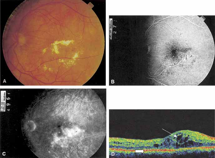
| Fig. 5 A. Circinate retinopathy inferotemporal to the center of the macula. B. The midphase of the fluorescein angiogram shows a cluster of microaneurysms in the center of the circinate ring. C. The late phase of the angiogram shows leakage of fluorescein. D. Optical coherence tomogram centered on the fovea of an eye with diabetic macular edema. The area of marked retina thickening contains numerous hyporeflective cystoid spaces (fine arrow). The outer retina is limited by the hyperreflective pigment epithelial band (thick arrow). |