|
|
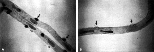
| Fig. 1 A. Trypsin digest preparation of early background retinopathy. Normal retinal capillaries, with one pericyte (closed arrows) per endothelial cell (open arrows). B. Retinal capillary of a patient with diabetes with necrotic pericytes (arrows). (Courtesy of Dr. Myron Yanoff) |