|
|
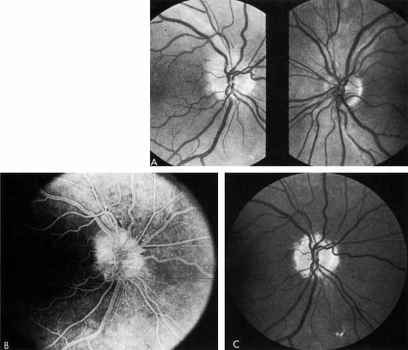
| Fig. 27 A. Right eye: ischemic optic neuritis. Note pale swelling of optic disc and blurring of disc margins. Left eye: normal disc. B. Fluorescein angiogram. Note poor filling on disc inferotemporally as compared with the rest of the disc. C. Right eye 6 months after optic neuritis. Note slight pallor. |