|
|
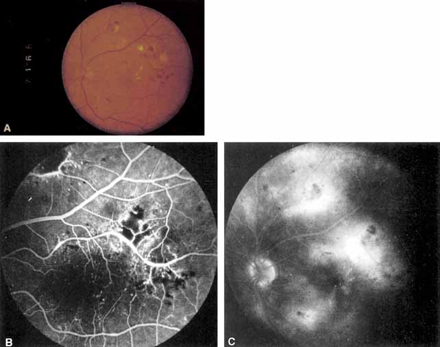
| Fig. 4 A Exudates surround an area of hypoperfused retina. Note soft exudate superiorly. The macular edema thickens the retina and obscures the normal choroidal appearance. B. In the center of the hard exudates the fluorescein angiogram shows capillary non perfusion surrounded by microaneurysms. C. The late phase of the angiogram shows leakage into the retina. |