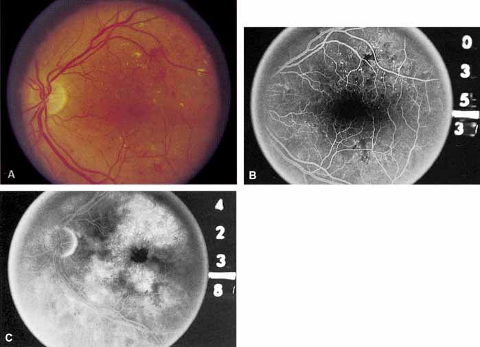
Fig. 6
A
. Background diabetic retinopathy.
B
. The midphase of the fluorescein angiogram shows multiple microaneurysms.
C
. Late phase of the angiogram shows cystoid macular edema.