|
|
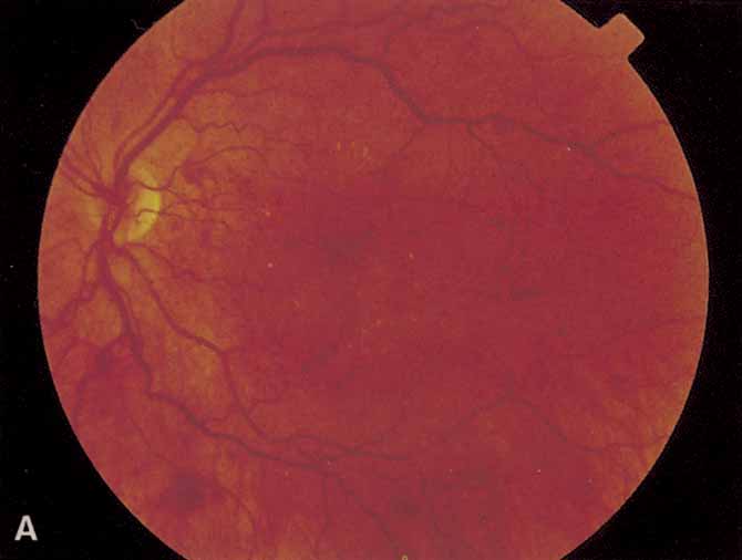
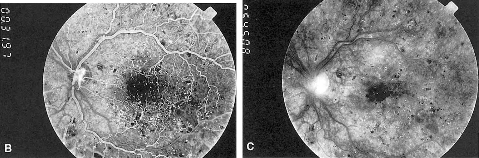
| Fig. 3 A. Diabetic retinopathy with multiple microaneurysms, dot hemorrhages, and early neovascularization of the optic disc (NVD). A small blot hemorrhage is seen inferiorly. B. Continued. Midphase of the fluorescein angiogram. Patent microaneurysms are seen as hyperfluorescent dots. Note that most microaneurysms cannot be seen ophthalmoscopically. There is some enlargement of the foveal avascular zone because of some occluded capillaries. Temporally there is a larger zone of capillary nonperfusion. The NVD is beginning to leak. C. Late phase of the fluorescein angiogram showing diffuse leakage of fluorescein into the macula. |