Chapter 18
Retinopathy of Blood Dyscrasias
ROBERT H. ROSA, JR and RICHARD D. CUNNINGHAM
Main Menu   Table Of Contents

Search

OPHTHALMOSCOPIC FINDINGS
THE BLOOD DYSCRASIAS
REFERENCES

The word dyscrasia comes from the Greek language and means “bad temperament.” In the older medical literature, the term dyscrasia was used to indicate disease. Currently, we use the phrase blood dyscrasia to indicate a pathologic condition of the blood, usually when referring to disorders of the cellular elements of the blood. An increase or decrease in the total number of red blood cells in a given patient is referred to as polycythemia or anemia, respectively. A large population of atypical or neoplastic white blood cells within the blood constitutes leukemia. A subnormal number of platelets in the circulating blood (thrombocytopenia) or loss of normal platelet function can lead to bleeding disorders or coagulopathies. Interestingly, disorders of the various cellular components of the blood often overlap (i.e., leukemia, anemia, and thrombocytopenia).

Clinical and pathologic studies indicate that the ocular manifestations of blood dyscrasias are frequent. Apart from the hemorrhagic and infiltrative complications observed in the skin and mucous membranes, the ocular fundus provides an unequaled direct view of the hematologic effects of blood dyscrasias in the living patient. The ophthalmologist may actually be the first member of the medical team to identify the hematologic effects of a blood dyscrasia by observing the characteristic changes in the retina.

Herein, the typical ophthalmoscopic findings in blood dyscrasias are reviewed, and general information regarding the most common blood dyscrasias is presented.

Back to Top
OPHTHALMOSCOPIC FINDINGS
The typical ophthalmoscopic findings in the various blood dyscrasias are not pathognomonic and may actually be observed in many different local and systemic diseases involving the eye (i.e., diabetes, hypertension, collagen vascular disease). However, the pattern and distribution of the retinal findings in blood dyscrasias are characteristic. If one identifies these characteristic findings on ophthalmoscopy, then ancillary testing may reveal a blood dyscrasia and allow for early referral for treatment by the appropriate medical subspecialist.

RETINAL HEMORRHAGES

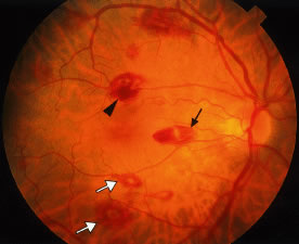
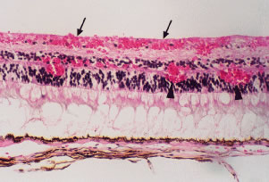
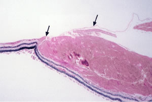
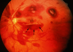
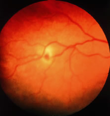
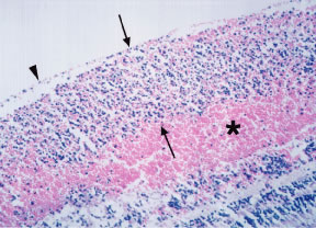
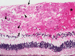
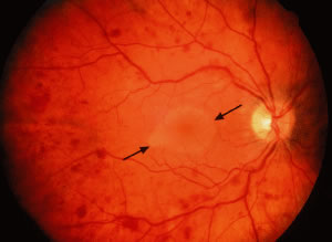
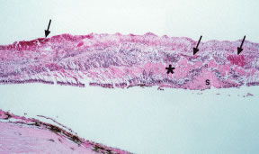
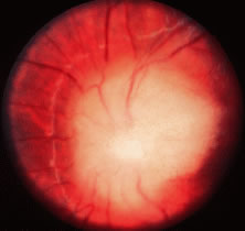
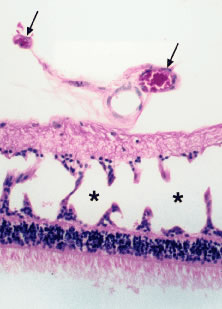
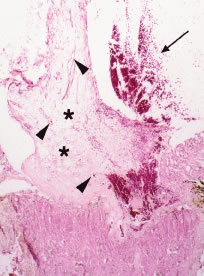
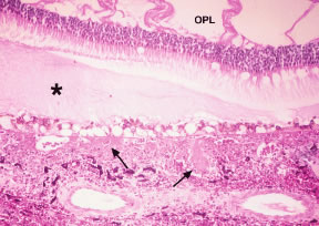
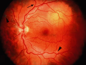
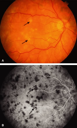
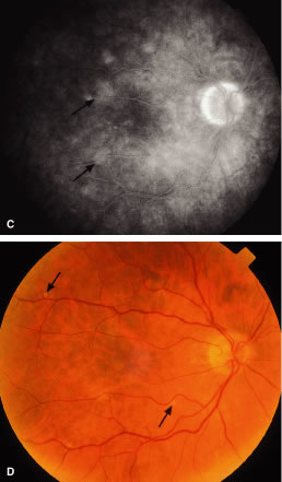
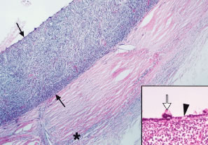
Retinal hemorrhages may take various configurations based on their location within the retina. Flame-shaped and dot-blot hemorrhages are the most commonly encountered intraretinal hemorrhages in blood dyscrasias (Fig. 1). Flame-shaped hemorrhages are located in the nerve fiber layer of the retina, and dot-blot hemorrhages are typically located in the inner nuclear and outer plexiform layers (Fig. 2). Large blot or boat-shaped hemorrhages may be observed and are present beneath the internal limiting membrane (ILM) of the retina (i.e., sub-ILM hemorrhage) (Fig. 3). These large superficial retinal hemorrhages may break through the ILM and extend into the vitreous cavity (Fig. 4). White-centered hemorrhages (see Fig. 1) and, much less commonly, red-centered infiltrates (Fig. 5) are seen in the retinae of patients with a blood dyscrasia. The white center may be associated with a leukemic embolus (Fig. 6) or more commonly a platelet-fibrin thrombus.1 The red center is blood, which may be associated with a cotton wool spot (CWS) or a leukemic retinal infiltrate.

Fig. 1. Intraretinal hemorrhages in acute myelocytic leukemia (AML) and anemia. Note the flame-shaped (arrow), blot (arrowhead), and white-centered (open arrows) hemorrhages.

Fig. 2. Histopathology of intraretinal hemorrhages. Flame-shaped hemorrhages are located in the nerve fiber layer (arrows) and dot-blot hemorrhages are typically located in the inner and outer plexiform and inner nuclear layers (arrowheads).

Fig. 3. Histopathology of sub-internal limiting membrane (sub-ILM) hemorrhage. Note the dense blood beneath the ILM (arrows) of the retina.

Fig. 4. Extensive intraretinal hemorrhages in acute myelocytic leukemia (AML). Note the flame-shaped, blot, and white-centered hemorrhages. A premacular subhyaloid hemorrhage (arrows) with a hyphema-like or boat-shaped configuration is present. (Courtesy Dr. Helmut Buettner.)

Fig. 5. Red-centered retinal infiltrate in acute lymphocytic leukemia (ALL). This retinal leukemic infiltrate with a central blot hemorrhage is localized to a branch retinal vein. (Courtesy Dr. Helmut Buettner.)

Fig. 6. Histopathology of a leukemic infiltrate associated with hemorrhage in chronic myelocytic leukemia (CML). Note the leukemic infiltrate (between arrows) beneath the internal limiting membrane (ILM) (arrowhead) in the nerve fiber layer and the associated intraretinal hemorrhage (asterisk). (Courtesy Dr. W. Richard Green.)

Factors that contribute to the development of retinal hemorrhages in the setting of blood dyscrasias include anemia, thrombocytopenia, elevated white blood cell count, and blood or serum hyperviscosity. In a study of 67 patients with anemia and/or thrombocytopenia, Rubenstein and coworkers2 found that retinal hemorrhage is much more likely to occur when anemia is accompanied by thrombocytopenia than when either is present alone. The same authors postulated that thrombocytopenia is an important causative factor in ocular bleeding in an anemic patient. In a study of 152 patients with blood diseases, Holt and Gordon-Smith3 reported that retinal hemorrhages occurred most often in leukemic patients with significantly more severe anemia and thrombocytopenia and a high percentage of circulating blast cells. In a prospective study of 117 patients with acute leukemia, Guyer and colleagues4 reported that thrombocytopenia is the most important factor in the pathogenesis of intraretinal hemorrhages. On the contrary, Jackson and coworkers found no association between intraretinal hemorrhages and the hemoglobin level or platelet count in 63 newly diagnosed acute leukemia patients.5 The same authors reported a higher median white blood cell count in patients with intraretinal hemorrhages than in those without intraretinal hemorrhages and concluded that a high white blood cell count may be at least as important as anemia and thrombocytopenia in the pathogenesis of retinopathy in acute leukemia.

In a 4-year prospective study of 82 patients with acute leukemia, Jackson and coworkers6 found that patients with a macular hemorrhage were at significantly greater risk for developing intracranial hemorrhage [particularly in the promyelocytic (M3) subtype of acute myeloid leukemia (AML)] within the first 30 days following diagnosis compared with patients without a macular hemorrhage. Based on these findings, the authors recommended that patients with a macular hemorrhage be monitored intensively for the development of intracranial hemorrhage and receive priority in the allocation of platelets when platelets are in short supply. Richards and colleagues7 also reported a high incidence of intraocular hemorrhage in patients with acute promyelocytic leukemia (M3 subtype of AML) and found no consistent detectable abnormalities in the hematologic parameters or coagulation studies predictive of ocular hemorrhage.

In a pathologic study of the eyes of 76 patients who died of leukemia and allied disorders, Allen and Straatsma8 found that retinal hemorrhage was the most frequent and serious ocular complication and that the most significant hemorrhages occurred in the acute forms of leukemia.

MICROANEURYSMS

Microaneurysms are small outpouchings in the retinal capillary wall that appear as tiny red dots in the retina on ophthalmoscopic examination. They are classically seen in the posterior fundus in diabetic retinopathy. Microaneurysms have also been reported in leukemia and plasma cell dyscrasias.9–12 Interestingly, the microaneurysms in blood dyscrasias tend to be located in the peripheral retina, in contrast to the location in the posterior retina in diabetic retinopathy. In a pathologic study employing trypsin digestion of flat mounts of the retina, Duke and coworkers10 emphasized the relative preservation of pericytes in patients with chronic leukemia as compared with the marked loss of pericytes in diabetic retinopathy. Figure 7 shows the typical microaneurysms in the retinal periphery in a flat mount preparation with trypsin digestion in a patient with a blood dyscrasia [multiple myeloma (MM)]. Common pathologic features between the microaneurysms of diabetes and blood dyscrasias include the globular shape of the lesion, the location predominantly on the venous side of the capillary, and the presence of intramural and intraluminal periodic acid-Schiff (PAS)-positive deposits.10 Factors that may play a role in the formation of microaneurysms in the setting of blood dyscrasias include increased venous pressure [i.e., central retinal vein occlusion (CRVO)], increased blood viscosity with a secondary increase in venous pressure (i.e., hyperviscosity syndrome associated with plasma cell dyscrasias), and anoxia (i.e., severe anemia).

Fig. 7. Retinal trypsin digest preparation showing a peripheral microaneurysm (arrow) in multiple myeloma. (Courtesy Dr. W. Richard Green.)

HARD EXUDATES

Hard exudates are refractile, yellowish deposits of proteins and lipids that are derived from incompetent or leaky retinal capillaries. Hyperpermeability of the retinal capillaries may be seen in hyperviscosity syndromes, retinal venous congestion, and CRVO secondary to plasma cell dyscrasias.13–15 The hard exudates that result from this capillary hyperpermeability are localized predominantly to the outer plexiform layer of the retina (Fig. 8).

Fig. 8. Histopathology of hard exudates. Note the thick retinal ganglion cell layer (more than 2 to 3 cells thick) (between brackets) identifying the macular region and the periodic acid-Schiff (PAS)-positive dense proteinaceous material (arrows) in the outer plexiform layer (nerve fiber layer of Henle).

RETINAL EDEMA

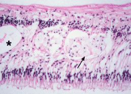
In addition to the formation of hard exudates, hyperpermeable or leaky retinal capillaries can lead to focal or generalized retinal edema. Mild opacification or graying of the normally transparent retina is noted in areas of retinal edema on ophthalmoscopic examination. Cystic degeneration of the retina may result from long-standing retinal edema. Retinal edema localized to the macular region may manifest as cystoid macular edema (CME). Histologically in CME, the cysts often contain eosinophilic proteinaceous material and are located in the outer plexiform and, to a lesser extent, in the inner nuclear layers of the retina (Fig. 9). Occasionally, aggregates of lipid-laden macrophages are observed in the regions of the cystic degeneration (see Fig. 9). As with hard exudates, retinal edema may be seen in hyperviscosity syndromes induced by the blood dyscrasias. The CME observed in blood dyscrasias may respond to specific treatment for CME (i.e., acetazolamide) and may resolve completely with treatment of the underlying disease process [i.e., after bone marrow transplantation in chronic myeloid leukemia (CML)].16

Fig. 9. Histopathology of cystoid macular edema. Note the intraretinal cysts (asterisk) partially filled with proteinaceous material and the focal aggregates of lipid-laden macrophages (arrow) in the inner nuclear and outer plexiform layers.

COTTON WOOL SPOTS

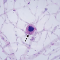
CWSs, also known as soft exudates, are microinfarctions in the nerve fiber layer. The normal flow of axoplasm is blocked in response to focal retinal ischemia caused by occlusion of a precapillary arteriole.17 Clinically, CWSs are superficial whitish retinal lesions with irregular feathery borders that may be associated with small retinal hemorrhages. With resolution, the CWSs will fade and an area of retinal depression may develop secondary to inner retinal ischemic atrophy. Histologically, CWSs are characterized by fusiform thickening of the nerve fiber layer with globular cytoid bodies (Fig. 10)—swollen ganglion cell axons with degenerated cellular organelles (i.e., mitochondria, neurofilaments, endoplasmic reticulum). CWSs are often encountered in the retinopathy associated with blood dyscrasias. In one report,18 the presence of a single CWS in each eye of a patient in the absence of diabetes and systemic hypertension led to the diagnosis of MM. In a prospective study of 54 newly diagnosed patients with acute leukemia, Abu El-Asrar and coworkers19 reported that patients with CWSs had significantly lower mean and median survival times than those patients without CWSs. Thus in this study, the presence of CWSs was a poor prognostic sign for survival in acute leukemia.

Fig. 10. Histopathology of cotton wool spot. Note the thickening of the nerve fiber layer (between arrows), the characteristic eosinophilic cytoid bodies (arrowheads), and the associated retinal hemorrhage (asterisk).

The presence of severe anemia may be a risk factor for the development of CWSs in blood dyscrasias. Holt and Gordon-Smith3 observed CWSs in 14 patients with severe anemia with a mean hemoglobin concentration of 5.6 g/100 ml. In contrast, Guyer and coworkers4 found no statistically significant association between the presence of CWSs and hematologic parameters (including the hematocrit) in their prospective series. CWSs may result from occlusion of the precapillary arterioles by leukemic cells or by platelet-fibrin thrombi.

RETINAL VASCULAR CHANGES

Retinal venous dilatation and tortuosity similar to, and in some cases indistinguishable from, the ophthalmoscopic findings in CRVO may be observed in blood dyscrasias, particularly the plasma cell dyscrasias [i.e., Waldenström's macroglobulinemia (WM), monoclonal gammopathies, and less commonly MM].9,13–15 With venous distention, arteriovenous nicking may become more apparent and give rise to the “sausage link” appearance of the retinal veins. The classical picture of venous stasis retinopathy associated with hyperviscosity is seen in WM (Fig. 11). In a prospective study of 120 patients with newly diagnosed leukemia of all cell types, Schachat and coworkers20 diagnosed a CRVO in 5 patients—all of whom had myeloid leukemia with extremely high white blood cell counts or platelet counts—and attributed the venous occlusion to blood hyperviscosity. The pathologic findings in venous stasis retinopathy associated with blood dyscrasias may be indistinguishable from those found in CRVO (Fig. 12).

Fig. 11. Venous stasis retinopathy in Waldenström's macroglobulinemia. Note the dilatation and tortuosity of the retinal veins, scattered intraretinal hemorrhages, and serous macular detachment (between arrows). (Courtesy Dr. Helmut Buettner.)

Fig. 12. Histopathology of central retinal vein occlusion (CRVO). Note the intraretinal hemorrhages in various layers of the retina (arrows) and the eosinophilic proteinaceous exudates in the outer plexiform layer (asterisk) and subretinal space (S).

Perivascular sheathing may be observed in the ocular fundus in patients with blood dyscrasias. The apparent opacification or loss of transparency of the retinal blood vessel wall may be due to infiltration by benign inflammatory cells in response to immune complex or other protein (e.g., amyloid, immunoglobulin heavy or light chains) deposition, direct infiltration by leukemic cells, or fibrosis secondary to hypertensive retinal vascular disease. Perivascular sheathing and a clinical picture of retinal vasculitis (with leakage from retinal vessels on fluorescein angiography) have been prominent ocular findings in patients presenting with certain blood dyscrasias, including human T-lymphotropic virus 1 (HTLV-1)-associated adult T-cell leukemia/lymphoma,21,22 cryoglobulinemia,23 hairy cell leukemia,24 and, rarely, MM.25 Kim and coworkers26 described a patient with relapsing acute lymphoblastic leukemia who presented with a retinal vasculopathy resembling frosted branch angiitis and an infiltrative optic neuropathy, which resolved with local radiation and intrathecal chemotherapy.

PALLOR OF THE OCULAR FUNDUS

The normal color of the ocular fundus is derived from the retinal pigment epithelium (RPE), the choroidal melanocytes, and the blood in the retinal and choroidal vasculature. The retina is normally transparent. In patients with severe anemia, the fundus may appear pale and the retinal vessels may be less red than normal.

OPTIC DISC EDEMA

In optic disc edema, nerve fiber swelling in the optic disc and peripapillary retina is present and often associated with flame-shaped hemorrhages, whitish punctate lesions (secondary to obstructed axoplasmic flow), and peripapillary CWSs. On ophthalmoscopy, the optic disc and peripapillary retina appear elevated and the normally well-defined margins of the optic disc are blurred. Optic disc edema is usually bilateral and may be due to elevated intracranial pressure (i.e., papilledema) (Fig. 13) secondary to hemorrhage or a mass effect associated with the blood dyscrasia (i.e., meningeal infiltration, granulocytic sarcoma). The disc edema may also be secondary to serum (blood) hyperviscosity and may resemble that seen in CRVO. Less commonly, direct infiltration of the optic nerve or optic disc may cause marked optic disc edema.

Fig. 13. Histopathology of papilledema. Note the marked swelling and elevation of the nerve fiber bundles in the optic disc (asterisks), lateral displacement of the peripapillary retina from the disc margin (bracket), nerve fiber layer hemorrhage (arrowhead), and subhyaloid hemorrhage (arrow).

Rosenthal27 emphasized that optic nerve infiltration occurs predominantly in children with acute lymphocytic leukemia (ALL) and must be differentiated clinically from papilledema. Patients with leukemic infiltration of the prelaminar optic nerve typically have marked swelling of the optic disc with a fluffy superficial infiltrate and variable hemorrhage (Fig. 14). The visual acuity may be minimally or severely affected. With retrolaminar optic nerve infiltration, moderate to marked disc elevation and edema with variable hemorrhage may be present and marked vision loss is usually observed. Optic nerve infiltration may occur despite prophylactic brain irradiation in leukemia because of the shields employed to protect the eyes.28 In general, optic nerve infiltration by leukemia responds well to radiotherapy (with or without intrathecal chemotherapy). Brown and coworkers29 reported the case of a patient with acute promyelocytic leukemia and optic disc infiltration who showed complete resolution with oral all trans-retinoic acid alone.

Fig. 14. Optic disc infiltration in a child with acute lymphocytic leukemia (ALL). (Courtesy Dr. Lawrence Frankel.)

Bilateral optic disc swelling is a common early sign (in up to 73% of cases) of the peripheral neuropathy, organomegaly, endocrinopathy, monoclonal gammopathy, and skin changes (POEMS) syndrome, which typically occurs in middle-aged men in association with a plasma cell dyscrasia.30,31 The optic disc edema may occur in the absence of elevated intracranial pressure and may be mediated by autoantibodies or cytokines.31 Cases of POEMS syndrome—also known as Crow-Fukase syndrome—have been reported worldwide, with the greatest concentration of cases in Japan. Other major features of the syndrome include widespread edema with ascites and pleural effusions, lymphadenopathy, lethargy, weakness, and shortness of breath.30

RETINAL AND OPTIC DISC NEOVASCULARIZATION

Retinal neovascularization with a sea fan configuration, as seen in sickle cell retinopathy, has been reported in four patients with chronic myelogenous leukemia.27 In most of these reports, the affected patients had extremely high white blood cell counts. Prolonged leukocytosis with or without an increased number of circulating platelets increases the blood viscosity, which may reduce blood flow and cause vascular stagnation.27 The hyperviscosity may lead to microaneurysm formation and retinal capillary nonperfusion or dropout. Retinal ischemia may ensue with the subsequent development of proliferative retinopathy (i.e., neovascularization of the retina and optic disc) (Figs. 15 and 16). More recently, Anderton and coworkers32 reported the case of a patient with chronic myelogenous leukemia who initially presented with a vitreous hemorrhage associated with bilateral retinal and optic disc neovascularization. Wiznia and colleagues33 reported the case of a patient with ALL who developed severe bilateral ischemic retinopathy during chemotherapy with cytosine arabinoside, which progressed to bilateral optic disc and retinal neovascularization despite panretinal photocoagulation. The patient had received low-dose irradiation to the brain within 2 months before presenting with bilateral severe vision loss. The toxic effects of the combination of irradiation and chemotherapy, as well as a hyperviscosity syndrome induced by the leukemia, may have contributed to the acute presentation with proliferative retinopathy.

Fig. 15. Histopathology of neovascularization of the retina. Note the blood vessels on the surface of the retina extending into the vitreous cavity (arrows) and the cystoid degeneration of the retina (asterisks) predominantly in the inner nuclear and outer plexiform layers.

Fig. 16. Histopathology of neovascularization of the optic disc. Note the condensed vitreous (asterisks) with small blood vessels (arrowheads) on the surface of the optic disc and associated vitreous hemorrhage (arrow).

In diabetic patients, the concomitant development of a blood dyscrasia may cause rapid progression from mild background diabetic retinopathy to severe proliferative retinopathy in an unusually short period.34,35 The hyperviscosity syndrome and anemia associated with blood dyscrasias may exacerbate and accelerate diabetic retinopathy. The combination of anemia and diabetes results in both reduced blood oxygen-carrying capacity and a reduced dissociation of oxygen from the blood causing greater tissue hypoxia (i.e., retinal ischemia).34

VITREOUS HEMORRHAGE AND INFLAMMATION

Vitreous hemorrhage may be an ophthalmic manifestation of blood dyscrasias, particularly in patients with the combination of anemia and thrombocytopenia. Vitreous hemorrhage may occur in 10% to 15% of patients with aplastic anemia.36 Schachat and coworkers20 reported 3 patients with vitreous hemorrhage in a prospective series of 120 patients with newly diagnosed leukemia of all cell types. Vitreous hemorrhage may occur more often in acute promyelocytic leukemia (M3 subtype of AML), which is characterized by severe hemorrhagic manifestations either at presentation or following commencement of cytotoxic therapy.7 In addition, patients with acute promyelocytic leukemia may develop Terson's syndrome (vitreous hemorrhage associated with intracranial bleeding) during induction therapy with all trans-retinoic acid.37 The vitreous hemorrhage may also occur as a complication of proliferative retinopathy32 from neovascularization of the retina (see Fig. 15) or disc (see Fig. 16).

Apparent vitreous inflammation may be caused by the presence of leukemic cells within the vitreous.21,38,39 Swartz and Schumann38 reported the case of a patient with acute lymphoblastic leukemia in apparent remission who presented with vitreous infiltration diagnosed by cytopathologic evaluation of a vitreous aspirate. The apparent vitreous inflammation was the first clinical sign of central nervous system involvement. Prompt institution of radiotherapy and chemotherapy produced a rapid reduction of cells in the vitreous and clearing of cells from the cerebrospinal fluid.

Vitreous deposits of amyloid are rarely observed in primary systemic nonfamilial amyloidosis.40 This systemic disorder is characterized by an aberrant deposition in various organ systems of insoluble polypeptides derived from a portion of the light chain of immunoglobulins.

RETINAL AND RETINAL PIGMENT EPITHELIUM DETACHMENT

Retinal and retinal pigment epithelium (RPE) detachments are not infrequently observed in blood dyscrasias. A clinical picture with fluorescein angiography resembling central serous retinopathy (i.e., serous retinal and RPE detachments) has been described in patients with plasma cell dyscrasias, including monoclonal gammopathies and cryoglobulinemia.41,42 Rarely, apparent serous macular detachments can occur, and fluorescein angiography shows macular hypofluorescence and no evidence of retinal vascular or RPE leakage associated with the macular elevation. Ho and coworkers43 reported three such cases in patients with MM, WM, and benign polyclonal gammopathy. The same authors hypothesized that the macular detachments were transudative rather than exudative with subretinal precipitates of immunoglobulin or other serum proteins. Ogata and colleagues44 described a patient with WM who presented with an apparent serous macular detachment with no abnormal hyperfluorescence on fluorescein angiography; however, with optical coherence tomography (OCT), a large occult RPE detachment was noted beneath the serous retinal detachment. In plasma cell dyscrasias, the choriocapillaris may be partly obstructed by light chain or other immunoglobulin deposits, and this may play an important role in the pathogenesis of the serous retinal and RPE detachments observed in these disorders.12,45

Rarely, visual impairment secondary to a retinal or RPE detachment may be the first clinical sign of a blood dyscrasia or a leukemic relapse during apparent remission.46–49 In leukemia, the serous retinal and RPE detachments may occur secondary to leukemic infiltration of the choroid.11,27,50 Solid detachments of the retina by leukemic cell infiltrates in the subretinal space have also been reported.11,51 In one such case, a hypopyon-like configuration of the leukemic cells was observed in the subretinal space.51

Serous retinal and RPE detachments may also be seen in platelet disorders and coagulopathies such as thrombotic thrombocytopenic purpura (TTP) and disseminated intravascular coagulation (DIC).52–55 The mechanism of retinal and RPE detachment in these disorders is associated with choriocapillary occlusion by platelet-fibrin thrombi (Fig. 17).

Fig. 17. Histopathology of disseminated intravascular coagulation (DIC). Note the platelet-fibrin thrombi in the choriocapillaris and inner choroidal vessels (arrows), the vacuolar changes in the overlying retinal pigment epithelium (RPE), the proteinaceous subretinal exudate (asterisk), the cysts in the outer plexiform layer (OPL), and the focal choroidal hemorrhage. (Courtesy Dr. W. Richard Green.)

RETINAL AND CHOROIDAL INFILTRATES

Retinal and choroidal infiltrates are often observed in patients with leukemia. A retinal leukemic infiltrate may appear as an elevated yellowish white mass of variable size with or without associated hemorrhage or vitreous inflammation and is most often located in the posterior pole or peripapillary region. Leukemic infiltration of the retina and/or choroid may simulate an infectious chorioretinitis.21,22,56 Gordon and coworkers56 emphasized the importance of distinguishing between infectious and neoplastic retinal infiltrates in patients with a history of leukemia. The same authors found that neoplastic (or leukemic) retinal infiltrates occurred in patients who had newly diagnosed leukemia and those patients in blast crisis. In contrast, two patients who were in complete remission after bone marrow transplantation had infectious retinal infiltrates. Thus the authors concluded that the systemic status (i.e., newly diagnosed leukemia vs. immunosuppression after bone marrow transplantation) ofthe patient is highly informative in determining whether infection or neoplasia is responsible for the retinal infiltration.

Retinal detachment may occur in the region of a choroidal leukemic infiltrate.8,11,27,49 RPE changes, including pigment clumping and pigment mottling, may be observed overlying the choroidal infiltrate.57 A choroidal leukemic infiltrate typically appears as an elevated, creamy, yellowish-white lesion (Fig. 18). Fluorescein angiography may reveal early multifocal punctate hyperfluorescence over the choroidal infiltrate with late pooling of fluorescein in the subretinal space resembling Harada's disease.49 Ophthalmoscopic and fluorescein angiographic findings resembling acute posterior multifocal placoid pigment epitheliopathy (APMPPE) have been described in patients with leukemia.58,59 The APMPPE-like features include deep, whitish, well-circumscribed fundus lesions that exhibit early blockage and late leakage on fluorescein angiography (Fig. 19).

Fig. 18. Choroidal infiltrate in chronic myeloid leukemia (CML). Note the creamy yellow-white lesion supertemporal to the central macula, few dot hemorrhages (arrow), and cotton wool spots (arrowheads). (Courtesy Dr. Helmut Buettner.)

Fig. 19. Choroidal infiltrates resembling acute posterior multifocal placoid pigment epitheliopathy (APMPPE) in acute myelocytic leukemia (AML). A. Note the deep yellowish-white lesions (arrows) in the posterior fundus. B. Laminar venous phase (24.8 seconds) of fluorescein angiogram showing early blockage (arrows) corresponding to the fundus lesions noted in A. C. Late phase (676 seconds) of fluorescein angiogram showing late leakage corresponding to the early hypofluorescence (arrows) in B and the yellow-white lesions in A. D. Four months after local irradiation. Note the resolution of the yellow-white lesions and few small punched-out atrophic chorioretinal scars (arrows) in areas corresponding to previous lesions as noted in A.

McManaway and Neely60 reported the case of a patient with acute lymphoblastic leukemia who presented with leukocoria and proptosis resulting from an extensive intraocular and orbital tumor mimicking advanced retinoblastoma. An elevated leukocyte blood count, blast forms on the peripheral blood smear, rubbery suboccipital nodules, and subcutaneous scalp masses all suggested leukemia, although the fundus picture was atypical and caused diagnostic confusion. No intratumoral calcification was observed on a computed tomography (CT) scan. The ocular and orbital infiltrates responded rapidly to chemotherapy and low-dose orbital irradiation.

The frequency of these infiltrates, particularly choroidal infiltrates, is much higher in pathologic studies (compared with clinical studies) of eyes from patients with acute and chronic leukemia and ranges between 28% and 80%.8,11,61 In a prospective study of 120 patients with newly diagnosed leukemia (all cell types), Schachat and coworkers20 found 4 patients (3%) with leukemic retinal infiltrates and none with choroidal infiltrates. Leukemic choroidal infiltrates may be difficult to diagnose clinically without special ancillary tests (i.e., fluorescein angiography, A- and B-scan ultrasonography). In a pathologic study of 135 autopsy eyes in patients with leukemia, Leonardy and coworkers61 reported choroidal involvement in 93% of eyes with leukemic infiltrates. Allen and Straatsma8 found a much higher incidence of ocular involvement, including choroidal infiltration, in eyes of patients with acute leukemia (80%) compared with chronic leukemia (29%). The same authors emphasized the diffuse pattern of leukemic infiltration of the posterior choroid (Fig. 20) and infrequent association with hemorrhage as opposed to the focal leukemic infiltration observed in the retina with associated hemorrhage in nearly every case.

Fig. 20. Histopathology of leukemic choroidal infiltrate. Note the diffuse thickening of the choroid by leukemic cell infiltration (between arrows) in acute myelocytic leukemia (AML). Leukemic cells are also present in the episclera (asterisk) and in scleral emissary canals. The inset shows retinal pigment epithelium (RPE) changes, including focal hyperplasia (open arrow) and depigmentation (arrowhead) overlying the leukemic choroidal infiltrate. (Courtesy Dr. W. Richard Green.)

Diffuse bilateral chorioretinal abnormalities including deep hemorrhages and pigment mottling in the posterior pole, diffuse areas of hypofluorescence on fluorescein and indocyanine green angiography in the posterior pole, and hypofluorescent lines or streaks in the midperipheral fundus were observed by Pece and coworkers62 in a patient with primary systemic nonfamilial amyloidosis. These authors postulated that the areas of hypofluorescence may represent either choroidal vascular occlusion or intravascular and/or perivascular amyloid deposits.

Back to Top
THE BLOOD DYSCRASIAS

ANEMIA

The term anemia refers to a reduction in the concentration of hemoglobin or red blood cells in the blood. Three concentrations—hemoglobin, hematocrit, and number of red blood cells—may be measured to establish the presence of anemia. Blood hemoglobin concentration is the preferred measure because of its accuracy, reproducibility, and pathophysiologic correlation. The morphologic classification of anemia divides the anemias into three broad groups on the basis of average cell size and hemoglobin content: the macrocytic anemias; the microcytic and hypochromic anemias; and the normocytic, normochromic anemias. The number of red blood cells in the circulation at any given time is a result of the production and delivery of cells into the circulation and their destruction or loss from the circulation. Iron-deficiency anemia and anemia of chronic disease are the most common causes of anemia and are characteristically of the microcytic, hypochromic type. In the blood dyscrasias, the bone marrow may be infiltrated and replaced by neoplastic white cells, resulting in pancytopenia. Retinal hemorrhages are common findings in anemic patients, particularly when the anemia is accompanied by thrombocytopenia.2,4 In profound anemia, CWSs3 and white-centered hemorrhages4 may be observed with greater frequency in addition to retinal hemorrhages.

Pernicious anemia—the prototype of megaloblastic (macrocytic) anemia—is a chronic illness resulting from the lack of Castle's “intrinsic factor” in gastric secretions. In the absence of intrinsic factor, a binding protein, the absorption of vitamin B12 is impaired, resulting in a vitamin deficiency. Pernicious anemia occurs in two forms. In the relatively common adult type, the lack of intrinsic factor is associated with gastric atrophy and a deficiency of many other gastric secretions. In the congenital form, only intrinsic factor is lacking, and levels of other gastric secretions are normal. Holt and Gordon-Smith3 reported the retinal abnormalities in 13 patients with pernicious anemia: 54% of the patients had intraretinal hemorrhages (flame-shaped, dot-blot, and white-centered) and 31% had CWSs. Only one patient was thrombocytopenic. The retinal abnormalities invariably cleared after the administration of vitamin B12.

Aplastic anemia is a life-threatening disorder characterized by pancytopenia and a hypoplastic bone marrow. Two deaths per million occur annually in patients with aplastic anemia secondary to cerebral hemorrhage, septicemia, or gastrointestinal bleeding. This type of anemia is especially prevalent in the first three decades of life. Exposure to insecticides and certain drugs have been implicated in the bone marrow suppression. Ocular fundus findings are often observed (up to 75%) in patients with aplastic anemia and include, in order of decreasing frequency, retinal hemorrhages, CWSs, vitreous hemorrhage, a CRVO-like picture, and papilledema.36 Preretinal and vitreous hemorrhages occur almost exclusively in patients with combined anemia and thrombocytopenia.

POLYCYTHEMIA

Polycythemia vera (PV) is a chronic, clonal, myeloproliferative disorder characterized by a striking absolute increase in the number of red blood cells and in the total blood volume and is usually accompanied by leukocytosis, thrombocytosis, and splenomegaly. The bone marrow is hypercellular and exhibits hyperplasia of myeloid, erythroid, and megakaryocyte lineages. PV occurs most commonly in older men (60 to 80 years of age). The signs and symptoms of PV can be attributed to the expanded total blood volume and to the slowing of blood flow as a result of increased blood viscosity.

Secondary causes of polycythemia include disorders that involve decreased tissue oxygen delivery, usually as a result of decreased arterial oxygen saturation. Such disorders include cyanotic congenital heart disease, right-sided heart failure, and chronic obstructive pulmonary disease. In these disorders, tissue hypoxia causes an increase in renal erythropoietin production, which stimulates the erythroid precursors in the bone marrow and increases the number of circulating red blood cells to increase tissue oxygen delivery. In PV and secondary polycythemia, the consequences of marked hyperviscosity that accompany a hematocrit greater than 60% include decreased cerebral blood flow, decreased cardiac output, and a tendency to thrombosis.

The ocular findings in polycythemia are actually manifestations of the associated hyperviscosity syndrome and similar to those observed in plasma cell dyscrasias. Ocular fundus findings include retinal venous dilatation and tortuosity and intraretinal hemorrhages.

LEUKEMIA

The acute leukemias are a heterogeneous group of neoplasms affecting hematopoietic stem cells. Some differentiation in these white blood cell precursors allows for phenotypic classification. Acute leukemias are divided into nonlymphocytic (commonly referred to as myeloid) and lymphoid categories depending on the cell of origin and are further subclassified by the French-American-British (FAB) system based on morphologic characteristics.

Myeloid and lymphoid leukemias differ in regard to their clinical presentation, course, and response to therapy. Current management with specific therapeutic regimens is based on phenotypic characterization of the leukemic cells at diagnosis [i.e., ALL vs. acute nonlymphocytic leukemia (ANLL) or AML]. In addition to morphologic assessment, cytochemical, immunologic, cytogenetic, and occasionally ultrastructural and molecular genetic analyses are performed to further characterize the specific subtype of acute leukemia.

Clinical features are useful but not pathognomonic in the differentiation of ALL and AML. ALL has a peak incidence in childhood, and AML has a peak incidence in adult age; however, some overlap exists between the two types of acute leukemia. Massive lymph node enlargement is much more common in ALL than in AML. Solid masses of leukemic cells (chloromas or granulocytic sarcomas) usually involving facial or intracranial structures are observed in AML. Extensive involvement of the gums is characteristically seen in acute monocytic leukemia.

Chronic lymphocytic leukemia (CLL) is characterized by the accumulation of nonproliferating, mature-appearing lymphocytes in the blood, marrow, lymph nodes, and spleen. In most cases, the cells are monoclonal B lymphocytes. CLL is the most common form of leukemia in North America and Europe, accounting for one third of all cases. CLL typically occurs in older patients, with a peak incidence at 50 to 55 years, and it affects men twice as often as women. The cause of CLL is unknown; however, there is an increased incidence in farmers, rubber manufacturing workers, asbestos workers, and tire repair workers. Genetic factors may play a role. Clinical manifestations include slowly enlarging lymph nodes and gradual enlargement of the liver and spleen resulting from the accumulation of neoplastic lymphocytes. As patients become symptomatic from these clinical manifestations or if progressive anemia and/or thrombocytopenia occur, alkylating agents (i.e., chlorambucil) and prednisone are beneficial. Radiotherapy to the spleen or to areas of bulky adenopathy may also be employed.

CML is a clonal stem cell disorder characterized by increased proliferation of myeloid elements at all stages of differentiation. The incidence of CML steadily increases with age (1 in 100,000 in Western countries), with a peak incidence at 53 years. Men are affected more often than women. The etiology of CML is unknown, although it may develop after radiation exposure. All marrow cell lines in CML express a marker chromosome—the Philadelphia chromosome—a reciprocal translocation of part of the long arm of chromosome 22 to chromosome 9. CML generally occurs in two distinct clinical phases. The first or chronic phase (usually lasting 3 to 5 years) is marked by a proliferation of myeloid cells showing a full range of maturation. Eventually, a decrease in myeloid differentiation typically occurs, and the disease enters an accelerated phase or blast crisis with a very poor prognosis. Clinically, leukocytosis with early myeloid precursors in the peripheral blood, thrombocytosis, and splenomegaly are commonly present at the time of diagnosis. Initially, patients are managed with periodic oral chemotherapy (alkylating agents or hydroxyurea) to normalize the blood count and reduce splenomegaly. Eventually, anemia and thrombocytopenia develop, and fever and increasing weakness may occur with transformation to the accelerated phase or blast crisis (usually lasting 3 to 6 months).

The ocular fundus manifestations of leukemia include intraretinal hemorrhages (flame-shaped, dot-blot, white-centered, and boat-shaped), microaneurysms, hard exudates, retinal edema, CWSs, venous stasis retinopathy, perivascular sheathing, papilledema, optic disc infiltration, retinal and optic disc neovascularization, vitreous inflammation and/or hemorrhage, retinal and RPE detachment, and retinal and choroidal infiltrates.11,27 The presence of intraretinal hemorrhages63 or other ophthalmic manifestations64 in acute leukemia, particularly in childhood leukemias, may be a poor prognostic sign for long-term survival. Reddy and Menon65 recommended routine ophthalmologic examination as part of a complete evaluation at the time of diagnosis in childhood acute leukemias because of the high prevalence (17%) of asymptomatic ocular lesions in their prospective series of 82 children. Treatment of the underlying cause of the ocular fundus manifestations (i.e., anemia, thrombocytopenia, retinal or choroidal leukemic infiltrate, hyperviscosity syndrome, circulating neoplastic white blood cells, and/or bone marrow infiltration) will often result in improvement or complete resolution of the ocular fundus findings.66

PLATELET DISORDERS AND COAGULOPATHIES

Thrombocytopenia is defined as a subnormal number of platelets in the circulating blood and is the most common cause of abnormal bleeding. Thrombocytopenia results from four basic processes: artifactual thrombocytopenia (falsely low platelet counts most commonly resulting from platelet clumping), deficient platelet production (secondary to bone marrow injury by drugs, irradiation, aplastic anemia, or an infiltrative process), accelerated platelet destruction (most commonly resulting from various immunologic factors), and abnormal pooling of platelets within the body (in disorders associated with splenomegaly or dilution of platelets with massive blood transfusions). Accelerated platelet destruction is the most common cause of thrombocytopenia and may be manifested as platelet consumption in intravascular thrombi or on damaged endothelial surfaces, as seen in TTP and DIC.

TTP is an acute, relapsing disease characterized by disseminated thrombotic occlusions of the microcirculation and a syndrome of hemolytic anemia, thrombocytopenia, neurologic symptoms, fever, and renal dysfunction. TTP is more common in women than in men by a 2:1 ratio and shows a peak incidence between 30 to 40 years. The thrombotic lesions of TTP typically involve terminal arterioles and capillaries and are composed of platelets, von Willebrand factor, and fibrin. The etiology of the thrombotic lesions is unknown; however, the primary process may involve endothelial damage caused by immune, infectious, or chemical agents. The lesions of TTP are observed most commonly in the brain, kidney, pancreas, heart, spleen, and adrenal glands. The clinical manifestations of this disorder result from the consumption of platelets within partial and complete vascular occlusions in different organs, which leads to secondary organ dysfunction. Treatment options include splenectomy, steroids, platelet inhibitory drugs (i.e., aspirin, dipyridamole), exchange transfusion, plasmapheresis, and plasma infusion. Ocular fundus manifestations of TTP include retinal hemorrhages, serous macular detachments secondary to choriocapillary occlusion by platelet-fibrin thrombi, optic disc edema, optic disc neovascularization, vitreous hemorrhage, and optic atrophy.54

DIC is a relatively common hemorrhagic disorder caused by the release of tissue thromboplastin into the circulation, allowing massive clotting to occur in blood vessels throughout the body. DIC may be caused by various conditions, including sepsis (especially with gram-negative bacteria), severe tissue injury (i.e., burns and head injuries), obstetric complications (i.e., amniotic fluid emboli, incomplete abortion, retained dead fetus, abruptio placentae), cancer (i.e., acute promyelocytic leukemia, mucinous adenocarcinoma), snakebites, and major hemolytic transfusion reactions. With consumption of the platelets and clotting factors during widespread clot formation, the blood becomes hypocoagulable, leading to hemorrhage, tissue hypoxia, and further release of thromboplastin (i.e., a vicious cycle of bleeding and thrombosis). The major ocular fundus manifestations of DIC include serous detachments of the retina (particularly in the macular and peripapillary regions), retinal and choroidal hemorrhages, and vitreous hemorrhage.52,53,55 In addition, histopathologic studies of eyes in patients with DIC revealed vacuolar disruption of the RPE and fibrin thrombi in the retina and in the choriocapillaris (see Fig. 17) and adjacent choroidal arterioles and venules beneath the areas of serous retinal detachment in the macular and peripapillary regions.52,53

PLASMA CELL DYSCRASIAS

The plasma cell dyscrasias are blood disorders that share two basic characteristics: (1) the proliferation or accumulation of cells normally involved in antibody production and (2) the synthesis and secretion of a structurally homogeneous gamma-globulin (“M-component”) and/or its constituent polypeptide subunits. Plasma cell dyscrasias include MM, WM, the heavy chain diseases, amyloidosis, and benign monoclonal hypergammaglobulinemia (or monoclonal gammopathies of undetermined significance). There are five classes of immunoglobulins: IgA, IgG, IgD, IgE, and IgM. Each immunoglobulin has two heavy chains and two light chains (designated kappa and lambda). In plasma cell dyscrasias, the neoplastic cell may produce a single chain, part of a chain or a complete monoclonal immunoglobulin, which can be defined by protein electrophoresis.

MM is the most common plasma cell dyscrasia. MM is a neoplasm of mature and immature plasma cells that produce a typical monoclonal (M) protein identified as a “spike” by serum protein electrophoresis. The clinical manifestations of this disorder result from the proliferation of plasma cells, bone marrow infiltration and replacement by these plasma cells, and the overproduction of certain proteins and polypeptide chains (M-components). The incidence of MM increases with age; it is most often encountered in middle-aged and older men (although some studies indicate a similar incidence in men and women). Systemic involvement includes osteolytic bone lesions, Bence Jones proteinuria, renal insufficiency, anemia, thrombocytopenia, neurologic sequelae, cryoglobulinemia, hyperviscosity syndrome, and secondary amyloidosis.13 Ocular fundus manifestations of MM include intraretinal hemorrhages (flame-shaped, dot-blot, white-centered), microaneurysms, venous stasis retinopathy with venous dilatation and tortuosity, retinal edema, CRVO, BRVO, vitreous hemorrhage, neovascularization of the disc and retina, serous retinal and RPE detachments, papilledema, and rarely optic nerve infiltration by plasma cells.9,12,13

WM is a disease seen typically in older adults, with a peak incidence in the sixth and seventh decades and a slight male preponderance. WM may follow a long period of benign monoclonal gammopathy. When signs and symptoms of WM develop, the disorder is often mild, with prolonged survival. Symptoms of vague ill health, weakness, a hemorrhagic tendency, and weight loss are common and may antedate more serious complications by many years. With progression of the disease, hepatomegaly, splenomegaly, and lymphadenopathy develop and the clinical picture becomes more consistent with lymphoma or CLL. Eventually, the lungs, gastrointestinal tract, kidneys, and central nervous system or meninges may be infiltrated by neoplastic cells. The characteristic ocular fundus findings are likely secondary to serum hyperviscosity9 and include serous macular and RPE detachments,43,44 retinal venous tortuosity and dilatation with a sausage-like configuration, microaneurysms, CRVO, intraretinal hemorrhages (flame-shaped, dot-blot, white-centered), retinal and optic disc edema, CWSs, and less often neovascularization of the retina and optic disc. Plasmapheresis can lower the serum viscosity and reverse the retinopathy.9

Cryoglobulins are serum proteins or protein complexes that undergo reversible precipitation at low temperatures. Several types of cryoglobulins have been recognized, including monoclonal immunoglobulins, light chains, polyclonal immunoglobulins, and antigen-antibody complexes. The most common signs and symptoms result from impaired blood flow secondary to cryoprecipitation within the capillaries of the skin and include Raynaud phenomenon; necrosis or gangrene of the tip of the nose, ears, fingers, toes, or legs; acrocyanosis; and vascular purpura. Reported ocular fundus findings include retinal hemorrhages, hard exudates, CWSs, CRVO, central retinal artery occlusion (CRAO), venous stasis retinopathy with venous dilatation and tortuosity, retinal edema, retinal and RPE detachments simulating central serous retinopathy, and optic atrophy.9,23,41,42

The term amyloidosis refers to a family of diseases characterized by the deposition of a homogeneous, eosinophilic material in various tissues throughout the body. The major component and hallmark of these deposits consists of fibrils with a twisted Β-pleated sheet configuration. The modern classification of the amyloid syndromes is based on the biochemical composition of the constituent protein chains. Primary amyloidosis is characterized by the deposition of light chain-derived fibrils (AL), and secondary amyloidosis is caused by the deposition of amyloid protein A (AA). Amyloidosis occurs in 5% to 15% of patients with MM. Systemic involvement in MM is similar to primary amyloidosis. Clinical manifestations may include renal insufficiency with proteinuria and nephrotic syndrome, congestive heart failure, cardiac arrhythmias, paresthesias, orthostatic hypotension, gastrointestinal obstruction and malabsorption, hepatosplenomegaly, and purpura. Ocular fundus findings include perivascular sheathing, vitreous veils, CWSs, retinal hemorrhages, retinal and choroidal infiltrates in a perivascular distribution, RPE mottling, and choroidal hypofluorescence on fluorescein and indocyanine green angiography.62,67

Back to Top
REFERENCES

1. Duane TD, Osher RH, Green WR. White centered hemorrhages: Their significance. Ophthalmology 1980;87:66

2. Rubenstein RA, Yanoff M, Albert DM. Thrombocytopenia, anemia, and retinal hemorrhage. Am J Ophthalmol 1968;65:435

3. Holt JM, Gordon-Smith EL. Retinal abnormalities in diseases of the blood. Br J Ophthalmol 1969;53:145

4. Guyer DR, Schachat AP, Vitale S et al. Leukemic retinopathy: Relationship between fundus lesions and hematologic parameters at diagnosis. Ophthalmology 1989;96:860

5. Jackson N, Reddy SC, Hishamuddin M et al. Retinal findings in adult leukaemia: Correlation with leukocytosis. Clin Lab Haematol 1996;18:105

6. Jackson N, Reddy SC, Harun MH et al. Macular haemorrhage in adult acute leukaemia patients at presentation and the risk of subsequent intracranial haemorrhage. Br J Haematol 1997;98:204

7. Richards EM, Marcus RE, Harper P et al. Intra-ocular haemorrhage, a frequent complication of acute promyelocytic leukaemia. Clin Lab Haematol 1992;14:169

8. Allen RA, Straatsma BR. Ocular involvement in leukemia and allied disorders. Arch Ophthalmol 1961;66:490

9. Carr RE, Henkind P. Retinal findings associated with serum hyperviscosity. Am J Ophthalmol 1963;56:23

10. Duke JR, Wilkinson CP, Sigelman S. Retinal microaneurysms in leukaemia. Br J Ophthalmol 1968;52:368

11. Kincaid MC, Green WR. Ocular and orbital involvement in leukemia. Surv Ophthalmol 1983;27:211

12. Khouri GG, Murphy RP, Kuhajda FP et al. Clinicopathologic features in two cases of multiple myeloma. Retina 1986;6:169

13. Knapp AJ, Gartner S, Henkind P. Multiple myeloma and its ocular manifestations. Surv Ophthalmol 1987;31:343

14. Hayasaka S, Ugomori S, Kodama T et al. Central retinal vein occlusion in two patients with immunoglobulin G multiple myeloma associated with blood hyperviscosity. Ann Ophthalmol 1993;25:191

15. Enzenauer RW, Brozetti JJ, Dragoo RA. Central retinal vein occlusion in a patient with IgG lambda monoclonal gammopathy. Arch Ophthalmol 1999;117:134

16. Horgan SE, Fraser SG, Ferrante PF et al. Cystoid macular oedema in chronic myeloid leukaemia: Treatment with acetazolamide and response to bone marrow transplantation. Eye 1996;10:394

17. Ashton N, Harry J. The pathology of cotton-wool spots and cytoid bodies in hypertensive retinopathy and other diseases. Trans Ophthalmol Soc U K 1963;83:91

18. Shami MJ, Uy RN. Isolated cotton-wool spots in a 67-year-old woman. Surv Ophthalmol 1996;40:413

19. Abu El-Asrar AM, Al-Momen AK, Kangave D et al. Prognostic importance of retinopathy in acute leukemia. Doc Ophthalmol 1996;91:273

20. Schachat AP, Markowitz JA, Guyer DR et al. Ophthalmic manifestations of leukemia. Arch Ophthalmol 1989;107:697

21. Kohno T, Uchida H, Inomata H et al. Ocular manifestations of adult T-cell leukemia/lymphoma: A clinicopathologic study. Ophthalmology 1993;100:1794

22. Kumar SR, Gill PS, Wagner DG et al. Human T-cell lymphotropic virus type I-associated retinal lymphoma: A clinicopathologic report. Arch Ophthalmol 1994;112:954

23. Talks SJ, Shah P, Willshaw HE et al. Cryoglobulinaemia masquerading as rheumatoid vasculitis: The retina provides the clue. Eye 1996;10:399

24. Di Maria A, Redaelli C, Canevari A et al. Unilateral retinal vasculitis associated with hairy cell leukaemia: Immunogenetic study. Ophthalmologica 1998;212:355

25. Keung YK, Yung C, Wong JW et al. Association of temporal arteritis, retinal vasculitis, and xanthomatosis with multiple myeloma: Case report and literature review. Mayo Clinic Proc 1998;73:657

26. Kim TS, Duker JS, Hedges TR. Retinal angiopathy resembling unilateral frosted branch angiitis in a patient with relapsing acute lymphoblastic leukemia. Am J Ophthalmol 1994;117:806

27. Rosenthal AR. Ocular manifestations of leukemia: A review. Ophthalmology 1983;90:899

28. Wallace RT, Shields JA, Shields CL et al. Leukemic infiltration of the optic nerve. Arch Ophthalmol 1991;109:1027

29. Brown DM, Kimura AE, Ossoinig KC et al. Acute promyelocytic infiltration of the optic nerve treated by oral trans-retinoic acid. Ophthalmology 1992;99:1463

30. Bolling JP, Brazis PW. Optic disk swelling with peripheral neuropathy, organomegaly, endocrinopathy, monoclonal gammopathy, and skin changes (POEMS syndrome). Am J Ophthalmol 1990;109:503

31. Enzenauer RW, Brozetti JJ, Dragoo RA. POEMS syndrome: An unusual cause of bilateral optic disc swelling. Am J Ophthalmol 1998;126:452

32. Anderton L, Amoaku WM, Noble BA. Retinal and optic disc neovascularization in leukaemia. Acta Ophthalmol 1994;72:752

33. Wiznia RA, Rose A, Levy AL. Occlusive microvascular retinopathy with optic disc and retinal neovascularization in acute lymphocytic leukemia. Retina 1994;14:253

34. Melberg NS, Grand MG, Rup D. The impact of acute lymphocytic leukemia on diabetic retinopathy. J Pediatr Hematol Oncol 1995;17:81

35. Raynor MK, Clover A, Luff AJ. Leukaemia manifesting as uncontrollable proliferative retinopathy in a diabetic. Eye 2000;14:400

36. Mansour AM, Salti HI, Han DP et al. Ocular findings in aplastic anemia. Ophthalmologica 2000;214:399

37. Abu El-Asrar AM, Al-Momen AK, Harakati MS. Terson's syndrome in a patient with acute promyelocytic leukemia on all-trans retinoic acid treatment. Doc Ophthalmol 1993;84:373

38. Swartz M, Schumann B. Acute leukemic infiltration of the vitreous diagnosed by pars plana aspiration. Am J Ophthalmol 1980;90:326

39. Robinson A, Eting E, Zeidman A et al. Ocular manifestation of hairy cell leukemia with dramatic response to 2-chloro-deoxy-adenosine. Am J Ophthalmol 1996;121:97

40. Schwartz MF, Green WR, Michels RG et al. An unusual case of ocular involvement in primary systemic nonfamilial amyloidosis. Ophthalmology 1982;89:394

41. Cohen SM, Kokame GT, Gass JD. Paraproteinemias associated with serous detachments of the retinal pigment epithelium and neurosensory retina. Retina 1996;16:467

42. Zamir E, Chowers I. Central serous chorioretinopathy in a patient with cryoglobulinaemia. Eye 1999;13:265

43. Ho AC, Benson WE, Wong J. Unusual immunogammopathy maculopathy. Ophthalmology 2000;107:1099

44. Ogata N, Ida H, Takahasi K et al. Occult retinal pigment epithelial detachment in hyperviscosity syndrome. Ophthalmic Surg Lasers 2000;31:248

45. Daicker BC, Mihatsch MJ, Strom EH et al. Ocular pathology in light chain deposition disease. Eur J Ophthalmol 1995;5:75

46. Tang G, Vila-Coro AA, Wall S et al. Acute leukemia presenting as a retinal pigment epithelium detachment. Arch Ophthalmol 1988;106:21

47. Primack JD, Smith ME, Tychsen L. Retinal detachment in a child as the first sign of leukemic relapse: Histopathology, MRI findings, treatment, and tumor-free follow up. J Pediatr Ophthalmol Strabismus 1995;32:253

48. Brody JM, Butrus SI, Ashraf MF et al. Multiple myeloma presenting with bilateral exudative macular detachments. Acta Ophthalmol Scand 1995;73:81

49. Miyamoto K, Kashii S, Honda Y. Serous retinal detachment caused by leukemic choroidal infiltration during complete remission. Br J Ophthalmol 2000;84:1318

50. Murphy JA, Pitts JF, Dudgeon J et al. Retinal detachments due to chronic lymphocytic leukaemia. Clin Lab Haematol 1991;13:217

51. Schworm HD, Nasemann JE, Schriever S. Subretinal hypopyon in prolymphocytic leukaemia. Br J Ophthalmol 1995;79:863

52. Cogan DG. Ocular involvement in disseminated intravascular coagulopathy. Arch Ophthalmol 1975;93:1

53. Ortiz JM, Yanoff M, Cameron JD et al. Disseminated intravascular coagulation in infancy and in the neonate: Ocular findings. Arch Ophthalmol 1982;100:1413

54. Jellie HG, Gonder JR, Canny CL et al. Ocular involvement in thrombotic thrombocytopenic purpura: The angiographic and histopathological features. Can J Ophthalmol 1984;19:279

55. Bjerknes T, Askvik J, Albrechtsen S et al. Retinal detachment in association with preeclampsia and abruptio placentae. Eur J Obstet Gynecol Reprod Biol 1995;60:91

56. Gordon KB, Rugo HS, Duncan JL et al. Ocular manifestations of leukemia: Leukemic infiltration versus infectious process. Ophthalmology 2001;108:2293

57. Clayman HM, Flynn JT, Kock K et al. Retinal pigment epithelial abnormalities in leukemic disease. Am J Ophthalmol 1972;74:416

58. Dosso AA, Kapetanios AD. Placoid chorioretinopathy in acute leukemia. Retina 1996;16:455

59. Verbraak FD, van den Berg W, Bos PJM. Retinal pigment epitheliopathy in acute leukemia. Am J Ophthalmol 1991;111:111

60. McManaway JW, Neely JE. Choroidal and orbital leukemic infiltrate mimicking advanced retinoblastoma. J Pediatr Ophthalmol Strabismus 1994;31:394

61. Leonardy NJ, Rupani M, Dent G et al. Analysis of 135 autopsy eyes for ocular involvement in leukemia. Am J Ophthalmol 1990;109:436

62. Pece A, Yannuzzi L, Sannace C et al. Chorioretinal involvement in primary systemic nonfamilial amyloidosis. Am J Ophthalmol 2000;130:250

63. Reddy SC, Quah SH, Low HC et al. Prognostic significance of retinopathy at presentation in adult acute leukemia. Ann Hematol 1998;76:15

64. Ohkoshi K, Tsiaras WG. Prognostic importance of ophthalmic manifestations in childhood leukaemia. Br J Ophthalmol 1992;76:651

65. Reddy SC, Menon BS. A prospective study of ocular manifestations in childhood acute leukaemia. Acta Ophthalmol Scand 1998;76:700

66. Mehta AB, Goldman JM, Kohner E. Hyperleucocytic retinopathy in chronic granulocytic leukaemia: The role of intensive leucapheresis. Br J Haematol 1984;56:661

67. Gass JDM. Stereoscopic Atlas of Macular Diseases: Diagnosis and Treatment. 4th ed. St Louis, Mosby-Year Book, 1997

Back to Top
GENERAL REFERENCE

Lee GR, Foerster J, Lukens J et al. Wintrobe's Clinical Hematology. 10th ed. Baltimore, Williams & Wilkins, 1999

Back to Top