|
|
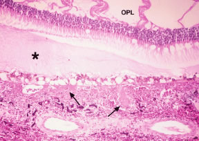
| Fig. 17. Histopathology of disseminated intravascular coagulation (DIC). Note the platelet-fibrin thrombi in the choriocapillaris and inner choroidal vessels (arrows), the vacuolar changes in the overlying retinal pigment epithelium (RPE), the proteinaceous subretinal exudate (asterisk), the cysts in the outer plexiform layer (OPL), and the focal choroidal hemorrhage. (Courtesy Dr. W. Richard Green.) |