|
|
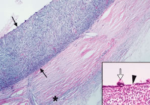
| Fig. 20. Histopathology of leukemic choroidal infiltrate. Note the diffuse thickening of the choroid by leukemic cell infiltration (between arrows) in acute myelocytic leukemia (AML). Leukemic cells are also present in the episclera (asterisk) and in scleral emissary canals. The inset shows retinal pigment epithelium (RPE) changes, including focal hyperplasia (open arrow) and depigmentation (arrowhead) overlying the leukemic choroidal infiltrate. (Courtesy Dr. W. Richard Green.) |