|
|
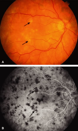
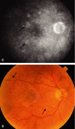
| Fig. 19. Choroidal infiltrates resembling acute posterior multifocal placoid pigment epitheliopathy (APMPPE) in acute myelocytic leukemia (AML). A. Note the deep yellowish-white lesions (arrows) in the posterior fundus. B. Laminar venous phase (24.8 seconds) of fluorescein angiogram showing early blockage (arrows) corresponding to the fundus lesions noted in A. C. Late phase (676 seconds) of fluorescein angiogram showing late leakage corresponding to the early hypofluorescence (arrows) in B and the yellow-white lesions in A. D. Four months after local irradiation. Note the resolution of the yellow-white lesions and few small punched-out atrophic chorioretinal scars (arrows) in areas corresponding to previous lesions as noted in A. |