|
|
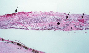
| Fig. 12. Histopathology of central retinal vein occlusion (CRVO). Note the intraretinal hemorrhages in various layers of the retina (arrows) and the eosinophilic proteinaceous exudates in the outer plexiform layer (asterisk) and subretinal space (S). |