|
|
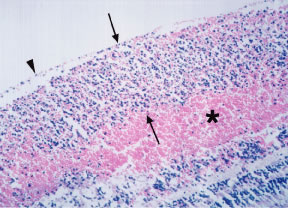
| Fig. 6. Histopathology of a leukemic infiltrate associated with hemorrhage in chronic myelocytic leukemia (CML). Note the leukemic infiltrate (between arrows) beneath the internal limiting membrane (ILM) (arrowhead) in the nerve fiber layer and the associated intraretinal hemorrhage (asterisk). (Courtesy Dr. W. Richard Green.) |