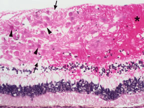
Fig. 10.
Histopathology of cotton wool spot. Note the thickening of the nerve fiber layer
(between arrows)
, the characteristic eosinophilic cytoid bodies
(arrowheads)
, and the associated retinal hemorrhage
(asterisk)
.