Chapter 12
Ocular Ischemic Syndrome
NEAL H. ATEBARA and GARY C. BROWN
Main Menu   Table Of Contents

Search

SYMPTOMS
SIGNS
DIFFERENTIAL DIAGNOSIS
STUDIES
SYSTEMIC ASSOCIATIONS
TREATMENT
CONCLUSION
REFERENCES

Ocular ischemic syndrome refers to the constellation of ophthalmic features that result from chronic hypoperfusion of the entire arterial supply to the eye, including the central retinal, posterior ciliary, and anterior ciliary arteries. In clinical practice, the occlusion is seen most frequently at the level of the carotid artery, but it may occur anywhere proximal to the point where the central retinal and ciliary arteries branch from the ophthalmic artery.

Ocular ischemic syndrome usually occurs as a result of atherosclerosis of the carotid artery. Less commonly, signs of ocular ischemia may be part of a larger clinical picture, such as aortic arch syndrome, which in turn is most often caused by Takayasu's arteritis or giant cell arteritis. Rarely, a chronic subtype of ophthalmic artery stenosis may result in ocular ischemic syndrome.

In the clinical presentation of ocular ischemic syndrome, the patient is typically more than 50 years old, is more likely to be male than female, and reports having a loss of vision in one or both eyes over a period of weeks to months.1 There may or may not be associated ocular or periocular pain, and one may elicit a history of difficulty adjusting from bright light to relative darkness. Additionally, the patient may relate a history of transient focal neurologic deficits.

The clinical signs of chronic ocular ischemia are legion, yet often subtle. Classic features include midperipheral fundus hemorrhages and dilated (but usually not tortuous) retinal veins.2,3 However, these classic features are not always present, and ocular ischemic syndrome can easily be confused with diabetic retinopathy, central retinal vein occlusion (CRVO), or even idiopathic iritis.4 Because of its broad presentation, ocular ischemic syndrome is probably underdiagnosed. Yet, its detection often has important implications for the patient, both ophthalmologically and systemically.5

In 1963, Kearns and Hollenhorst3 reported ocular symptoms and signs that occurred secondary to severe carotid artery obstruction. They called the disease “venous stasis retinopathy,” presumably because of the venous dilation and intraretinal hemorrhages, which in some ways resemble a mild case of CRVO. This term is confusing, however, because certain authors later used it to indicate mild, or nonischemic, CRVO,6 an entity that is different in origin and clinical manifestations from ocular ischemic syndrome. We therefore recommend that the term “venous stasis retinopathy” be avoided altogether. Other terms that have been applied to describe the ocular condition occurring secondary to marked carotid artery stenosis include ischemic ocular inflammation4,7,8 and ischemic oculopathy.8

Because atherosclerosis accounts for most cases of ocular ischemic syndrome, it is not surprising that most patients are first diagnosed in their fifties or sixties. The mean age at the time of presentation is approximately 65 years.1,4,7–10 In fact, manifestations of ocular ischemia occurring in a patient younger than 50 years of age should raise the suspicion of some other underlying systemic condition, such as large vessel arteritis or an abnormality of lipid metabolism causing premature atherosclerosis.

The true incidence of ocular ischemic syndrome is unknown. The ocular signs are not always obvious and, when present, may mimic other ocular disorders, such as diabetic retinopathy and retinal vein obstruction. Kearns and Hollenhorst3 noted this entity in 5% of their patients with internal carotid artery obstruction. Sturrock and Mueller7 found six affected patients over a 2-year period in an English hospital serving a population of approximately 400,000, an estimated incidence of 7.5 per million.

Histopathologic findings in eyes with ocular ischemic syndrome due to carotid occlusion are consistent with retinal capillary endothelial damage caused by chronic hypoperfusion. Histologic examination demonstrates intraretinal hemorrhages and numerous microaneurysms, particularly in the retinal midperiphery. The retinal capillaries demonstrate a decrease of the pericyte-endothelial cell ratio.11 The ophthalmoscopic appearance of midperipheral intraretinal hemorrhages was reproduced in a rat model after bilateral carotid artery ligation.12

Back to Top
SYMPTOMS
Visual loss is the most frequent symptom encountered in ocular ischemic syndrome; it is seen in more than 90% of affected persons at the time of presentation (Table 1).1 In most cases, this loss of vision is gradual, occurring over a period of weeks to months. It is caused by chronic posterior segment ischemia, macular edema, and/or cataract occurring secondary to anterior segment ischemia.


TABLE 1. Symptoms of Ocular Ischemic Syndrome and the Frequency of Their Occurrence1


SymptomPercentage of Cases
Visual loss90
Amaurosis fugax10
Ocular or periocular pain40


Less commonly, abrupt loss of vision may occur as a result of severe hypoperfusion of the retinal arterial system, which is marked by a cherry-red spot in the macula. In some cases, this hypoperfusion is caused by an embolus from a carotid atherosclerotic plaque that occludes the central retinal artery. In other cases, the perfusion pressure of the central retinal artery falls below that of the intraocular pressure because of an abnormally low arterial perfusion pressure caused by carotid artery occlusion and/or an abnormally high intraocular pressure due to neovascular glaucoma.

Sometimes the cause of reduced vision is not immediately obvious. There is no cataract, no macular edema, and no signs of retinal ischemia, and the intraocular pressure is normal. In these eyes, fluorescein angiography usually reveals impaired choroidal perfusion, which is the probable cause of reduced vision. Color Doppler studies of the orbital vessels have correlated poor visual acuity with absence of blood flow in the posterior ciliary arteries,13 supporting the theory that choroidal hypoperfusion is the cause of poor vision in many patients.

A history of amaurosis fugax may be elicited in up to 10% of patients.1 These episodes of transient loss of vision are caused by fibrin-platelet emboli originating from the atherosclerotic carotid plaques, which may temporarily occlude part of the retinal arterial circulation before being resorbed.

In 10% of patients diagnosed with ocular ischemic syndrome, there is an absence of any visual symptoms.1 In some cases, the detection of ocular ischemic syndrome may be the first indication of the patient's underlying systemic problems (e.g., atherosclerosis, arterial hypertension, hypercholesterolemia), as well as the first indication of the patient's risk for cerebrovascular accident and myocardial infarction.

Persons with ocular ischemic syndrome often report having difficulty seeing after being exposed to bright light.10 Typically, the patient will complain of prolonged difficulty in adjusting to a darker indoors when moving from a brightly lit outdoors. This subjective complaint can be more objectively tested with the photopic stress test, which measures the amount of time the patient requires to recover visual function after being exposed to a bright light source.

The symptom of pain is found in approximately 40% of persons with ocular ischemic syndrome.1 Typically, the pain is described as a dull ache over the eye or brow. Ischemia to the anterior segment structures, or ocular angina, is thought to be the cause of this pain in some cases. In more advanced cases, an elevated intraocular pressure due to neovascular glaucoma may cause pain because of an increased perfusion gradient to the anterior segment causing ischemia, or because of rupture of a corneal epithelial microcyst of acute glaucoma. Ipsilateral dural ischemia has also been proposed as a cause of the pain.

Back to Top
SIGNS
Patients with ocular ischemic syndrome may present with any of a constellation of signs in both anterior and posterior segments of the eye (Table 2). The classic triad includes midperipheral dot hemorrhages, dilated retinal veins, and iris neovascularization. Yet, in its early stages, none of these features may be present, and the diagnosis may rely on the recognition of other clues, such as the elicitation of central retinal artery pulsations with gentle digital pressure on the globe.


TABLE 2. Signs of Ocular Ischemic Syndrome and the Frequency of Their Occurrence1


SignPercentage of Cases
Anterior Segment 
Rubeosis iridis67
Neovascular glaucoma35
Iritis18
Posterior Segment 
Narrowed retinal arteriesMost
Dilated retinal veinsMost
Retinal hemorrhages80
Neovascularization of the disc35
Neovascularization of the retina8
Cherry-red spot12
Cotton-wool spots6
Spontaneous retinal artery pulsations4
Cholesterol emboli2
Anterior ischemic optic neuropathy2


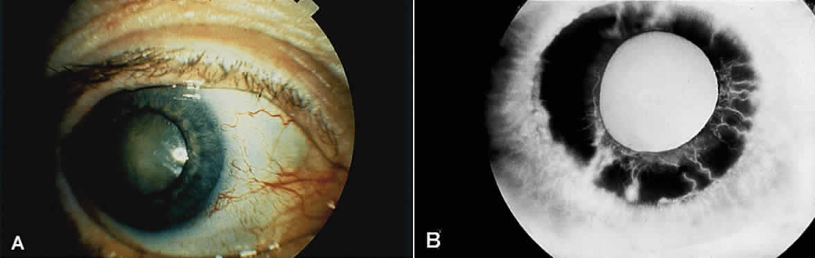
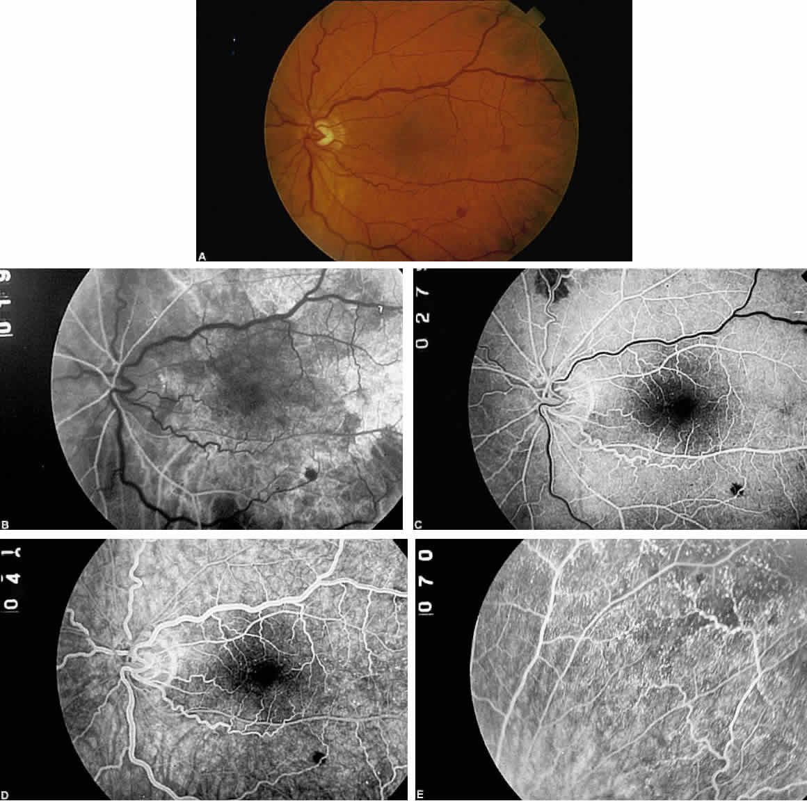
Eyes with ocular ischemic syndrome appear to be at high risk for rubeosis iridis, which is found in 67% of eyes at the time of presentation1 (Figs. 1 and 2). By comparison, only 2.8% of nonischemic CRVOs, 58% of ischemic CRVOs,14 and 15% to 20% of central retinal artery occlusions will develop neovascularization of the iris.15,16 When clinicians are faced with a patient with rubeosis iridis and no obvious etiology (i.e., there are no signs of diabetic retinopathy or retinal vascular occlusion), they should consider the possibility of ocular isch-emic syndrome. The high incidence of rubeosis iridis is probably related to the combined ischemia of both anterior and posterior segments of the globe.

Fig. 1. A. External photograph of an eye with ocular ischemic syndrome displaying features of conjunctival injection, cataract, and rubeosis irides. B. Iris fluorescein angiogram of an eye with ocular ischemic syndrome showing leakage from iris neovascularization.

Fig. 2. Gonioscopic appearance of neovascularization of the iridocorneal angle due to ocular ischemic syndrome.

The risk of neovascular glaucoma is likewise very high. At the time of presentation, 35% of patients will have neovascularization of the iris and an intraocular pressure greater than 22 mmHg.1 It is interesting to note that in some cases, because of poor ciliary body perfusion and decreased aqueous production, the intraocular pressure remains low, even when fibrovascular tissue completely occludes the iridocorneal angle. Eyes with normal or well-controlled intraocular pressures have been reported to have a dramatic rise in intraocular pressure after carotid endarterectomy, and it is therefore recommended that eyes be followed closely immediately after carotid surgery.

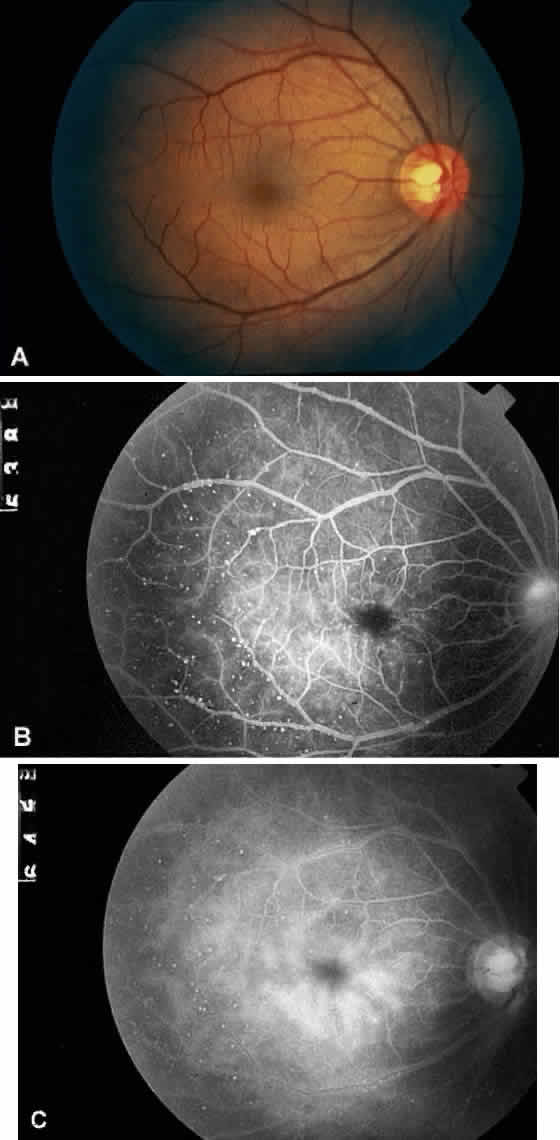
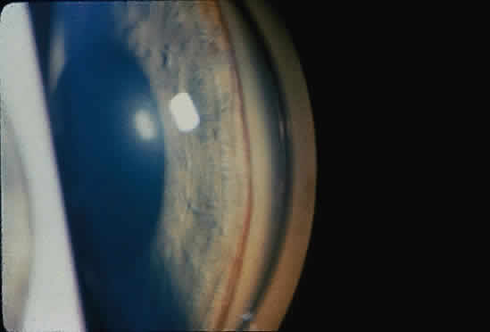
Anterior segment ischemia may cause conjunctival and episcleral vessel injection, corneal edema, and folds in Descemet's membrane (Fig. 1A).2 Aqueous flare is seen in most eyes with iris neovascularization, but a mild anterior chamber reaction is also seen in 18% of eyes with ocular ischemic syndrome, even in the absence of rubeosis iridis.1 In some cases, this anterior chamber inflammation may be the only obvious feature of the disease, and it may be easily mistaken for anterior uveitis.4 Progressive cataractous changes are often seen (Fig. 1A), presumably related to anterior segment ischemia and/or anterior chamber inflammation.

The retinal arteries are generally narrowed and straightened in eyes with ocular ischemic syndrome (Figs. 3A and 4A).3 Although such arterial narrowing can be seen in systemic hypertension or as a normal variation in the elderly, it is important to note that retinal arterial narrowing can also be caused by ocular ischemic syndrome. A significant asymmetry in retinal arterial narrowing between the two eyes may be seen in unilateral cases of ocular ischemic syndrome.

Fig. 3. A. Fundus photograph of an eye with ocular ischemic syndrome showing features of venous dilation, midperipheral intraretinal hemorrhages, and attenuated arterioles. B. Fluorescein angiogram at 19 seconds (arterial phase). There is a prolonged arm-to-retina circulation time and a patchy choroidal filling pattern. C. Early laminar-venous phase at 27 seconds. Areas of incomplete choroidal filling persist. D. At 41 seconds, laminar-venous phase is almost complete, and the arteriovenous transit time is approximately 22 seconds. E. Fluorescein angiogram of the midperipheral fundus, revealing numerous microaneurysms that are not obvious on ophthalmoscopic examination.

Fig. 4. A. Fundus photograph of an eye with ocular ischemic syndrome showing features of venous dilation, attenuated arterioles, and midperipheral intraretinal hemorrhages. B. Fluorescein angiogram at 18 seconds, displaying a visible leading edge of fluorescein dye within the inferotemporal arteriole. C. Numerous microaneurysms along the inferotemporal arcade. At 33 seconds, the arteriolar dye front is still apparent. D. Retinal capillary nonperfusion and microaneurysms at 58 seconds. E. Prominent arterial staining in the late phase of the angiogram.

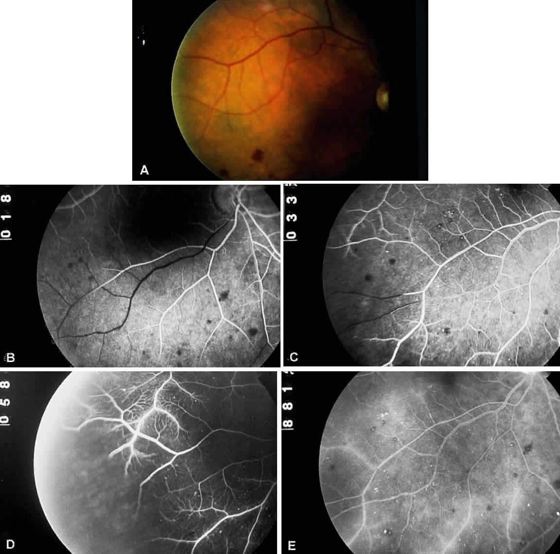
Dilated retinal veins are frequently seen in eyes with ocular ischemic syndrome (see Figs. 3A and 4A). Irregularity of the vessel caliber may be present, similar to the venous beading seen in preproliferative or proliferative diabetic retinopathy. In contrast to CRVO, the veins in eyes with ocular ischemic syndrome are generally not tortuous. The fact that ocular ischemic syndrome occurs secondary to impaired inflow, whereas CRVO is usually associated with compromised outflow, may account for the difference.

Intraretinal dot and blot hemorrhages are seen in approximately 80% of eyes with ocular ischemic syndrome (see Figs. 3A and 4A).1 As with diabetic retinopathy, these hemorrhages probably arise secondary to the rupture of microaneurysms, which, in turn, are caused by endothelial damage. Unlike background diabetic retinopathy, in which the intraretinal hemorrhages are distributed throughout the posterior pole, the hemorrhages of ocular ischemic syndrome tend to be located mainly in the midperipheral fundus. The hemorrhages of CRVO tend to be more numerous and larger than those of ocular ischemic syndrome.

Neovascularization of the retina or optic disc may occur in ocular ischemic syndrome. Optic disc neovascularization is quite common, seen in 35% of patients.1 This incidence is much higher than that seen in ischemic CRVO (5%)14 and central retinal artery occlusion (2%).15 This high incidence of disc involvement may be reflective of the generalized hypoxic status of the eye in ocular ischemic syndrome. In an older patient with no history of diabetes mellitus or vascular occlusive disease, the presence of neovascularization of the optic disc should arouse the clinical suspicion of ocular ischemic syndrome.

Neovascularization of the retina is seen in 8% of patients with ocular ischemic syndrome, and it is usually seen concurrently with neovascularization of the disc.1 Secondary vitreous hemorrhage may occur as a result of traction upon the new vessels by the vitreous gel.

A cherry-red spot is seen at presentation in approximately 12% of eyes with ocular ischemic syndrome.1 It may occur acutely because of ischemia of the inner retinal layer and whitening from emboli to the central retinal artery, or it may occur more chronically. The chronic form occurs when the intraocular pressure encroaches upon the perfusion pressure of the central retinal artery, and blood flow to the inner layer of the retina is compromised. The visual prognosis is generally grim when a cherry-red spot develops.

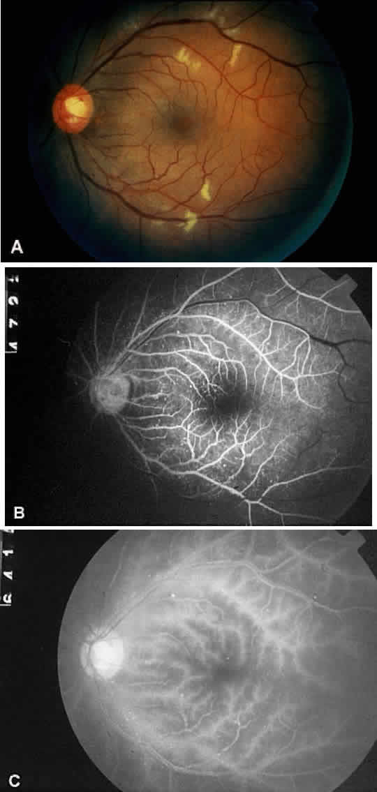
Cotton-wool spots are observed in approximately 5% of eyes with ocular ischemic syndrome (Fig. 5A).1 These spots are generally located in the posterior pole.

Fig. 5. A. Fundus photograph of the left eye of a patient diagnosed with Takayasu's arteritis, showing attenuated arterioles and venule dilation. There are scattered dot hemorrhages and microaneurysms along the vascular arcades and extending outward to the equator. Large cotton-wool spots along the arcades are apparent. B. Fluorescein angiogram, revealing prominent venous dilation and beading, with microaneurysms along the vascular arcades and throughout the posterior pole. C. Late arterial phase, showing marked arterial staining. Choroidal filling time is also delayed to more than 17 seconds. The arteriovenous transit time in this study is delayed to more than 36 seconds. (Courtesy Travis A. Meredith, MD, St. Louis, MO)

Spontaneous pulsations of the central retinal artery are highly suggestive of ocular ischemic syndrome. When the diastolic pressure within the central retinal artery falls below the intraocular pressure, the retinal arteries collapse during diastole, and spontaneous pulsation of the central retinal artery is observed. This phenomenon is present in 5% of eyes with ocular ischemic syndrome.1

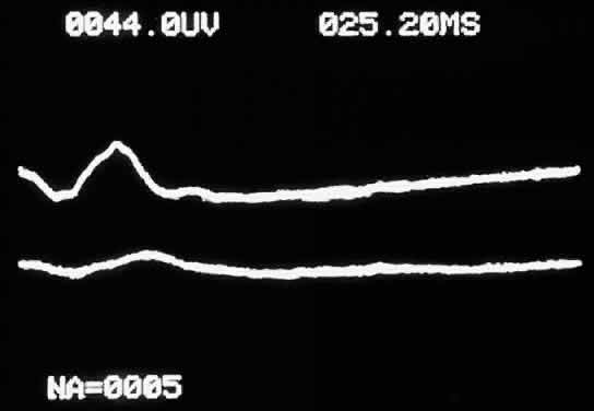
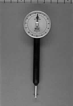
When not present spontaneously, retinal arterial pulsations may be induced by light digital pressure to the globe, thereby artificially raising the intraocular pressure above the diastolic retinal arterial pressure. With slightly more pressure, the retinal arterial tree may even be seen to collapse as the intraocular pressure is raised above the systolic retinal arterial pressure. Correspondingly, as would be expected, ophthalmodynamometry and oculoplethysmography reveal decreased readings in eyes with ocular ischemic syndrome.

Ischemic optic neuropathy has been reported in an eye with ocular ischemic syndrome.17 Hypoperfusion of the posterior ciliary circulation to the optic disc due to impaired carotid blood flow or emboli of carotid origin is the probable cause of this complication.

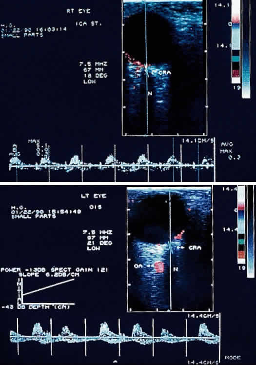
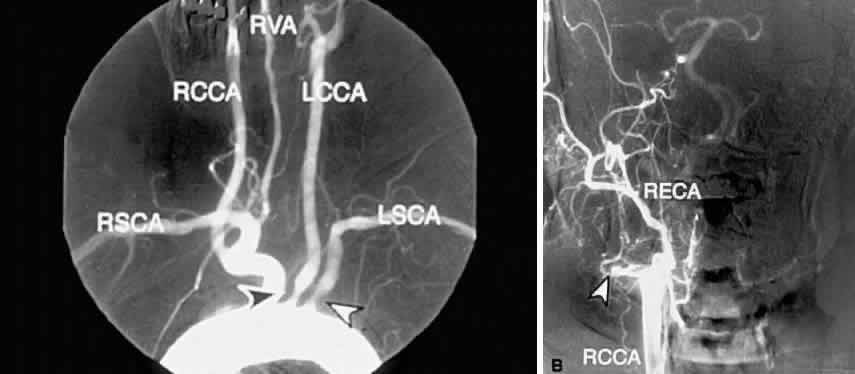
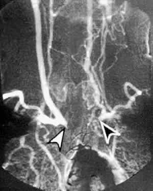
The carotid pulse is often diminished or absent in the presence of carotid occlusive disease. The finding of a cervical bruit is significant, although its absence does not exclude a significant or even a complete carotid occlusion.18,19

Back to Top
DIFFERENTIAL DIAGNOSIS
Diabetic retinopathy and nonischemic CRVO are the two entities most likely to be confused with ocular ischemic syndrome. Differentiation from diabetic retinopathy can be especially difficult, since many persons with ocular ischemic syndrome have diabetes mellitus as well, and the retinal manifestations of each may be superimposed. Both ocular ischemic syndrome and diabetic retinopathy tend to display dilated and beaded retinal venules, dot and blot intraretinal hemorrhages, and microaneurysms.2,20 Diabetic retinopathy tends to have bilateral involvement, whereas ocular ischemic syndrome is more often unilateral than bilateral. Ocular ischemic syndrome generally affects older persons in their 50s to 80s, whereas diabetic retinopathy may affect any age group. Retinal hemorrhages and microaneurysms tend to be midperipheral in ocular ischemic syndrome, but mainly involve the posterior pole in diabetic retinopathy. Hard exudates are common in diabetic retinopathy, but are usually absent in ocular ischemic syndrome unless the patient has concomitant diabetic retinopathy. On fluorescein angiography, ocular ischemic syndrome typically displays delayed and patchy choroidal filling, prolonged arteriovenous transit time, and arterial staining; in diabetic retinopathy, these features are usually absent.

CRVO and ocular ischemic syndrome both tend to affect an older patient population and display dilated and tortuous venules with dot and blot intraretinal hemorrhages.21,22 Bilateral involvement is uncommon in CRVO and occurs in a minority of cases of ocular ischemic syndrome. In contrast to ocular ischemic syndrome, CRVO may display optic disc swelling and more pronounced macular edema. On fluorescein angiography, both CRVO and ocular ischemic syndrome may display a prolonged arteriovenous transit time, but in CRVO there is no defect in the choroidal filling pattern. In ocular ischemic syndrome, vessel staining is predominantly arterial; in CRVO, it is mostly venous. On ophthalmodynamometric examination, the retinal arterial perfusion pressure is decreased in ocular ischemic syndrome but normal in CRVO.

A fundus appearance similar to that found in ocular ischemic syndrome was reported to have occurred secondary to an embolus within the central retinal artery.23 Presumably, this occurred on the basis of chronic hypoperfusion.

Back to Top
STUDIES

FLUORESCEIN ANGIOGRAPHY

Fluorescein angiography is a valuable tool for the clinical diagnosis and management of ocular ischemic syndrome. Characteristic fluorescein angiographic features reflect the chronic hypoperfusion of the retinal and choroidal circulations, as well as ischemic damage to the neurosensory retina and retinal vessels (Table 3).


TABLE 3. Fluorescein Angiographic Signs of the Ocular Ischemic Syndrome and the Frequency of Their Occurrence1


SignPercentage of Cases
Prolonged arteriovenous transit time95
Retinal vascular staining85
Delayed choroidal filling60
Macular edema17


Prolonged arm-to-retina circulation times are frequently observed (Fig. 3B).24 This reflects the slower flow of blood from the carotid artery to the central retinal artery due to carotid occlusive disease. In normal persons, fluorescein dye is first detected in the retinal vasculature 12 to 15 seconds after dye is injected into the arm vein. Uncommonly, dye may take up to 20 seconds to reach the retinal arteries in a normal person. In ocular ischemic syndrome, arm-to-retina circulation times well over 20 seconds are not uncommon.

The arm-to-choroid circulation time is also frequently prolonged in ocular ischemic syndrome because of the slow flow of blood from the carotid artery to the short posterior ciliary arteries. In pronounced cases, complete choroidal filling may still be lacking at 1 minute after injection. Accurate measurement of the arm-to-retina or arm-to-choroid circulation times requires experienced fluorescein angiography personnel, and these measurements depend on whether the dye was injected in the antecubital fossa or the hand, the rate of injection, and a reliable timer start time.

Delayed or patchy choroidal filling may be seen in 60% of eyes affected with ocular ischemic syndrome (see Fig. 3B; Fig. 3C).1 Normally, the choroid is completely filled within 5 seconds after the initial appearance of dye in the choroid. Patchy choroidal filling greater than 5 seconds reflects sluggish blood flow through the short posterior ciliary arteries. In some cases, one or more quadrants of the choroid may display significantly slower filling than the other quadrants, indicating a relatively lower perfusion of individual short posterior ciliary arteries.

Retinal arteriovenous transit time is measured from the initial appearance of dye within the retinal arteries in the temporal vascular arcade until the corresponding veins are completely filled. In normal patients, this occurs within 11 seconds; in ocular ischemic syndrome, the arteriovenous transit time is prolonged in approximately 95% of affected eyes (see Fig. 3B and C; Fig. 3D).1 Although this finding is relatively sensitive for a retinal vascular flow anomaly, it may also be seen in other vascular occlusive disorders, such as arterial and venous occlusive disease. Sometimes, a well-demarcated, leading edge of fluorescein dye within a retinal artery may be seen (Fig. 4B and C). This distinctly abnormal finding is seen in retinal arterial occlusion and ocular ischemic syndrome. In extreme cases of ocular ischemic syndrome, the retinal veins may fail to fill throughout the entire study.

Staining of the retinal vessels can be seen in approximately 85% of ocular ischemic syndrome eyes in the later phases of fluorescein angiography (Figs. 4E, 5C, and 6C).1 The arterioles are usually involved to a greater extent than the venules. Hypoxic damage to the endothelial cells and pericytes may account for this phenomenon.11,25 In contrast to ocular ischemic syndrome, fluorescein angiography of eyes with central retinal artery obstruction rarely show late vascular staining. In CRVO, the retinal veins typically stain more than the retinal arteries.

Fig. 6. A. Fundus photograph of the right eye in the same patient as in Figure 5 diagnosed with Takayasu's arteritis, displaying features of arterial sclerosis, venous dilation, dot hemorrhages, and microaneurysms. B. Fluorescein angiogram of the same eye, revealing prominent venous dilation and beading, with microaneurysms concentrated along the venous arcades. C. Late phase of the angiogram revealing staining of the arterioles, increasing prominence of the microaneurysms, and diffuse staining of the perifoveal microaneurysms. (Courtesy Travis A. Meredith, MD, St. Louis MO)

Macular edema, as evidenced by intraretinal leakage on fluorescein angiography, can be demonstrated in 15% of eyes with ocular ischemic syndrome (see Fig. 6C).26 Hypoxia and endothelial damage to the small retinal vessels, as well as microaneurysmal changes, probably account for this leakage.1,27 The ophthalmoscopic signs of macular edema are generally not as pronounced. Although small central foveal cysts have been described, they are generally not as prominent as those seen in CRVO or ocular inflammation. Late staining of the optic disc on fluorescein angiography often accompanies leakage in the macula, but disc swelling is generally absent.

Microaneurysms are most commonly seen in the midperipheral and peripheral retina (see Figs. 3A and 4A). They become hyperfluorescent during the venous phase of the angiogram, and they leak as the study progresses. Rupture of these microaneurysms and leakage from small retinal vessels account for the midperipheral dot and blot intraretinal hemorrhages seen in ocular ischemic syndrome.

Retinal capillary nonperfusion can be seen in some cases of ocular ischemic syndrome (Fig. 4D). These areas may contribute to the development of retinal or iris neovascularization. Histologically, these areas of capillary dropout are marked by an absence of endothelial cells and pericytes within the retinal capillaries.11,25

PHOTOPIC STRESS TEST

A characteristic symptom of patients with ocular ischemic syndrome is difficulty in visual adjustment to relative darkness after exposure to bright light. This subjective symptom can be more objectively measured with the photopic stress test.

After a prolonged period of normal ambient illumination, best-corrected visual acuity is measured in each eye separately. With one eye covered, the other eye is then subjected to 10 seconds of direct illumination from a bright light source, such as a direct ophthalmoscope. The amount of time required for the patient to recover his or her best-corrected visual acuity is recorded. The same procedure is performed on the other eye, and the recovery time is again recorded.

A marked difference in photopic stress recovery times between the two eyes is suggestive of macular disease, such as ocular ischemic syndrome; diseases of the optic nerve would be less affected.

ELECTRORETINOGRAPHY

Electroretinography in eyes with ocular ischemic syndrome reveals a decreased amplitude of both the a- and b-waves (Fig. 7).1,9 The b-wave, which corresponds to the inner retinal layer, probably reflects the function of the bipolar and/or Müller cells, and it is diminished by compromised perfusion of the central retinal artery. The a-wave correlates with photoreceptor function and is affected by choroidal ischemia. Thus, the electroretinographic features of decreased a- and b-waves in ocular ischemic syndrome reflect the pathophysiology of combined retinal and choroidal hypoperfusion.

Fig. 7. Electroretinogram showing normal a- and b-waves in the unaffected right eye (upper tracing) and marked reduction in both a and b waves in the eye with ocular ischemic syndrome (lower tracing).

ORBITAL COLOR DOPPLER IMAGING

Orbital color Doppler imaging is a noninvasive ultrasound test that permits simultaneous imaging of small orbital vessels and recording of quantitative data on blood flow velocity. Unlike duplex carotid ultrasonography, which images and measures flow velocity in the large carotid vessels, orbital color Doppler imaging can image the smaller ophthalmic, central retinal, and short posterior ciliary arteries.28

With the use of orbital color Doppler imaging, the central retinal and posterior ciliary artery peak systolic velocities have been reported to be markedly reduced in ocular ischemic syndrome, as would be expected (Fig. 8).13 Reversal of ophthalmic artery blood flow, which probably represents collateral blood flow to lower resistance vascular beds, was detected in 75% of eyes with ocular ischemic syndrome. It is interesting to note that although high-grade carotid stenosis, reversal of ophthalmic artery flow, and decreased central retinal artery flow have been associated with ocular ischemic syndrome, this combination in and of itself has not been sufficient to routinely produce decreased vision. Absence of detectable posterior ciliary arterial blood flow was the only reliable indicator of poor vision.

Fig. 8. Color Doppler imaging study of the central retinal artery in an eye with ocular ischemic syndrome (left). Areas in red superimposed on the B-scan image depict blood flow moving toward the transducer, mostly representing arterial flow. Areas in blue depict flow, mostly venous, away from the transducer. Notice the reduced systolic peaks of the time-velocity waveform (graph below the B-scan image) in contrast to the normal pattern of the uninvolved contralateral eye (right). (Ho AC, Lieb WE, Flaharty PM et al: Color Doppler imaging of the ocular ischemic syndrome. Ophthalmology 99:1453, 1992)

One limitation of orbital color Doppler imaging is its inability to measure volumetric blood flow directly; rather, this study modality relies on measurements of peak systolic blood flow velocity and vascular resistance, which in most physiologic situations are gauges of blood flow. Currently, orbital color Doppler imaging is most useful as an investigational technique for studying the hemodynamics of ophthalmic vascular disorders. In the future, it may provide useful information for the diagnosis and management of ocular ischemic syndrome.

OPHTHALMODYNAMOMETRY AND OCULOPLETHYSMOGRAPHY

In ophthalmodynamometry, the sterilized footplate of an ophthalmodynamometer is applied to the sclera after administration of topical corneal anesthesia (Fig. 9).29 The central retinal artery is observed at the slit lamp using a Hruby lens. Gentle pressure on the sclera is gradually increased by the footplate until arterial pulsations are observed. The measurement on the dial of the ophthalmodynamometer at this point is taken to be the relative retinal arterial diastolic pressure. The point at which the retinal artery collapses is taken to be the relative retinal arterial systolic pressure.

Fig. 9. Photograph of a hand-held analog ophthalmodynamometer. After topical corneal anesthesia, the sterilized footplate (bottom) is gently applied to the sclera. The central retinal artery is observed at the slit lamp with the use of a Hruby lens. Increasing force is gradually applied until pulsations of the central retinal artery are observed. The measurement on the dial at this point is taken to be the relative diastolic pressure of the central retinal artery. The pressure at which the retinal artery is collapsed is taken to be the relative systolic pressure.

In oculoplethysmography, instead of a mechanical footplate applied to the globe, a suction device is used to achieve a gradual increase in the intraocular pressure.30,31 In a fashion similar to that used for the ophthalmodynamometer, the intraocular pressure at which arterial pulsations are induced is taken to be the relative diastolic pressure of the central retinal artery, and the intraocular pressure at which the retinal artery collapses is taken to be its relative systolic pressure.

The normal ophthalmic artery has a systolic pressure of approximately 100 mmHg and a diastolic pressure of approximately 60 mmHg.32 In ocular ischemic syndrome, the systolic pressure is often less than 40 mmHg, and the diastolic pressure may be less than 10 mmHg.33 It is often useful to measure an unaffected contralateral eye for comparison: a significantly lower retinal arterial diastolic or systolic pressure in the affected eye, compared with the uninvolved eye, is suggestive of ocular ischemic syndrome.

A significant limitation of these two indirect measurements of carotid blood flow is that they cannot reliably detect a carotid artery stenosis of less than 80% in area because there is no significant drop in the carotid artery pressure until the lumen of the vessel is reduced by at least 80%.34–36 This fact, in addition to the inherent degree of inaccuracy of the instruments, limits their usefulness in the evaluation of carotid disease, even as screening tools. However, for the ophthalmologist who needs only to rule out a carotid obstruction significant enough to cause ocular ischemic syndrome, ophthalmodynamometry is a quick, inexpensive, and noninvasive test that can help confirm a suspicion of ocular ischemic syndrome or help differentiate it from other similar funduscopic appearances, such as nonischemic CRVO and background diabetic retinopathy.

CAROTID DUPLEX (B-MODE AND DOPPLER) SCANNING

Although not as accurate as carotid angiography, ultrasonographic carotid tests can determine whether the carotid arteries are normal or abnormal, and they can in many cases adequately determine the degree of stenosis.37 Opinions differ regarding the accuracy of ultrasonography in the evaluation of atherosclerotic carotid disease, and its utility is highly dependent on the experience of the operator.

B-mode ultrasonography can produce real-time anatomic detail of the bifurcation of the common carotid artery. It can reliably identify most atherosclerotic lesions, and the newer machines may be able to visualize plaque ulceration and hemorrhage.38–41 The Doppler-shift signal of the returning echo from flowing blood can be used to calculate blood flow velocity. Duplex ultrasonography combines B-mode images of the artery with Doppler analysis of blood flow velocity at each point on the image.

Carotid duplex scanning, although safe and inexpensive, has limitations. In general, it is less reliable than carotid angiography in distinguishing between a completely occluded artery and one that is nearly occluded. Because noninvasive tests such as carotid duplex scanning pose minimal risk to the patient, they are generally used as screening techniques for atherosclerotic disease.

MAGNETIC RESONANCE ANGIOGRAPHY

Magnetic resonance imaging has been applied to the study of blood flow in the carotid arteries. In this technique, called magnetic resonance angiography (MRA), areas of flowing blood display increased signal intensity according to relative proton excitation. The collected data are stored as three-dimensional images, which can be displayed as cross-sectional two-dimensional images or as planar images that resemble conventional carotid angiograms. The brain parenchyma and the carotid arteries can be imaged in the same study.

The accuracy of MRA in measuring carotid stenosis is steadily improving. Accuracy in the detection of carotid artery stenosis greater than 50% ranges from 69% to 91% in reported series, with MRA techniques tending to overestimate the degree of carotid stenosis compared with standard carotid angiography.42–45 Currently, MRA is less accurate and more expensive than duplex ultrasonography by an experienced operator, and it is not nearly as accurate as conventional carotid angiography. Therefore MRA should currently be considered a screening test for carotid artery disease.

CAROTID ANGIOGRAPHY

Carotid angiography is performed by selective common carotid arterial injection of a radiopaque contrast dye via transfemoral catheterization. In the traditional technique, the image of the inner luminal silhouette is produced on x-ray emulsion (Fig. 10A and B). More recently, digital images and computerized image enhancement have improved resolution and reduced the amount of contrast dye required.46,47 This method can detect ulcerative lesions, severe stenosis, and formation of mural thrombus. It can also demonstrate collateral circulatory patterns.

Fig. 10. A. Digital subtraction aortogram revealing a 90% atherosclerotic stenosis of the proximal left common carotid artery (black arrowhead) and 80% stenosis of the proximal left subclavian artery (white arrowhead). RSCA, right subclavian artery; RCCA, right common carotid artery; RVA, right vertebral artery; LCCA, left common carotid artery; LSCA, left subclavian artery. B. Digital subtraction angiogram with a right common carotid injection from the same patient. There is complete occlusion of the right internal carotid artery due to atherosclerosis (white arrowhead). The right external carotid artery (RECA) and the right common carotid artery (RCCA) fill normally.

The most serious complications of carotid angiography are aortic or carotid artery dissection, embolic stroke, and myocardial infarction.48 Other complications include dye reactions, renal injury, hematoma, pseudoaneurysm, and arterial thrombosis. Carotid angiography carries a 1% risk of morbidity and 0.06% risk of mortality. As with all tests, the advantages of selective carotid angiography must be weighed against the possible risks.

Despite its expense and low but definite morbidity rate, carotid angiography remains the most reliable method of assessing atherosclerotic carotid disease,38 and it serves as the “gold standard” in the evaluation of all other tests of the carotid arteries.

Back to Top
SYSTEMIC ASSOCIATIONS

ATHEROSCLEROSIS

Atherosclerotic disease, because of its occlusive effects on the carotid arteries, is by far the most common cause of ocular ischemic syndrome. Histologically, there is thickening and hardening of the carotid artery caused by lipid-rich lesions within the innermost vessel layer.49,50 These lesions may partially or completely occlude the artery, either by slow growth or by a more rapid occlusion from hemorrhage into a plaque or thrombus formation.

The most common site of atherosclerosis in the neck vessels is at the origin of the internal carotid artery. From this point, the atherosclerosis often propagates downward in a retrograde fashion into the common carotid artery. Less commonly, lesions may occur at the siphon, the S-shaped portion of the internal carotid artery that lies in the cavernous sinus. Rarely, the origin of the common carotid artery is the region affected.

There is an association between atherosclerosis and increased levels of plasma lipoprotein, principally low-density lipoproteins.51 Risk factors for atherosclerosis also include cigarette smoking, systemic hypertension, diabetes mellitus, advancing age, male sex, obesity, stress, and type-A personality.52

In addition to its ischemic effects on the eye via carotid occlusion, atherosclerosis is responsible for the majority of cases of cerebrovascular accident and myocardial infarction, and it is therefore the leading cause of death in developed countries. Early diagnosis of ocular ischemic syndrome can therefore reveal underlying atherosclerotic disease in the carotid arteries or other parts of the body. Conversely, evidence of atherosclerotic disease in other parts of the body should raise the possibility of occlusive atherosclerotic carotid disease as well.

CEREBROVASCULAR ACCIDENT

Cerebrovascular accident, the third leading cause of death in developed countries, is most commonly caused by embolism or thrombus from an atherosclerotic carotid vessel. Thus, the ophthalmologist, in recognizing ocular ischemic syndrome, can play an important role in preventing a stroke by recognizing the effects of atherosclerotic carotid disease on the eye. Conversely, a history of stroke, especially of the middle cerebral circulation, may support a suspicion of ocular ischemic syndrome.53

Internal carotid emboli most commonly affect the middle cerebral artery or its branches, and they may result in paralysis and sensory impairment of the contralateral face, arm, and leg due to damage to the somatic sensory and motor cortices. Central aphasia, word deafness, anomia, jargon speech, sensory agraphia, acalculia, alexia, right-left confusion, and hemineglect may also occur. Homonymous hemianopsia may occur because of damage to the optic radiation deep to the temporal lobe. Paralysis of conjugate gaze to the opposite side may also be seen.54

AORTIC ARCH SYNDROME

Aortic arch syndrome describes the clinical picture that results when multiple branches of the aortic arch are affected by a chronic occlusive narrowing of any etiology. Compromised blood flow through the brachial artery causes arm weakness, coldness, and claudications; involvement of the carotid arteries causes symptoms of cerebral or ocular ischemia. When the ophthalmologist encounters signs of ocular ischemia accompanied by symptoms of brachial insufficiency, aortic arch syndrome should be considered as the cause. The most common causes of aortic arch syndrome are Takayasu's arteritis, giant cell arteritis, and atherosclerotic disease affecting multiple branches of the aorta.

TAKAYASU'S ARTERITIS

Takayasu's arteritis is a rare, chronic inflammatory disease of unknown etiology affecting the large arteries of the body, namely the aortic arch, the descending aorta, the pulmonary artery, and their primary branches. It occurs mainly in young women, and the typical age of onset is 10 to 40 years.55 It appears in all populations, but is more frequently seen in Asians.

The early signs and symptoms of Takayasu's arteritis include fatigue, weight loss, low-grade fever, arthralgias, myalgias, and headaches, and it may be mistaken for juvenile rheumatoid arthritis in a young female patient. The later clinical manifestations of Takayasu's arteritis relate to the anatomic site of vascular obstruction. Decreased blood flow to the brachial arteries results in coldness of the hands, paresthesias, and asymmetric or absent brachial pulses.56 Renovascular hypertension develops in half of patients because of involvement of the renal arteries of the abdominal aorta.57 Occlusion or narrowing of the carotid or vertebral arteries may result in cerebrovascular accident.58

Ophthalmic symptoms, due to ocular hypoperfusion from occlusion of the carotid artery, are seen in 15% of cases.59 These ophthalmic changes may be identical to those of ocular ischemic syndrome caused by carotid atherosclerotic disease (see Figs. 5 and 6).60,61 Arteriography of the aortic arch region, showing smooth-walled areas of stenosis and dilation, is usually necessary to confirm the diagnosis (Fig. 11). Collateral circulation, due to the chronicity of the stenosis, is usually prominent.

Fig. 11. Digital subtraction thoracic angiogram of the aortic arch and its major branches in the same patient with Takayasu's arteritis as shown in Figures 5 and 6. The right common carotid artery shows nearly complete occlusion (white arrowhead). The left common carotid artery shows complete occlusion with extensive collateral flow (black arrowhead). (Courtesy Travis A. Meredith, MD, St. Louis MO)

In cases of rubeosis iridis in which the iridocorneal angle is open, laser photoablative or cryoablative procedures may be considered.9 Takayasu's arteritis in its active stage typically responds well to corticosteroid therapy. In early cases, arterial stenosis may actually reverse and ischemic symptoms improve; however, after fibrous tissue has formed or thrombosis has already occurred, steroids may no longer help. Angioplasty or bypass grafts may be considered in late cases in which irreversible artery stenosis has occurred and significant ischemic symptoms persist.

GIANT CELL ARTERITIS

Giant cell arteritis is an idiopathic, granulomatous, inflammatory condition of patients 50 years of age or older that can affect any size vessel of the body, although the temporal arteries are most frequently affected. Autopsy studies have estimated the frequency of this condition at 1.1% of the population.62 The systemic clinical features of giant cell arteritis include headache, fever, weight loss, anorexia, malaise, polymyalgia rheumatica, jaw claudication, and tenderness, nodularity, and pulselessness of the temporal artery.63 Although the most common ocular manifestations of giant cell arteritis are ischemic optic neuropathy, retinal artery occlusion,64 and diplopia due to a cranial nerve palsy,63 ocular ischemic syndrome has been reported as a rare manifestation as well.

Suspicion of giant cell arteritis relies on a careful clinical history and physical examination, supported by an elevated erythrocyte sedimentation rate. Diagnosis of this disease is generally made by temporal artery biopsy, revealing occlusion of the vessel lumen, fragmentation of the internal elastic lamina, and granulomatous inflammation of the vessel wall.

Giant cell arteritis is treated with systemic corticosteroids.65 Treatment is generally initiated at a high dose and then slowly tapered, with the erythrocyte sedimentation rate and clinical symptoms used to monitor the effectiveness of treatment.

RARE ASSOCIATIONS

Rare causes of ocular ischemic syndrome include moyamoya disease,66 fibromuscular dysplasia,67 and radiation damage to the carotid arteries.68

Back to Top
TREATMENT

RETINAL ABLATIVE PROCEDURES

Although data on the natural course of untreated, full-blown ocular ischemic syndrome is lacking, these eyes in all probability do poorly. The presence of rubeosis iridis portends a very poor visual prognosis.69 In cases of rubeosis iridis in which the iridocorneal angle is open, panretinal photocoagulation may be considered.70–72 Although regression of these new vessels may occur in some instances, the response is generally not as dramatic as that observed when the same treatment is given for iris neovascularization secondary to diabetic retinopathy or CRVO. If the iridocorneal angle becomes closed by fibrovascular tissue and intraocular pressure rises, cycloablation or glaucoma-filtering procedures may be considered.

SYSTEMIC ANTICOAGULATION

Perhaps as important to the patient with newly diagnosed ocular ischemic syndrome is the management of the underlying systemic atherosclerotic disease, which should be carried out by an experienced neurologist or internist. The treatment of atherosclerotic carotid disease accompanied by cerebral transient ischemic attacks (TIAs) with systemic anticoagulation is controversial because of the risk of bleeding complications.73–77 Nevertheless, because of the often devastating effect of middle cerebral artery occlusion, some authorities advocate anticoagulation in patients with TIAs of carotid origin who are not surgical candidates, either because of medical conditions or because the intracranial location of the carotid lesion precludes carotid endarterectomy.78,79 Systemic anticoagulation is also helpful in temporarily managing patients at high risk for stroke until surgery can be performed.

SYSTEMIC ANTIPLATELET THERAPY

There have been randomized studies that have suggested that aspirin may be helpful in preventing further TIAs or stroke in symptomatic patients.80–82 Ticlopidine, a newer antiplatelet agent, has been associated with a significantly lower 3-year risk of stroke and death (17%) compared with aspirin (19%).83 Although the magnitude of this difference is not large, it suggests that future antiplatelet agents may be even more effective in reducing the risk of stroke and death in patients with atherosclerotic carotid occlusive disease. Despite these intriguing results, most authorities agree that systemic antiplatelet therapy alone is usually reserved for patients who have inoperable carotid disease, nonstenotic carotid disease, or a medical contraindication to surgery. In most cases, carotid endarterectomy remains the treatment of choice for symptomatic patients with severe stenotic atherosclerotic disease of the internal carotid artery.

CAROTID ENDARTERECTOMY: EFFICACY IN TREATING OCULAR ISCHEMIC SYNDROME

Despite a generally poor visual prognosis, reversing the carotid stenosis may be the most important factor in maintaining or improving vision in eyes with ocular ischemic syndrome.24,69,84 In one study, stabilization or amelioration of vision was seen in approximately 25% of eyes after carotid endarterectomy.69 In many cases, however, carotid endarterectomy will fail to improve or stabilize vision because of damage caused by neovascular glaucoma, vascular occlusive disease, retinal capillary dropout, or permanent hypoxic damage to the retina and retinal pigment epithelium.

The ophthalmologist should realize that in eyes with iridocorneal angle closure and a normal intraocular pressure, reversal of the carotid obstruction may improve ciliary body perfusion, increase aqueous formation, and cause a marked rise in intraocular pressure.

CAROTID ENDARTERECTOMY: EFFICACY IN PREVENTING STROKE

American Symptomatic Carotid Endarterectomy Trial

Perhaps more important than its effects on improving blood flow to the eye, carotid endarterectomy has been shown to be efficacious in preventing further TIAs and strokes in cases of symptomatic severe internal carotid stenosis. Recently, there have been prospective, controlled, randomized clinical trials designed to assess the efficacy of carotid endarterectomy in the prophylaxis against stroke. The North American Symptomatic Carotid Endarterectomy Trial (NASCET)85 included patients who had a hemispheric TIA, amaurosis fugax, or a nondisabling stroke within the previous 120 days. A total of 659 patients with stenosis of 30% to 99% in the ipsilateral internal carotid artery, as assessed by selective carotid angiography, were included in the study. Patients were randomized to receive either aspirin alone or aspirin plus carotid endarterectomy.

For patients with high-grade stenosis (70% to 99%), at 2 years' follow-up, 13.1% of medically treated patients suffered a major or fatal ipsilateral stroke versus 2.5% of surgically managed patients (p < 0.001), a relative risk reduction of 81%. In the perioperative period, 2.1% of surgically treated patients versus 0.9% of medically treated patients experienced major stroke or death (Table 4). The NASCET concluded that carotid endarterectomy is highly beneficial to patients with recent amaurosis fugax, TIA, or stroke with 70% to 99% stenosis of the internal carotid artery.


TABLE 4. Risk of Severe Ipsilateral Stroke or Death in Medically Managed Versus Surgically Managed Patients*


TrialMedical PatientsSurgical PatientsRisk ReductionPerioperative ComplicationsMean Follow-up (y)
NASCET13.1% (n = 331)2.5% (n = 328)81%2.1%2
ECST8.4% (n = 323)4.8% (n = 455)43%3.7%3

* From the North American Symptomatic Carotid Endarterectomy Study (NASCET)85 and the European Carotid Surgery Trial (ECST).86 The relative risk reduction seen with carotid endarterectomy and the incidence of perioperative severe stroke or death are summarized.


With regard to symptomatic moderate stenosis (30% to 69%), definite conclusions are not yet possible. The investigators in the NASCET are continuing to study this subset of patients in order to determine whether they will benefit from carotid endarterectomy.

European Carotid Surgery Trial

Similarly results were reported from the European Carotid Surgery Trial (ECST).86 A total of 778 symptomatic patients with severe stenosis (70% to 99%) were randomly assigned to carotid endarterectomy versus medical treatment alone. With 3 years of follow-up, the risk of major or fatal ipsilateral stroke amounted to 8.4% for medical patients versus 4.8% for surgical patients (p < 0.0001), a relative risk reduction of 43%. In the perioperative period, 3.7% of surgical patients experienced a disabling stroke or death (see Table 4). The ECST concluded that the benefits of surgery far outweighed the risks for symptomatic patients with 70% to 99% carotid stenosis.

For symptomatic patients with mild (0% to 29%) stenosis, the risk of ipsilateral ischemic stroke was 3.3% for medically treated patients versus 5.9% for surgically treated patients, with 4.6% of patients experiencing perioperative stroke. The ECST concluded that the 3-year risk of ipsilateral ischemic stroke in medically managed mild carotid stenosis was small, and that the risks of carotid endarterectomy outweigh its benefits. Thus, surgery was not recommended for this group of patients.

With regard to symptomatic moderate stenosis (30% to 69%), definite conclusions from the ECST are not yet possible, and the trial is being continued. Investigators in both the ECST and NASCET are continuing to study symptomatic patients with moderate stenosis. It is hoped that together these trials will determine whether patients with this degree of stenosis will benefit from carotid endarterectomy and, if so, will identify the point at which the risks of surgery outweigh its benefits.

Asymptomatic Carotid Atherosclerotic Study

Although the ophthalmologist is much more likely to diagnose a symptomatic rather than an asymptomatic patient with carotid occlusive disease, there are occasions where ocular signs alone will direct the observant clinician toward a carotid workup before any visual loss, TIA, or stroke. In these instances, there is evidence that patients with severe carotid artery stenosis, even when asymptomatic, may benefit from carotid endarterectomy, although this indication is somewhat more controversial than for symptomatic carotid artery stenosis. The Asymptomatic Carotid Atherosclerotic Study (ACAS)87 was a prospective, randomized, multicenter trial to determine whether carotid endarterectomy can reduce the incidence of stroke in patients with asymptomatic carotid artery stenosis. A total of 1662 patients with asymptomatic carotid artery stenosis of 60% or greater were randomized to either daily aspirin management or surgical treatment and followed for a median of 2.7 years.

The ACAS investigators reported that the risk of ipsilateral stroke and any perioperative stroke or death was 5.1% for surgically treated patients versus 11% for patients treated medically, using an estimate of 5-year event risk from the Kaplan-Meier extrapolation. The study concluded that patients with asymptomatic carotid artery stenosis of 60% or greater and whose general health makes them good candidates for elective surgery will have a reduced 5-year risk of ipsilateral stroke if carotid endarterectomy is performed, assuming that the perioperative morbidity and mortality is less than 3%. It should be noted that the conclusions of this study are controversial. If only observed data (i.e., no Kaplan-Meier extrapolation) are considered, only 2.9% of the medically treated patients had major ipsilateral stroke or death versus 2.6% of surgically treated patients, a difference that is not statistically significant.

Surgical Risk

For carotid endarterectomy to be beneficial, the morbidity and mortality of the procedure must be less than the incidence of neurologic events in the nonoperated patients. Operative complications reported in the NASCET included stroke, death, cranial nerve injury, wound hematoma, wound infection, and cardiovascular problems. The perioperative risk of major stroke and death within 30 days of surgery in the NASCET was 2.1%, and the risk of mortality was 0.6%.85 In the ECST, 3.7% of surgical patients had a disabling stroke or died within 30 days of surgery.86 In the ACAS, perioperative morbidity and mortality was 2.7%, which included a carotid angiographic complication rate of 1.2%.87

It is extremely important that carotid endarterectomy be performed by experienced surgeons with a low rate of surgical complications. Study surgeons in the NASCET were selected only after audits of their endarterectomy results confirmed a high level of expertise. Each of the 50 study centers in the United States and Canada was required to have a rate of less than 6% for perioperative stroke and death in the previous 2 years. In fact, if the perioperative risk of major stroke and death in the NASCET approaches 10%, the benefit of surgery vanishes entirely. It is similarly important that patients are carefully evaluated preoperatively to consider their individual medical conditions and surgical risk.

EXTRACRANIAL-INTRACRANIAL ARTERIAL BYPASS SURGERY

Carotid endarterectomy is usually ineffective for total (100%) carotid occlusion because of the high incidence of postoperative carotid thrombosis. In these cases, extracranial-to-intracranial (EC-IC) bypass procedures have been attempted to ameliorate the problem.

Of all eyes diagnosed with ocular ischemic syndrome, carotid workup will reveal a total ipsilateral carotid stenosis in about half of cases.69 Although data on visual results are scarce, one study suggests that there may be little long-term visual benefit from EC-IC bypass procedures.69

To assess the effect of bypass surgery on preventing stroke, the Extracranial-Intracranial Bypass Surgery Trial88 was organized. This study was a prospective, randomized clinical trial of patients with symptomatic atherosclerotic disease of the internal carotid artery. Patients were randomized to medical therapy versus EC-IC bypass surgery joining the superficial temporal artery and middle cerebral artery; the average follow-up period was more than 4 years. Although the study concluded that EC-IC anastomosis was not effective in preventing stroke in patients with atherosclerotic disease in the carotid and middle cerebral arteries, these conclusions are currently under debate. It is possible that on further analysis of these data, certain subgroups of these patients may yet be found to benefit from this procedure.

MANAGEMENT OF OTHER SYSTEMIC ASSOCIATIONS OF OCULAR ISCHEMIC SYNDROME

Besides atherosclerotic carotid disease, ocular is-chemic syndrome is associated with a multitude of other systemic diseases. Management of a patient with multiple medical problems is often challenging, and the ophthalmologist must always appreciate the larger picture of the patient's care beyond the eye.

By the time occlusive carotid disease progresses to the point where ocular ischemic syndrome becomes manifest, atherosclerotic disease is often present in other vessels of the body as well. In fact, nearly half of patients diagnosed with ocular is-chemic syndrome have concurrent ischemic heart disease, the leading cause of death in these patients (the 5-year mortality in patients with ocular is-chemic syndrome is approximately 40%).53 Patients diagnosed with ocular ischemic syndrome should be questioned about a past medical history of ischemic heart disease and referred to a medical internist or cardiologist for further management.

Approximately 25% of patients with ocular is-chemic syndrome have a history of a previous cere-brovascular accident, the second leading cause of death in these patients.53 To help prevent further cerebrovascular accidents in this region, endarterectomy may be recommended for patients with a high degree of carotid stenosis who present with a minor stroke in the territory of the internal carotid artery.

Diabetes mellitus is encountered in more than half of patients diagnosed with ocular ischemic syndrome.53 These patients may or may not be aware that they have diabetes; thus, diabetes screening should be initiated for all patients diagnosed with ocular ischemic syndrome who are not known to be diabetic. Diabetes should be managed carefully by a medical internist or endocrinologist.

Predisposing factors to atherosclerosis are also prevalent in patients with ocular ischemic syndrome. Systemic arterial hypertension is found in more than two thirds of patients diagnosed with ocular ischemic syndrome.53 These patients may be unaware that they have elevated blood pressure, or their pressure may be inadequately controlled. Hypercholesterolemia may similarly be undiagnosed or inadequately managed. Cigarette smoking is a risk factor for both atherosclerosis and stroke.89 These patients should be informed that cessation of cigarette smoking will not only help slow the progression of atherosclerosis, but also have other beneficial effects on their health.

In diagnosing patients with ocular ischemic syndrome, the ophthalmologist makes a significant contribution to their medical care by revealing underlying systemic disease and referring them to the appropriate medical specialists.90,91

Back to Top
CONCLUSION
Ocular ischemic syndrome is a relatively uncommon condition with a broad presentation. Patients may complain of visual loss, eye pain, and difficulty with light adaptation, or they may be completely asymptomatic. Suspicion of this condition may be aroused by recognizing any of the constellation of often subtle signs, including anterior segment inflammation, iris neovascularization, retinal venous dilation, retinal arteriolar attenuation, central retinal artery pulsation, midperipheral intraretinal hemorrhages, macular edema, and posterior segment neovascularization.

The diagnosis of carotid artery occlusion is usually made by retinal fluorescein angiography and imaging studies of the carotid arteries. In the Western world, atherosclerotic carotid artery disease is by far the most common cause. Less commonly, granulomatous arteritides such as Takayasu's arteritis and giant cell arteritis may produce ocular is-chemic syndrome alone or combined with aortic arch syndrome.

Ophthalmic management of ocular ischemic syndrome involves retinal ablative procedures for iris and posterior segment neovascularization,70,71 as well as medical and surgical management of neovascular glaucoma. The prognosis for visual recovery in full-blown ocular ischemic syndrome is unfortunately quite poor, especially in the presence of rubeosis iridis and poor initial vision.69

The ophthalmologist plays an important role in the diagnosis and management of ocular ischemic syndrome by referring these patients for appropriate evaluation of the carotid and cardiac arteries. Results from the North American Carotid Endarterectomy Study85 and the European Carotid Surgery Trial86 have shown that carotid endarterectomy for symptomatic severe carotid stenosis reduces these patients' risk of stroke and death.

Back to Top
REFERENCES

1. Brown GC, Magargal LE: The ocular ischemic syndrome: Clinical, fluorescein angiographic and carotid angiographic features. Int Ophthalmol 11:239, 1988

2. Kearns TP: Ophthalmology and the carotid artery. Am J Ophthalmol 88:714, 1979

3. Kearns TP, Hollenhorst RW: Venous-stasis retinopathy of occlusive disease of the carotid artery. Proc Mayo Clin 38:304, 1963

4. Knox DL: Ischemic ocular inflammation. Am J Ophthalmol 60:995, 1965

5. Brown GC: The ocular ischemic syndrome. In Ryan SJ (ed): Retina, Vol 2, p 547. St. Louis, CV Mosby, 1989

6. Hayreh SS: So-called central retinal vein occlusion: II. Venous-stasis retinopathy. Ophthalmologica 172:14, 1976

7. Sturrock GD, Mueller HR: Chronic ocular ischaemia. Br J Ophthalmol 68:716, 1984

8. Young LHY, Appen RE: Ischemic oculopathy: a manifestation of carotid artery disease. Arch Neurol 38:358, 1981

9. Brown GC, Magargal LE, Simeone FA et al: Arterial obstruction and ocular neovascularization. Ophthalmology 89:139, 1982

10. Jacobs NA, Ridgeway AEA: Syndrome of ischaemic ocular inflammation: six cases and a review. Br J Ophthalmol 69:681, 1985

11. Kahn M, Green WR, Knox DL et al: Ocular features of carotid occlusive disease. Retina 6:239, 1986

12. Spertus AD, Slakter JS, Wessman SS, Henkind P: Experimental carotid occlusion: funduscopic and fluorescein angiographic study. Br J Ophthalmol 68:47, 1984

13. Ho AC, Lieb WE, Flaharty PM et al: Color Doppler imaging of the ocular ischemic syndrome. Ophthalmology 99:1453, 1992

14. Hayreh SS, Rojas P, Podhajsky et al: Ocular neovascularization with retinal vascular occlusion: III. Incidence of ocular neovascularization with retinal vein occlusion. Ophthalmology 90:488, 1983

15. Duker J, Brown GC: Neovascularization associated with obstruction of the central retinal artery. Ophthalmology 95:1244, 1988

16. Hayreh SS, Podhajsky P: Ocular neovascularization with retinal vascular occlusion: II. Occurrence in central and branch retinal artery occlusion. Arch Ophthalmol 100: 1585, 1982

17. Brown GC: Anterior ischemic optic neuropathy occurring in association with carotid artery obstruction. J Clin Neuro Ophthalmol 6:39, 1986

18. Riles TS, Lieberman A, Kopelman I et al: Symptoms, stenosis, and bruit: interrelationships in carotid artery disease. Arch Surg 116:218, 1981

19. David TE, Humphries AW, Young JR et al: A correlation of neck bruits and arteriosclerotic carotid arteries. Arch Surg 107:729, 1973

20. Benson WE, Brown GC, Tasman WS: Diabetes and its ocular complications, p 64. Philadelphia, WB Saunders, 1988

21. Brown GC: Central retinal vein obstruction: Diagnosis and management. In Reinecke RD (ed): Ophthalmology Annual, p 65. East Norwalk, CT, Appleton-Century-Crofts, 1985

22. Kearns TP: Differential diagnosis of central retinal vein obstruction. Ophthalmology 90:475, 1983

23. Magargal LE, Sanborn GE, Zimmerman A: Venous stasis retinopathy associated with embolic obstruction of the central retinal artery. J Clin Neuro-Ophthalmol 2:113, 1982

24. Ridley M, Walker P, Keller A et al: Ocular perfusion in carotid artery disease. Poster presentation, American Academy of Ophthalmology Annual Meeting, New Orleans, 1986

25. Michelson PE, Knox DL, Green WR: Ischemic ocular inflammation: a clinicopathologic case report. Arch Ophthalmol 86:274, 1971

26. Brown GC: Macular edema in association with severe carotid artery obstruction. Am J Ophthalmol 102:442, 1986

27. Campo RV, Reeser FH: Retinal telangiectasia secondary to bilateral carotid artery occlusion. Arch Ophthalmol 101:1211, 1983

28. Williamson TH, Harris A: Color Doppler ultrasound imaging of the eye and orbit. Surv Opthalmol 40:255, 1996

29. Sanborn GE, Miller NR, McGuire M et al: Clinical-angiographic correlation of ophthalmodynamometry in suspected carotid artery disease: prospective study. Arch Ophthalmol 99:1811, 1981

30. Kartchner MM, McRae LP, Morrison FD: Noninvasive detection and evaluation of carotid occlusive disease. Arch Surg 106:528, 1973

31. Gee W, Smith CA, Hinsen CE et al: Ocular pneumoplethysmography in carotid artery disease. Med Instrum 8:244, 1974

32. Gee W, Oller DW, Wylie EJ: Noninvasive diagnosis of carotid occlusion by ocular pneumoplethysmography. Stroke 7:18, 1976

33. Hu HH, Sheng WY, Yen MY et al: Color Doppler imaging of orbital arteries for detection of carotid occlusive disease. Stroke 24:1196, 1993

34. Mann FC, Herric JF, Essex HE et al: The effect on the blood flow of decreasing the lumen of a blood vessel. Surgery 4:249, 1938

35. Rand RW: Retinal arterial pressure studies associated with cervical carotid artery occlusion in the treatment of cerebral aneurysms. Bull Los Angeles Neurol Soc 21:175, 1956

36. May AG, DeWeese JA, Rob CG: Hemodynamic effects of arterial stenosis. Surgery 53:513, 1963

37. Call G, Abbott WM, Macdonald NR et al: Correlation of continuous-wave Doppler spectral flow analysis with gross pathology in carotid stenosis. Stroke 19:584, 1988

38. Health and Public Policy Committee, American College of Surgeons: Position paper: diagnostic evaluation of the carotid arteries. Ann Intern Med 109:835, 1988

39. Acker JD, Cole CA, Mauney KA et al: VI. Duplex carotid ultrasound. Neuroradiology 28:608, 1986

40. O'Donnell TF, Erdoes L, Mackey WC et al: Correlation of B-mode ultrasound imaging and arteriography with pathologic findings at carotid endarterectomy. Arch Surg 120:443, 1985

41. Feussner JR, Matchar DB: When and how to study the carotid arteries. Ann Intern Med 109:805, 1988

42. Mattle HP, Kent CK, Edelman RR et al: Evaluation of the extracranial carotid arteries: correlation of magnetic resonance angiography, duplex ultrasonography, and conventional angiography. J Vasc Surg 13:838, 1991

43. Pan XM, Anderson CM, Reilly LM et al: Magnetic resonance angiography of the carotid artery combining two and three dimensional acquisitions. J Vasc Surg 16:609, 1992

44. Wesbey GE, Bergen JJ, Moreland SI et al: Cerebrovascular magnetic resonance angiography: a critical verification. J Vasc Surg 16:619, 1992

45. Turnipseed WD, Kennell TW, Turski PA et al: Combined use of duplex imaging and magnetic resonance angiography for evaluation of patients with symptomatic ipsilateral carotid stenosis. J Vasc Surg 17:832, 1993

46. Little JR, Furlan AJ, Modic MT et al: Digital subtraction angiography in cerebrovascular disease. Stroke 13:557, 1982

47. Brant-Zawadzki M, Gould R, Norman D et al: Digital subtraction cerebral angiography by intraarterial injection: comparison with conventional angiography. AJNR 3:593, 1982

48. Faught E, Trader SD, Hannah GR: Cerebral complications of angiography for transient ischemia and stroke: prediction of risk. Neurology 29:4, 1979

49. Ross R, Glomset JA: The pathogenesis of atherosclerosis. N Engl J Med 295:369, 1976

50. Ross R: The pathogenesis of atherosclerosis—an update. N Engl J Med 314:488, 1986

51. Steinberg D: Metabolism of lipoproteins and their role in the pathogenesis of atherosclerosis. Atherosclerosis Rev 18:1, 1988

52. Gordon T, Castelli WP, Hjortland MC et al: Diabetes, blood lipids, and the role of obesity in coronary heart disease risk for women: The Framingham Study. Ann Intern Med 87:393, 1977

53. Sivalingam A, Brown GC, Magargal LE et al: The ocular ischemic syndrome: II. Mortality and systemic morbidity. Int Ophthalmol 13:187, 1990

54. Kistler JP, Ropper AA, Martin JB: Cerebrovascular diseases. In Wilson JD (ed): Harrison's Principles of Internal Medicine, 12th ed, p 1977. New York, McGraw-Hill, 1991

55. Lupi-Herrera E, Sanchez-Torres G, Marcushamer J et al: Takayasu's arteritis: clinical study of 107 cases. Am Heart J 93:94, 1977

56. Lie JT, members and consultants of the American College of Rheumatologic Subcommittee on Classification of Vasculitis: Illustrated histopathologic classification criteria for selected vasculitis syndromes. Arthritis Rheum 33:1074, 1990

57. Sharma S, Kamalakar T, Tajani M: The incidence and patterns of pulmonary involvement in Takayasu's arteritis. Clin Radiol 42:177, 1990

58. Subramanyan R, Joy J, Bolokrishnan KG: Natural history of aortoarteritis (Takayasu's disease). Circulation 80:429, 1989

59. Hall S, Marr W, Lie JT et al: Takayasu's arteritis: a study of 32 North American patients. Medicine (Baltimore) 64:89, 1985

60. Bodker FS, Tessler HH, Shapiro MJ: Ocular complications of Takayasu's disease in a Hispanic woman. Am J Ophthalmol 115:676, 1993

61. Tanaka T, Shimizu K: Retinal arteriovenous shunts in Takayasu disease. Ophthalmology 94:1380, 1987

62. Osberg G: Temporal arteritis in a large necropsy series. Ann Rheum Dis 30:224, 1971

63. Goodman BW: Temporal arteritis. Am J Med 67:839, 1979

64. Ellis CJ, Hamer DB, Hunt RW et al: Medical investigation of retinal vascular occlusion. Br Med J 2:1093, 1964

65. Rosenfeld SI, Kosmorsky GS, Klingele TG et al: Treatment of temporal arteritis with ocular involvement. Am J Med 80:143, 1986

66. Noda S, Hayasaka S, Setogawa T et al: Ocular symptoms of moyamoya disease. Am J Ophthalmol 103:812, 1987

67. Effeney DJ, Krupski WC, Stoney RJ et al: Fibromuscular dysplasia of the carotid artery. Aust NZ J Surg 53:527, 1983

68. Murros KE, Toole JF: The effect of radiation on carotid arteries: a review article. Arch Neurol 46:449, 1989

69. Sivalingam A, Brown GC, Magargal LE: The ocular ischemic syndrome: III. Visual prognosis and the effect of treatment. Int Ophthalmol 15:15, 1991

70. Carter JE: Panretinal photocoagulation for progressive ocular neovascularization secondary to occlusion of the common carotid artery. Ann Ophthalmol 16:572, 1984

71. Eggleston TF, Bohling CA, Eggleston HC et al: Photocoagulation for ocular ischemia associated with carotid artery occlusion. Ann Ophthalmol 12:84, 1980

72. Johnston ME, Gonder JR, Canny CLB: Successful treatment of the ocular ischemic syndrome with panretinal photocoagulation and cerebrovascular surgery. Can J Ophthalmol 23:114, 1988

73. Kistler JP, Ropper AH, Heros RC: Therapy of ischemic cerebral vascular disease due to atherothrombosis. N Engl J Med 311:27, 1984

74. Phillips SJ: An alternative view of heparin anticoagulation in acute focal brain ischemia. Stroke 20:295, 1989

75. Hirsh J, Poller L, Deykin D et al: Optimal therapeutic range for oral anticoagulants. Chest 95(suppl 2):5S, 1989

76. Genton E, Barnett HJM, Fields WS et al: Cerebral ischemia: the role of thrombosis and of antithrombotic therapy: Study Group on Antithrombotic Therapy. Stroke 8:150, 1977

77. Brust JCM: Transient ischemic attacks: natural history and anticoagulation. Neurology (Minneapolis) 27:701, 1977

78. Fisher CM: The use of anticoagulants in cerebral thrombosis. Neurology (Minneapolis) 8:311, 1958

79. Millikan CH, McDowell FH: Treatment of transient ischemic attacks. Stroke 9:299, 1978

80. Antiplatelet Trialist' Collaboration: Secondary prevention of vascular disease by prolonged antiplatelet treatment. Br Med J 296:320, 1988

81. Bousser MG, Eschwege E, Haguenau M et al: “AICLA” controlled trial of aspirin and dipyridamole in the secondary prevention of athero-thrombotic cerebral ischemia. Stroke 14:5, 1983

82. Canadian Cooperative Study Group: A randomized trial of aspirin and sulfinpyrazone in threatened stroke. N Engl J Med 299:53, 1978

83. Hass WK, Easton JD, Adams HP Jr et al: A randomized trial comparing ticlopidine hydrochloride with aspirin for the prevention of stroke in high-risk patients. N Engl J Med 321:501, 1989

84. Kearns TP, Younge BR, Piepgras DG: Resolution of venous stasis retinopathy after carotid artery bypass surgery. Mayo Clin Proc 55:342, 1980

85. North American Symptomatic Carotid Endarterectomy Trial Collaborators: Beneficial effect of carotid endarterectomy in symptomatic patients with high-grade stenosis. N Engl J Med 325:445, 1991

86. European Carotid Surgery Trial: Interim results for symptomatic patients with severe (70-99%) or with mild (0-29%) carotid stenosis. Lancet 337:1235, 1991

87. Executive Committee for the Asymptomatic Carotid Atherosclerotic Study: Endarterectomy for asymptomatic carotid artery stenosis. JAMA 273:1421, 1991

88. The EC/IC Bypass Study Group: Failure of extracranial-intracranial arterial bypass to reduce the risk of ischemic stroke. N Engl J Med 313:1191, 1985

89. Shinton R, Beevers G: Meta-analysis of relation between cigarette smoking and stroke. BMJ 298:789, 1989

90. Klag MJ, Whelton PK, Seidler AJ: Decline in US stroke mortality: demographic trends and antihypertensive treatment. Stroke 20:14, 1989

91. Garraway WM, Whisnant JP: The changing pattern of hypertension and the declining incidence of stroke. JAMA 258:214, 1987

Back to Top