|
|
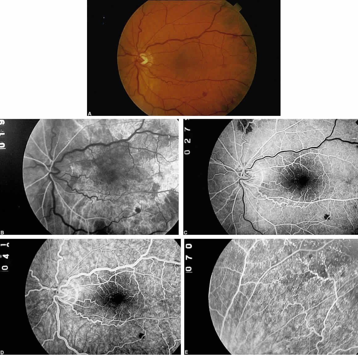
| Fig. 3. A. Fundus photograph of an eye with ocular ischemic syndrome showing features of venous dilation, midperipheral intraretinal hemorrhages, and attenuated arterioles. B. Fluorescein angiogram at 19 seconds (arterial phase). There is a prolonged arm-to-retina circulation time and a patchy choroidal filling pattern. C. Early laminar-venous phase at 27 seconds. Areas of incomplete choroidal filling persist. D. At 41 seconds, laminar-venous phase is almost complete, and the arteriovenous transit time is approximately 22 seconds. E. Fluorescein angiogram of the midperipheral fundus, revealing numerous microaneurysms that are not obvious on ophthalmoscopic examination. |