|
|
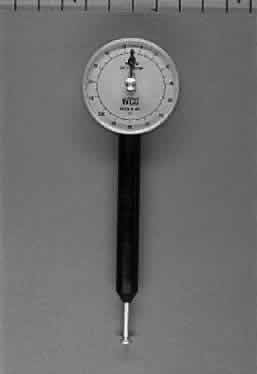
| Fig. 9. Photograph of a hand-held analog ophthalmodynamometer. After topical corneal anesthesia, the sterilized footplate (bottom) is gently applied to the sclera. The central retinal artery is observed at the slit lamp with the use of a Hruby lens. Increasing force is gradually applied until pulsations of the central retinal artery are observed. The measurement on the dial at this point is taken to be the relative diastolic pressure of the central retinal artery. The pressure at which the retinal artery is collapsed is taken to be the relative systolic pressure. |