|
|
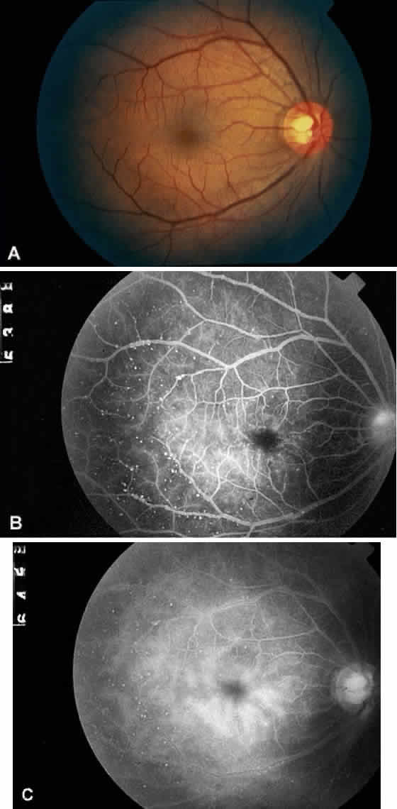
| Fig. 6. A. Fundus photograph of the right eye in the same patient as in Figure 5 diagnosed with Takayasu's arteritis, displaying features of arterial sclerosis, venous dilation, dot hemorrhages, and microaneurysms. B. Fluorescein angiogram of the same eye, revealing prominent venous dilation and beading, with microaneurysms concentrated along the venous arcades. C. Late phase of the angiogram revealing staining of the arterioles, increasing prominence of the microaneurysms, and diffuse staining of the perifoveal microaneurysms. (Courtesy Travis A. Meredith, MD, St. Louis MO) |