|
|
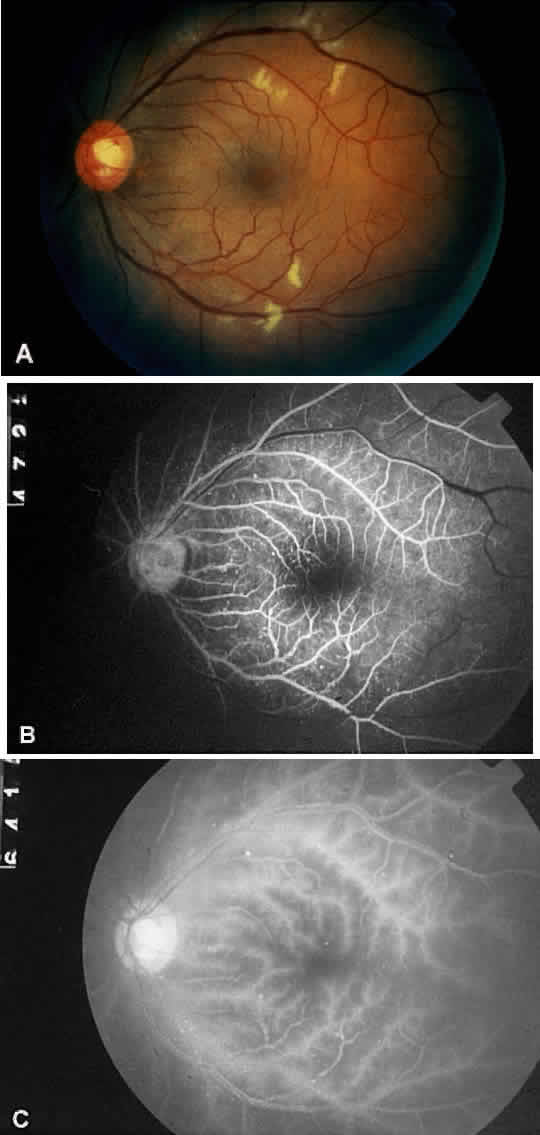
| Fig. 5. A. Fundus photograph of the left eye of a patient diagnosed with Takayasu's arteritis, showing attenuated arterioles and venule dilation. There are scattered dot hemorrhages and microaneurysms along the vascular arcades and extending outward to the equator. Large cotton-wool spots along the arcades are apparent. B. Fluorescein angiogram, revealing prominent venous dilation and beading, with microaneurysms along the vascular arcades and throughout the posterior pole. C. Late arterial phase, showing marked arterial staining. Choroidal filling time is also delayed to more than 17 seconds. The arteriovenous transit time in this study is delayed to more than 36 seconds. (Courtesy Travis A. Meredith, MD, St. Louis, MO) |