|
|
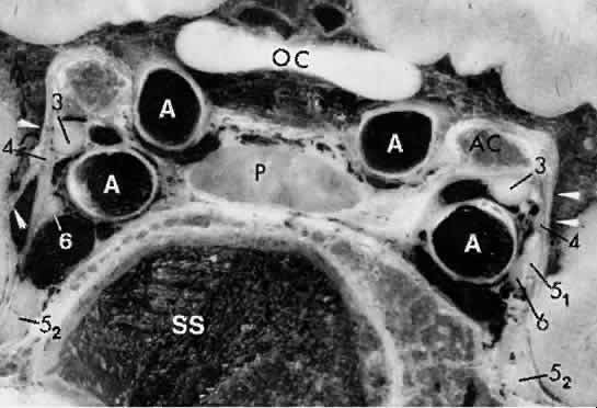
| Fig. 21. Microtomic preparation, coronal section through cavernous sinuses. SS, sphenoid sinus; P, pituitary gland; AC, anterior clinoids; OC, optic chiasm; 3, 4, 6, 5 1 (ophthalmic), and 5 2 (maxillary, cranial nerves). Siphons of intracavernous carotid arteries (A) cut in cross-section. White arrowheads indicate dura of lateral wall of cavernous sinuses. (Courtesy of Dr. David Daniels, Medical College of Wisconsin, Milwaukee) |