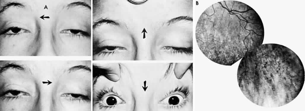
Fig. 32.
Progressive external ophthalmoplegia (
A
). Almost complete absence of eye movements in all fields of gaze.
B.
Peripheral fundus of patient shows mottled degeneration of retinal pigment epithelium with minimal pigment clumping.