|
|
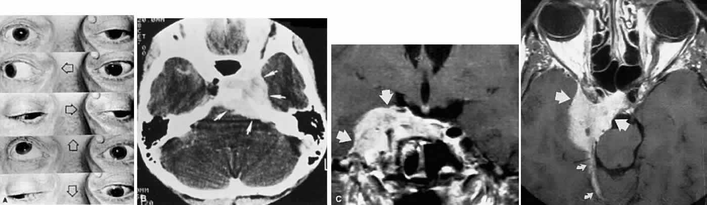
| Fig. 23. A. A 68-year-old man with progressive left ptosis and diplopia. Left pupil slightly larger than right, with sluggish light reaction. Note pseudo-vonGraefe lid retraction in downgaze. B. CT shows enhancing soft tissue mass involving left cavernous sinus, petrous ridge, dorsum, and sella, compatible with meningioma. C. Enhanced T-1 weighted MRI. Axial (top) and coronal (bottom) sections show medial sphenoidal (“cavernous”) meningioma (large arrows). Note tentorial extension (small arrows). |