|
|
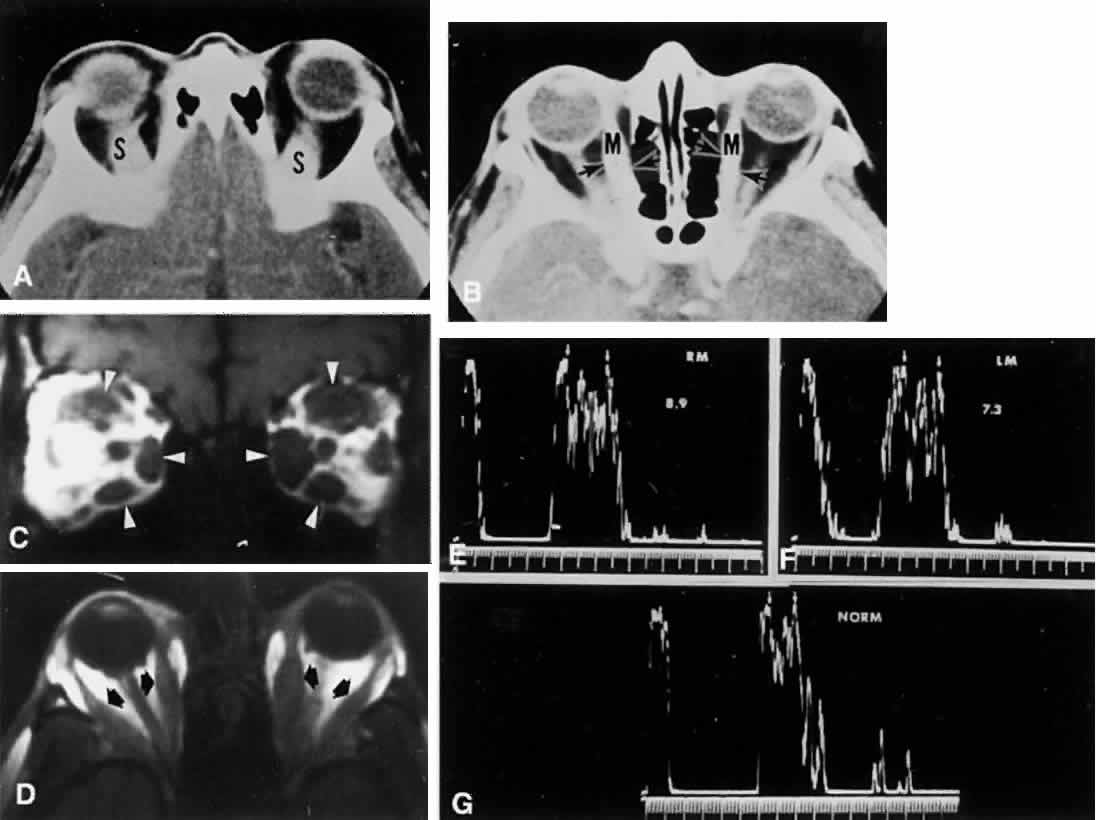
| Fig. 30. Graves ophthalmopathy. A. CT axial section through superior aspect of orbits shows symmetrically enlarged superior rectus muscles (S). B. Midorbital plane shows enlarged medial rectus muscles (M, and arrows). C. Coronal MRI shows hypertrophied ocular muscles (arrowheads) in both orbits. D. Axial section of MRI reveals enlarged medial and horizontal recti (arrows). E. Orbital ultrasonography (A-scan) provides sensitive measurement of muscle belly diameters. Enlarged right medial rectus shows interspike interval (small arrows) corresponding to 8.9 mm. F. Left medial rectus, 7.3 mm. G. Normal muscle diameter, 4.5 mm. |